


6Part I – High-Fluid Content Liver Lesions
3 Cyst I – Typical Small
Hepatic cysts are common lesions (they may occur in up to 20 % of the general population). These lesions are usually divided into unilocular (95 %) and multilocular varieties. Although the pathogenesis of these cysts is not clear, most hepatic cysts are considered to be developmental in origin. Currently, most hepatic cysts are discovered as incidental findings on cross-sectional imaging. Smaller cysts (< 10 mm) at US and CT may be difficult to distinguish from solid lesions, and may cause diagnostic problems especially in patients with an underlying (colorectal) malignancy.
MR Imaging
At MR imaging, cysts are low in signal intensity on T1-weighted images, high in signal intensity on T2-weighted images, and retain signal intensity on longer echo time (e.g., > 120 ms) T2-weighted images. After injection of contrast, cysts do not show any enhancement. On delayed post-gadolinium images (up to 5 min), cysts remain unenhanced. The latter findings may be useful to ensure that lesions are cysts and not poorly vascularized metastases that show gradual enhancement. T2-weighted sequences with a long echo time (TE) in combination with delayed phase images are especially effective at showing small ( 5 mm) cysts. MRI is particularly valuable when lesions are small and the patient has a known primary malignancy (Figs. 3.1, 3.2). Typical and relatively larger cysts can be characterized at CT as well as US (Figs. 3.3A, B).
Differential Diagnosis
Typical simple cysts at MR imaging do not cause any diagnostic problems and can be distinguished from other cystic lesions with confidence. Atypical cysts or cysts with septa may show overlapping features with other entities such as: (1) mucinous or cystic metastases (look for multiple septa and perilesional enhancement); and (2) ciliated foregut cysts (congenital lesions, frequently located at the anterior margin of the liver with mucinous content and a slightly bulging liver contour). The presence of a cystic lesion with an enhancing wall and extension beyond the contour of the liver may also be observed in some forms of metastatic disease such as hepatic metastasis from ovarian malignancies. For this reason, a diagnosis of foregut cyst on imaging studies should only be made in the absence of peritoneal disease and a clinical history of malignancy.
Pathology
Pathologically, the lining of the cyst shows a single layer of cuboidal to columnar epithelial cells. Lining epithelium rests on an underlying fibrous stroma (Fig. 3.3C, D).
Management
No treatment or follow-up with imaging is required for typical small hepatic cysts.
Literature
1.Rossai J (1995) Ackerman’s surgical pathology, vol 1, 8th edn. Mosby, St. Louis, p 898
2.Mortele KJ, Ros PR (2001) Cystic focal liver lesions in the adult: differential CT and MR imaging features. Radiographics 21:895 – 910
3.Shoenut JP, Semelka RC, Levi C, Greenberg H (1994) Ciliated hepatic foregut cysts: US, CT, and contrast-enhanced MR imaging. Abdom Imaging 19:150 – 152

3 Cyst I – Typical Small 7
Fig. 3.1. Cyst, drawings. T2 fatsat: cyst is very bright (fluid-like) compared to the liver with smooth and sharp margins; T1 in-phase: cyst is hypointense to
Fig. 3.2. Cyst, typical MRI findings. A Axial fat-suppressed T2-w TSE image (T2 fatsat) shows a small sharply marginated bright cyst. B Axial in-phase image (T1 in-phase): The cyst has low signal intensity. C Axial gadoliniumenhanced 3D GRE image in the arterial phase (ART): The cyst shows no enhancement. D Axial delayed phase (DEL): The cyst remains unenhanced. E Axial T2-w SSTSE image with longer TE of 120 ms (SSTSE): The cyst (arrow) retains its high signal intensity due to high fluid content (typical sign of non-solid liver lesions). F Axial opposed-phase image (T1 opposed-
the liver; ART: cyst shows no enhancement; DEL: cyst remains unenhanced
phase): The cyst (arrow) has low signal intensity. G Coronal SSTSE image (SSTSE): The bright cyst is well recognizable (arrow). H Coronal delayed phase image (DEL): The cyst is clearly visible as a small non-enhancing lesion with excellent correlation and confirmation of the T2 information (arrow). The ability of MRI to combine the information on various T1, T2, gadolinium-enhanced sequences is unparalleled and allows a highly confident and reliable diagnosis for sound medical and surgical decision making
Fig. 3.3. Cyst, CT, US, and histology. A CT (another patient) shows a small unenhanced cyst. B Ultrasound (another patient) shows a typical cyst with sharp margins and increased sound transmission (arrow). C Photomicro-
graph shows a typical cyst that is surrounded by the liver tissue with inflammatory infiltrates. H&E, × 100. D A detailed view of the previous photomicrograph shows the epithelial lining more closely (arrow)
8Part I – High-Fluid Content Liver Lesions
4 Cyst II – Typical Large with MR-CT Correlation
Large hepatic cysts are less common than the smaller or multiple cystic lesions in the liver. Such lesions may be detected incidentally but patients are more likely to present with symptoms such as upper abdominal discomfort, pain, or even obstructive jaundice due to the biliary compression by the cyst. Intracystic hemorrhage may also occur and cause symptoms.
MR Imaging
Very large liver cysts have a similar MR imaging appearance to smaller cysts. Cysts are low in signal intensity on T1-weighted images, high in signal intensity on T2-weighted images, and retain signal intensity on longer echo time (e.g., > 120 ms) T2-weighted images. Cysts, as non-solid liver lesions, share this T2 characteristic with hemangiomas in the liver. Because cysts do not enhance with gadolinium on MR images, delayed post-gadolinium images (up to 5 min) may be useful to ensure that lesions are simple cysts without any solid nodules or thickened septa. In addition, MR imaging can reliably exclude hemorrhage on T1-weighted GRE with fat suppression as well as any biliary dilatation (Figs. 4.1, 4.2). CT is also well able to visualize these lesions (Fig. 4.3). CT may be less accurate for small amounts of hemorrhage or solid components.
Differential Diagnosis
Differential diagnosis of a large cystic liver lesion includes: (1) infectious cystic lesions, such as abscess (clinical history and enhancing wall at imaging), intrahepatic hydatid cyst (clinical history and daughter cysts in 75 % at MRI and CT and calcifications in 50 % at CT); and (2) neoplastic lesions, such as undifferentiated embryonal sarcoma, biliary cystadenoma or cystadenocarcinoma, and cystic metastases. Hallmarks of neoplastic lesions are solid components with intralesional or perilesional enhancement. Such findings should not be present in large simple developmental liver cysts.
Management
Several types of treatment have been advocated for large (symptomatic) hepatic cysts, including: (1) percutaneous drainage and sclerosis using alcohol, tetracycline, doxycycline, or a combination (this method is associated with recurrence of cysts and symptoms);
(2) drainage in combination with various types of surgery; and (3) drainage in combination with minocycline injection (a more recent type of treatment). Percutaneous treatments may be contraindicated in patients with bleeding tendency, echinococcal cysts, and cysts communicating with the biliary tree.
Literature
1.vanSonnenberg E, et al. (1994) Symptomatic hepatic cysts: percutaneous drainage and sclerosis. Radiology 190:387 – 392
2.Cellier C, Cuenod CA, Deslandes P, et al. (1998) Symptomatic hepatic cysts: treatment with single-shot injection of minocycline hydrochloride. Radiology 206:205 – 209
3.Regev A, Reddy KR, Bercho M, et al. (2001) Large cystic lesions of the liver in adults: a 15-year experience in a tertiary center. J Am Coll Surg 193:36 – 45
4 Cyst II – Typical Large with MR-CT Correlation 9
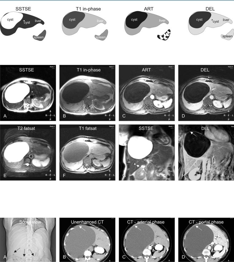
Fig. 4.1. Cyst, drawings. T2 long TE: a large cyst with smaller cysts is much brighter with sharp demarcation to the liver; T1 in-phase: the large cyst is hypointense to the liver; the smaller cyst is less well visible; ART: the large
Fig. 4.2. Cyst, large and small. A Axial T2-w SSTSE image (SSTSE) shows a very large and a smaller cyst with sharp margins and high signal to the liver. B Axial in-phase image (T1 in-phase): Cysts are hypointense to the liver. C Axial arterial phase GRE image (ART): Cysts show no enhancement. D Axial delayed phase image (DEL): Cysts remain unenhanced. E Axial fat-sup- pressed TSE image (T2 fatsat): Bright signal of the larger cyst causes ghost-
cyst shows no enhancement in the arterial phase after contrast; DEL: the large cyst shows no enhancement in the delayed phase after contrast; the smaller cyst is also visible
ing on this respiratory-triggered image. F Axial fat suppressed GRE image (T1 fatsat): Cysts show signal that is comparable to fluid (simple cysts). Note the perilesional liver tissue with a rim of higher signal due to compression. G Coronal SSTSE image (SSTSE): The larger cyst is in a subphrenic location with the smaller cyst adjacent to it (arrow). H Coronal delayed phase postGd 2D T1-w GRE image (DEL): The larger cyst shows a thin wall (arrow)
Fig. 4.3. Cyst, large, CT findings (another patient). A Scout view shows the space-occupying effect of the large hepatic cyst with displacement of the bowel loops (arrows). B Unenhanced CT of a large cyst in the right liver with
thin wall. C CT in the arterial phase shows no enhancement of the cyst with the thin wall (arrow). D CT in the portal phase shows no enhancement of any other structures within the cyst with the thin wall (arrow)