


44 Part IIA – Metastases: Colorectal
21Liver Metastases V – Large, Mucinous, Mimicking a Primary Liver Lesion
Colorectal metastases are typically small or medium-sized (multiple) liver lesions with target-like configuration and irregular ringshaped enhancement. Some (colo)rectal liver metastases may have unusual size and aspect, and may cause difficulty in proper diagnosis. For instance, some patients may present with a large, single lesion with compressed surrounding liver parenchyma and mimicking primary liver lesions such as cholangiocarcinomas or hepatocellular carcinomas.
Literature
1.Semelka RC, Cance WG, Marcos HB, et al. (1999) Liver metastases: comparison of current MR techniques and spiral CT during arterial portography for detection in 20 surgically staged cases. Radiology 213:86 – 91
2.Outwater E, Tomaszewski JE, Daly JM, et al. (1991) Hepatic colorectal metastases: Correlation of MR imaging and pathologic appearance. Radiology 180:327 – 332
3.Yoon SS, Tanabe TK (1999) Surgical treatment and other regional treatments for colorectal cancer liver metastases. Oncologist 4:197 – 208
MR Imaging Findings
At MR imaging, large mucinous colorectal liver metastases may not have a target-like appearance, which is a characteristic feature of most non-mucinous colorectal liver metastases. Such lesions usually will have predominantly heterogeneous increased signal intensity on T2-weighted images with sharp margins to the surrounding liver; on the T1-weighted images the lesions will mainly have low signal intensity to the liver. Parts of the lesion may have high signal intensity on T1-weighted images due to the presence of mucin. After injection of gadolinium, the lesion shows heterogeneous enhancement in the arterial phase and some washout with enhancement of a pseudocapsule (Figs. 21.1 – 21.3).
Differential Diagnosis
Some of the features, including (1) a single lesion, (2) large size, (3) heterogeneous enhancement, and (4) some washout, may suggest a primary malignant liver lesion such as cholangiocarcinoma or hepatocellular carcinoma in a non-cirrhotic liver. Clinical information of the underlying (colo)rectal malignancy and possibly mucinous histology may direct the reader in the right direction. If the patient has no underlying colorectal tumor, tumor markers including carcinoembryonic antigen (CEA) and alpha-fetoprotein (AFP) should be determined. If these tumor markers are also negative, a somatostatin-receptor scan should be performed to exclude a neuroendocrine tumor metastasis.
Management
Management will depend on the exact nature of the lesion. In the case of a solitary colorectal metastasis, curative resection should be attempted.
21 Liver Metastases V – Large, Mucinous, Mimicking a Primary Liver Lesion 45
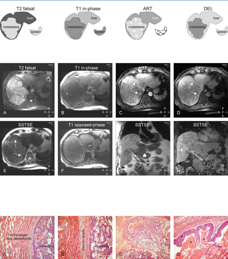
Fig. 21.1. Metastasis, colorectal, mucinous, drawings. T2 fatsat: metastasis is predominantly hyperintense to the liver with a large size and ragged edges; T1 in-phase: metastasis is predominantly hypointense to the liver; ART: me-
Fig. 21.2. Metastasis, colorectal, mucinous, large, MRI findings. A Axial TSE image (T2 fatsat): Metastasis is predominantly hyperintense to the liver with a large size and ragged edges. B Axial in-phase image (T1 in-phase): Metastasis is predominantly hypointense with some brighter areas (*). C Axial arterial phase image (ART): Metastasis shows intense heterogeneous enhancement, with some edge enhancement (arrows). D Axial delayed phase image (DEL): Metastasis shows washout with enhancement of a discontinuous
tastasis shows predominantly heterogeneous intense enhancement; DEL: metastasis shows washout with enhanced discontinuous pseudocapsule
pseudocapsule (arrows). E Axial SSTSE image (SSTSE): Metastasis shows a partial pseudocapsule with high signal (arrows), which is most likely caused by vessels and compressed liver parenchyma. F Axial opposed-phase image (T1 opposed-phase) shows no fatty infiltration. G Coronal SSTSE image (SSTSE) shows the metastasis bulging into the right hemidiaphragm. H Coronal delayed phase image (DEL) shows heterogeneously enhanced metastasis with encased intrahepatic bile ducts (arrow)
Fig. 21.3. Metastasis, colorectal, large lesion with pseudocapsule and direct MR pathology correlation. A, B Photomicrographs show the metastasis surrounded by compressed liver parenchyma with atrophic hepatocytes, mimicking
a thin fibrous capsule. H&E, × 100. C Photomicrograph of a large gland surrounded by compressed liver parenchyma. H&E, × 100. D Photomicrograph shows the gland that is in part filled with mucin (*). H&E, × 200

46 Part IIA – Metastases: Colorectal
22Colorectal Metastases VI – with Portal Vein and Bile Duct Encasement
Unresectable colorectal liver metastases carry a dismal prognosis. In fact, only 10 – 25 % of colorectal liver metastases are candidates for surgical resection, and the 5-year survival rate following resection of isolated colorectal liver metastases can be as high as 38 %. Without any treatment, the survival rate is less than 1 %. For the remaining 75 – 90 % of patients with liver metastases who are not amenable to surgery, several new minimally invasive therapies (MIT) have been developed. These include radiofrequency ablation (RFA), percutaneous ethanol injection, percutaneous acetic acid injection, transcatheter arterial chemoembolization, and stereotactic radiotherapy (SRT). There is a large body of data indicating that RFA can reliably treat tumors less than 3 cm in diameter. However, the modality by itself is less effective when applied to larger tumors and tumors in close proximity of vessels. SRT is a novel technique that may be used in patients who cannot be treated with interventional techniques or in conjunction with the other MIT.
Literature
1.Weeks SM, Burke C (2005) Local therapeutic treatments for focal liver disease. Radiol Clin N A 43:899 – 914
2.Mendez Romero A, Wunderink W, Hussain SM, et al. Stereotactic body radiation therapy for primary and metastatic liver tumors: A single institution phase I-II study. Acta Oncol. 2006;45:831 – 837
3.Yoon SS, Tanabe TK (1999) Surgical treatment and other regional treatments for colorectal cancer liver metastases. Oncologist 4:197 – 208
MR Imaging Findings
At MR imaging, unresectable liver metastases may present with a number of findings, which include (1) coincidental extrahepatic metastases; (2) all or most segments of the liver affected; (3) vascular invasion or vascular encasement (with or without biliary encasement); and (4) certain anatomic locations rendering the resection technically impossible. Particularly, T2-weighted diffusionweighted, black-blood echoplanar MR imaging is useful for detection of vascular as well as biliary encasement in combination with a high liver-to-lesion contrast (Figs. 22.1, 22.2). MR imaging is also an excellent modality for follow-up after minimally invasive therapies. The T2-weighted sequences in combination with the dynamic gadolinium-enhanced images allow accurate distinction from nonmalignant parenchymal changes within the liver (Figs. 22.2, 22.3).
Management of Unresectable Liver Lesions
Patients with unresectable liver lesions, usually metastases, may be amenable to systemic chemotherapy (goals: palliation or downstaging); isolated liver perfusion with chemotherapeutic agents (goals: local control or curative-in-attempt); RFA (goals: palliation; downstaging for possible resection; local control while on waiting list for liver transplantation); and stereotactic radiation therapy (palliation or downstaging usually if other MIT options are not applicable).
22 Colorectal Metastases VI – with Portal Vein and Bile Duct Encasement 47
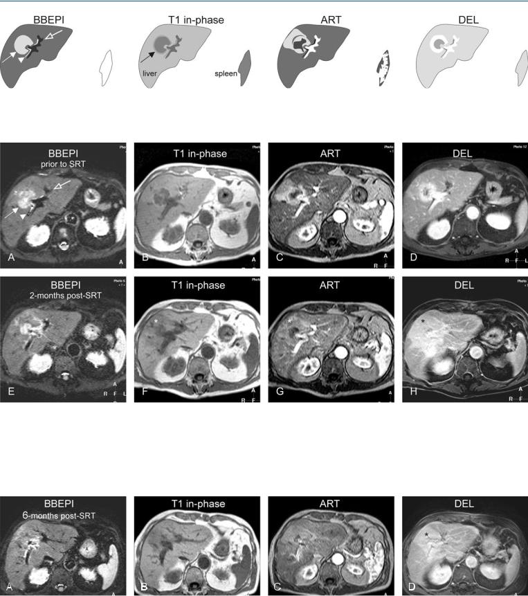
Fig. 22.1. Metastasis, inoperable, drawings. BBEPI: metastasis (solid arrow) shows encasement of the bile duct (arrowhead) and the portal vein (open arrow); T1 in-phase: metastasis is surrounded by a bright rim of compressed
Fig. 22.2. Metastasis, inoperable and treated with stereotactic radiation therapy (SRT), MRI findings. A Axial black-blood EPI (BBEPI): Metastasis (solid arrow) shows encasement of the (bright) bile ducts (arrowhead) and portal vein with signal void (open arrow). B Axial in-phase image (T1 in-phase): Metastasis is surrounded by a rim of compressed liver parenchyma. C Axial arterial phase GRE image (ART): Metastasis shows ring-shaped and perilesional enhancement. D Axial delayed phase image (DEL): Metastasis shows
liver parenchyma (arrow); ART: metastasis shows ring-shaped and perilesional enhancement; DEL: metastasis shows persistent ring-shaped enhancement
persistent ring enhancement. E Axial BBEPI (BBEPI) 2 months after SRT: Metastasis has decreased in size. F Axial in-phase image (T1 in-phase): Metastasis is surrounded by a wedge-shaped area of edema (*). G Axial arterial phase image (ART): Metastasis shows faint ring-shaped and a large wedgeshaped (*) enhancement. H Axial delayed phase image (DEL): Metastasis shows persistent wedge-shaped enhancement (*)
Fig. 22.3. Metastasis, 6 months after SRT, MRI findings. A BBEPI: Metastasis has almost disappeared. B Axial in-phase image (T1 in-phase): Metastasis cannot be distinguished from edema. C Axial arterial phase image (ART): Me-
tastasis shows negligible enhancement. D Axial delayed phase image (DEL): No metastasis is visible within the enhanced wedge-shaped area (*)
48 Part IIA – Metastases: Colorectal
23Colorectal Metastases VII – Recurrent Disease Versus RFA Defect
Increasing numbers of patients with (colorectal) liver metastases undergo surgical resection or minimally invasive therapy and need follow-up with imaging after treatment. There are no strict guidelines for follow-up of such patients. Currently, many centers use laboratory tests (liver function and serum carcinoembryonic antigen, CEA), with or without abdominal ultrasound, for follow-up of patients. Other centers use computed tomography as their main tool for follow-up. In our experience, MR imaging is highly reliable and a versatile imaging technique for detecting and characterizing residual or recurrent liver lesions. In addition, MR imaging allows the differentiation of relevant focal abnormalities from benign tissue changes within the liver.
Literature
1.Braga L, Semelka RC (2005) Magnetic resonance imaging features of focal liver lesions after intervention. Top Magn Reson Imaging 16:99 – 106
2.Outwater E, Tomaszewski JE, Daly JM, et al. (1991) Hepatic colorectal metastases: Correlation of MR imaging and pathologic appearance. Radiology 180:327 – 332
3.Abdalla EK, Vauthey JN, Ellis V, et al. (2004) Recurrence and outcomes following hepatic resection, radiofrequency ablation, and combined resection/ablation for colorectal liver metastases. Ann Surg 239:818 – 825
MR Imaging Findings
At MR imaging, recurrent or residual (colorectal) liver metastases have a distinct appearance. On (fat-suppressed) T2-weighted sequences, these lesions are predominantly higher in signal intensity than the surrounding liver, and often have a target-like appearance which is typical for most colorectal metastases. The lesions show continuous ring-shaped enhancement in the arterial phase, with or without concomitant perilesional or wedge-shaped subsegmental parenchymal enhancement. The latter finding may indicate bile duct and/or portal compression or invasion. In addition, perilesional enhancement with colon cancer may be caused by variable degrees of hepatic parenchymal compression, desmoplastic reaction, and inflammatory infiltrates. Based on the T2 aspect and the enhancement pattern, MR imaging allows reliable differentiation of recurrent or residual disease from benign conditions, for instance after radiofrequency ablation (RFA). In this respect, it should be kept in mind that tissue-specific contrast media may provide confusing results because metastases and non-cancerous tissues behave similarly on post-SPIO images (Figs. 23.1 – 23.3).
Differential Diagnosis
Differential diagnosis may include scar tissue, coagulative necrosis, benign liver lesions such as focal fatty infiltration or focal non-stea- tosis. State-of-the-art MR imaging combines the findings on the T2-weighted images, chemical shift imaging, and routine arterial and delayed phases after contrast injection. Therefore, MR imaging is more reliable than other imaging modalities.
23 Colorectal Metastases VII – Recurrent Disease Versus RFA Defect 49
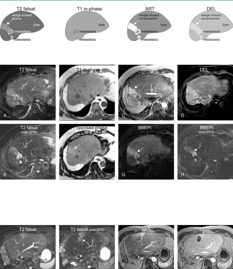
Fig. 23.1. Metastasis, colorectal, recurrent after hemihepatectomy, drawings.
T2 fatsat: two metastases and wedge-shaped edema are visible; T1 in-phase: larger metastasis is better visible than the smaller one; ART: metastases
Fig. 23.2. Metastasis, colorectal, recurrent after hemihepatectomy, MRI findings. A Axial TSE image (T2 fatsat): Two metastases and wedge-shaped edema are visible. B Axial in-phase image (T1 in-phase): Metastasis is hypointense to the liver. C Axial arterial phase image (ART): Metastases show irregular ring-shaped enhancement with wedge-shaped enhancement (solid arrow). Note the fold-over (SENSE) artifact (open arrow). D Axial delayed phase image (DEL): Metastases become less enhanced with decreased conspicuity. E Axial TSE image after SPIO uptake (T2 fatsat post-SPIO): Metastases show
show irregular ring-shaped enhancement with wedge-shaped enhancement; DEL: metastases become less enhanced
improved conspicuity due to the decreased liver signal; note that the wedgeshaped area also shows SPIO uptake and confirms that it is liver tissue. F Axial opposed-phase image (T1 opposed-phase) shows mild diffuse fatty infiltration with fatty sparing (arrow). G Axial BBEPI before and H after uptake of SPIO shows small dilated bile ducts within the wedge-shaped area suggesting segmental bile duct obstruction with decreased portal circulation, fatty sparing, and edema
Fig. 23.3. Metastasis, colorectal, radiofrequency ablation (RFA) defect (same patient as above), MRI findings. A Axial TSE image before and B after uptake of SPIO shows the oval-shaped RFA defect (arrow). C Axial arterial phase im-
age: Wedge-shaped enhancement is present around the RFA defect. D Axial delayed phase image: RFA defect is well demarcated with a smooth regular rim enhancement due to hyperemia, indicating successful local treatment

Part II/IIB
Solid Liver Lesions |
II |
Metastases: Non-Colorectal |
IIB |