

Schluesseltech_39 (1)
.pdf1.4 |
Th. Brückel |
1 Å for studies on such a microscopic scale. They also have a large penetration power as everybody knows from the medical x-ray images.
Classical physics describes electromagnetic radiation as propagation of electromagnetic waves. For a scattering experiment, we select waves of a certain wavelength and propagation direction, so-called plane waves, since all points on a plane in space have the same phase. If such a wave impinges on two point-like scattering centers (in a solid these could be atoms), spherical waves are being emitted from these scattering centers. This is nothing but Huygens principle for wave propagation. The emitted waves can superimpose and lead to either enhancement or cancellation of the signal in certain directions as depicted in figure 1.2.
Fig. 2: Moiré pattern for concentric circles with equal distances representing a planar cut through spherical waves emitted from two scattering centers. The circles represent surfaces of constant phase relationship. Linear superposition of the waves gives enhancement or cancellation of the wave amplitudes along certain directions. This interference effect is mimicked by the depicted Moiré pattern. If the distance between the scattering centers is increased, the distance in the interference maxima decreases and vice versa: distances in the image created by scattering are inverse proportional – or reciprocal - to distances in the original objects which motivates the introduction of a reciprocal space to describe scattering events compared to the real space of the object under investigation.
As becomes clear from figure 1.2, scattering can be described as an interference phenomenon of the radiation waves. However, since de Broglie and Einstein, we know that quantum objects have a dual nature: the particle-wave-dualism. In the case of electromagnetic waves, the quanta carrying certain energy are called photons and in the detector, which registers the scattering pattern, we count single x-ray photons. This is characteristic for the quantum mechanical description: during propagation of radiation a wave picture is appropriate, while for the interaction with matter a particle is the description of choice. Wave and particle picture are connected by the fact that the magnitude square of the wave at a certain position in space
Introduction |
1.5 |
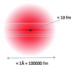
gives the probability density of finding the quantum particle at the corresponding position. Within this particle-wave-dualism it therefore becomes natural to use elementary particles as probes for scattering investigations of condensed matter systems. This was realised for the first time by Rutherford in 1909 in his famous experiment, where he directed a beam of - particles onto a gold foil and registered the transmitted and scattered particles. He found that many particles were backscattered and from the ratio between transmitted and backscattered-particles he could conclude on the model of an atom, which is now generally accepted, namely consisting of a positively charged nucleus of size about 10 femtometer = 10 fm = 10 x 10-15 m surrounded by a cloud of negatively charged electrons with an extension of about 1 Å = 10-10 m = 100000 fm, see figure 1.3.
Fig. 3: Schematic model of an atom with the atomic nucleus consisting of neutrons and protons having a size of about 10 fm surrounded by electrons in a cloud of a size of about 1 Å.
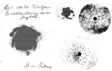
The real breakthrough for structure studies of condensed matter systems came with the idea of Max von Laue to use x-rays as scattering probes. Wilhelm Conrad Röntgen discovered x-rays in 1895 and soon it was concluded that x-rays were electromagnetic waves. Arnold Sommerfeld suggested that the wavelength of x-rays was about 1 Å. At the time of Max von Laue, after the experiments of Rutherford, it was accepted that matter consisted of atoms but their periodic arrangement in crystals was maybe suggested by the regular facets of the crystals but could not be really proven by experiment. Max von Laue was a theoretician, who derived the famous Laue equation describing scattering from a regular three-dimensional periodic arrangement of scattering centers. He convinced the two experimentalists Friedrich and Knipping to perform an x-ray diffraction experiment. The result is shown in figure 1.4.
1.6 |
Th. Brückel |
Fig. 4: Early x-ray diffraction diagrams recorded by a film from copper sulphite single crystals [1].
While the first transmission Laue photograph showed more or less just a fat plop, the quality of these images was soon refined and clear so-called Laue-spots could be identified. The impact of this discovery cannot be over-emphasized: it was the definite proof that solids consist of atoms, which are arranged in a regular three-dimensional periodic array and that x- rays were scattered as electromagnetic waves from such an arrangement of atoms. It is therefore natural that Max von Laue received the Nobel prize in 1914 for this breakthrough discovery. However, the experimentalists Friedrich and Knipping were left empty-handed.
Nearly everything we know today about the atomic structure of matter is based on this discovery which took place 100 years ago. Of course the techniques were significantly refined and nowadays x-ray diffraction is heavily being used to resolve complex structures of biological macromolecules in the field of protein crystallography. Such investigations need very intense and bright x-ray beams, which are provided from large accelerators, so-called synchrotron radiation sources. Many thousands of reflections are being recorded in a few seconds. As electromagnetic waves, x-rays are mainly scattered from the electronic charge distribution around the atoms and thus x-ray diffraction allows one to determine the electron density in solids.
1.3 Impact of Scattering in other Fields of Science
It should be pointed out that scattering is a much more general method in science, which is not only used by condensed matter scientists. The world’s largest accelerator is located close
to Geneva at the border between Switzerland and France in the CERN research center. CERN stands for Centre Européenne pour la Recherche Nucléaire, i. e. the European organisation
Introduction |
1.7 |
for nuclear research. Many accelerators are located on the CERN site of which the LHC, the Large Hadron Collider, is the world’s largest and highest energy particles accelerator. The LHC lies in a tunnel 27 km in circumference as deep as 175 m beneath ground level. This huge accelerator serves nothing but a scattering experiment, where opposing particle beams e. g. protons at energy of 7 TeV collide in certain interaction points, which are surrounded by huge detectors built by large international collaborations. In inelastic scattering events, new particles can be created and the hope is that this huge investment helps us to address some of the most fundamental questions of physics advancing the understanding of the deepest laws of nature. At Research Centre Jülich we have a smaller version of such a particle accelerator, the so-called COSY synchrotron for Hadron physics. These large accelerators are needed to achieve high particles energies corresponding to short wavelengths, which allow one to study fine structures within nucleons. Large detectors are needed because at these scales no imaging is possible but if all scattered particles are being traced a reconstruction of the scattering event in the computer can take place. While at the LHC new particles are being created during deep inelastic scattering events, the connection to x-ray diffraction is more evident for the former HERA accelerator, which had been in operation at DESY in Hamburg until a few years ago. There, electrons were being scattered from protons in head-on collisions and the inner structure of the proton consisting of quarks and gluons could be resolved.
1.4 Why Neutrons?
Coming back to condensed matter science: if x-rays are so successful for structure determination, why do we need neutrons? Neutrons have some very specific properties which make them extremely useful for condensed matter studies:
1.Neutrons are neutral particles. They are thus highly penetrating, can be used as nondestructive probes and to study samples in severe environment such as cryomagnets or furnaces.
2.The wavelengths of neutrons are similar to atomic spacings - just as is the case for x- rays. Therefore they can provide structural information from the picometer to the 100 μm range.
3.The energies of thermal neutrons are similar to the energies of elementary excitations in solids. Therefore neutrons can determine molecular vibrations, lattice excitations and the dynamics of atomic motion.
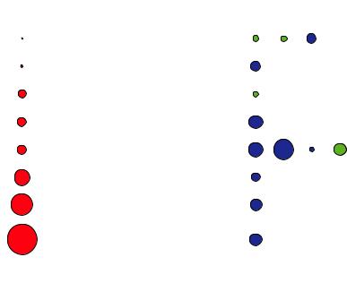
4.Neutrons interact with the nuclei in contrast to x-rays or electrons which interact with the electron cloud, see Figure 1.5. They are very sensitive to light atoms like hydrogen, which is difficult to detect by x-rays since hydrogen in bonds has often less than one surrounding electron. They can also distinguish between neighbouring elements in the periodic table like manganese, iron and chromium, for which x-rays are insensitive since these elements have nearly the same number of electrons. Also one can exploit isotopic substitution. A famous example is contrast variation in soft matter or biological macromolecules by replacing deuterium for hydrogen in certain molecules or functional groups. Similar to tinting in light microscopy, the location and
1.8 |
Th. Brückel |
5.movement of these functional groups can then be observed on the background of the other molecules.
6.Neutrons have a magnetic moment. This dipolar moment is due to the nuclear spin. Therefore neutrons can be used to study microscopic magnetic structures but also the magnetic excitations in solids, which have similar energies than the neutrons.
x10-1 |
[barn] |
|
[barn] |
|
-28 2 |
coh |
|
coh |
1 barn = 10 m |
||
|
0.66 |
1H |
1.76 |
2 |
|
|
|
6C |
1 |
|
|
|
24 |
5.55 |
|
|
|
|
416 |
25Mn |
1.75 |
|
|
|
450 |
26Fe |
11.22 |
|
|
|
522 |
28Ni |
13.30 |
|
|
|
1408 |
46Pd |
58 |
60 |
62 |
|
4.39 |
|
|
||
|
|
|
|
||
|
2986 |
67Ho |
8.06 |
|
|
|
5631 |
92U |
8.90 |
|
|
x-ray |
|
element |
neutrons |
|
|
|
|
Z |
|
|
|
Fig. 5: Comparison of x-ray and neutron scattering from single atoms for a few elements of the periodic table. The filled circles represent a measure of the total cross section, i,e, of the probability for scattering. For x-rays, which are scattered from the electron cloud, this probability goes up with the number square of electrons. Therefore Hydrogen is hardly visible for x-rays in the presence of heavier atoms. The situation is quite different for neutrons, which are scattered from the atomic nucleus. Here the scattering varies not monotonically throughout the periodic table and is different for different isotopes of the same atom. Blue and green circles distinguish scattering with and without 180° phase shift, respectively.
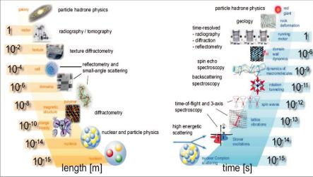
Figure 1.6 shows the extreme range of applicability of neutrons for condensed matter studies based on these special properties. Different scattering techniques have to be used for different applications, as indicated in the figure.
Introduction |
1.9 |
Fig. 6: Huge range of length (left side) and time (right side) scales covered by research with neutrons. Also indicated is the corresponding neutron technique.
Due to the huge impact of neutron scattering for condensed matter studies, it is no surprise that the Nobel prize in physics was awarded to two of the pioneers of neutron diffraction and inelastic neutron scattering, which Clifford G. Shull and Bertram Brockhouse received in 1994. The famous quote “neutrons tell us where atoms are and how they move” is due to Clifford Shull.
If you got the impression so far that neutrons are the ideal and most universal probe for condensed matter studies on an atomic scale, you are right in principle. However, as with everything in life, there are also some drawbacks. While neutrons are everywhere - without neutrons we would not exist - they are extremely difficult to produce as free particles not bound in nuclei. Free neutrons are produced by nuclear physics reactions, which require rather large and high-tech installations. Two main routes to produce free neutrons are being followed today:
(1)Fission of the uranium 235 nuclei in a chain reaction; this process happens in research reactors.
(2)Bombarding heavy nuclei with high energetic protons; the nuclei are “heated up” when a proton is absorbed and typically 20 - 30 neutrons are being evaporated. This process is called spallation and requires a spallation source with a proton accelerator and a heavy metal target station.
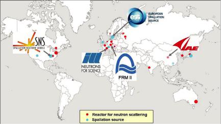
Since installations to produce free neutrons are rather expensive to build and to operate, there exist only a few sources worldwide. JCNS is present in some of the world best sources as shown in figure 1.7.
1.10 |
Th. Brückel |
Fig. 7: Major neutron research centres worldwide which have sources of appreciable flux and a broad instrumentation suite for condensed matter research. JCNS is present at four of the leading sources worldwide: the neutron research reactor FRM II in Garching, Germany, the Institute Laue-Langevin ILL in Grenoble, France, the Spallation Neutron Source SNS in Oak Ridge, USA and the Chinese Advanced Research Reactor CARR close to Beijing, China. JCNS also has a leading involvement in the European Spallation Source project, Lund, Sweden.
The fact that there are only a few sources worldwide implies that neutron scattering experiments have to be organised quite different from normal lab-based experiments. Users have to be trained in special schools (our JCNS school is one of them) and access to the experiments has to be organised (see below).
Not only the neutron research centres are rare but also free neutrons by themselves are rare. In a high flux reactor the neutron flux i. e. the number of neutrons passing through a given area in a given time is in the order of 1015 neutrons/cm2·s. If one compares this value with particle fluxes in gases, the neutron density in high flux sources corresponds to high vacuum conditions of about 10-6 mbar pressure. The neutrons have to be transported from the source to the experimental areas, which can either be done by simple flight tubes or so called neutron guides. These are evacuated tubes with glass walls (often covered with metal layers to increase the performance), where neutrons are transported by total reflection from the side, top, and bottom walls in a similar manner like light in glass fibers. The neutron flux downstream at the scattering experiments is then even much lower than in the source itself and amounts to typically 106 - 108 neutrons/cm2·s. This means that long counting times have to be taken into account to achieve reasonable statistics in the neutron detector. Just for comparison: the flux of photons of a small Helium-Neon laser with a power of 1 mW (typical for a laser pointer) amounts to some 1015 photons/s in a beam area well below 1 mm2.
Introduction |
1.11 |
However, it is not only the low flux that limits neutron scattering experiments, but also the fact that neutron sources are not very bright, i. e. neutron beams are rather large in the order of a few cm2 and therefore require in general rather large samples. Typical sample sizes are again in the order of a few cm2 and have masses of a few grams. However, this does not mean that we cannot study nanosized objects with neutrons as you will see in the subsequent lectures. However, for neutron scattering techniques, we have to have many of such objects and we will obtain ensemble averages.
1.5 The Social Practice of Neutron Scattering
The fact that neutron sources are rare leads to a particular social practice for neutron scattering: there are only a few major sources in Europe and worldwide and the operation of each one of these sources costs several million Euro per year. Therefore efforts have to be made to use the existing sources as efficient as possible. This means (i) continuous and reliable operation of the source during a large fraction of the year; (ii) many highly performing instruments, which can run in parallel, located around every source; (iii) professional instrument operation with highly qualified staff and a stringent risk management to keep the downtime of instruments and auxiliary equipment as low as possible; (iv) and access for as many scientists as possible.
There is no commercial market for neutron scattering instruments. Therefore these instruments are being built by research centres, where usually one or a few staff scientists work closely with engineers and technicians to realise an instrument for a certain application of research with neutrons. These highly experienced scientists will then later-on operate the instruments located at a certain neutron source. The Jülich Centre for Neutron Science JCNS has such staff scientists located at the outstations at FRM II, ILL and SNS. However, neutron facilities are way too expensive to be operated just for a small number of scientists. Beamtime is offered to external users from universities, research organisations (such as Max-Planck or Fraunhofer in Germany) and industry. In order for these users to obtain access to a neutron scattering instrument, the user will obtain information from the internet on available instruments, contact the instrument scientist and discuss the planned experiments with the instrument scientist. Once a clear idea and strategy for an experiment has been worked out, the user will write a beamtime proposal where he describes in detail the scientific background, the goal of the planned experiment, the experimental strategy and the prior work. The facility issues a call for proposals in regular intervals, typically twice a year. The proposals received are distributed to members of an independent committee of international experts, which perform a peer review of the proposals and establish a ranking. Typically overload factors between 2 to 3 on the neutron instruments exist, i. e. 2 to 3 times the available beam time is being demanded by external users. Once the best experiments have been selected, the beamtime will be allocated through the facility, where the directors approves the ranking of the committee, the beamline scientist schedules the experiments on her or his instrument and the user office sends out the invitations to the external users. Many facilities will pay travel and lodging for 1 up to 2 users per experiment. It is now up to the user to prepare his experiment as well as possible. If the experiment fails because it was not well prepared, it will be very difficult to get more beamtime for the same scientific problem.
1.12 |
Th. Brückel |
Typical experiments last between 1 day and up to 2 weeks. In this time lots of data will be collected which users take home and usually spend several weeks or months to treat the data and model it.
A typical neutron scattering facility will run about 200 days a year with a few hundred visits of user from all over the world. This is also what makes research with neutrons so attractive to young scientists: early-on in their career they will learn to work in large international collaborations, get the opportunity to work on state-of-the-art high-tech equipment and learn to organise their research as efficient as possible. You have therefore chosen well to attend this laboratory course!
After this simple introduction, you can now look forward to many interesting lectures, where more details will be explained and where you will learn the basic principles to enable you to perform neutron experiments. Have lots of fun and success working with this special gift of nature, the free neutron!
References
[1]W. Friedrich, P. Knipping, M. von Laue(1912). "Interferenz-Erscheinungen bei Röntgenstrahlen". Sitzungsberichte der Mathematisch-Physikalischen Classe der Königlich-Bayerischen Akademie der Wissenschaften zu München 1912: 303.
Some useful links for research with neutrons:
[2]German Committee for Research with Neutrons: www.neutronenforschung.de
[3]European Neutron Portal: http://www.neutron-eu.net/
[4]Neutron scattering reference material: http://www.neutron.anl.gov/reference.html
[5]Neutron scattering web: http://www.neutron.anl.gov/
[6]Jülich Centre for Neutron Science: http://www.fz-juelich.de/jcns/
Introduction |
1.13 |
Exercises
A E1.1 Multiple Choice
Electromagnetic radiation with a wavelength of 500 nm corresponds to:
microwaves
visible light
ultraviolet
X-rays
The typical distance between atoms in a solid amounts to:
10 nm
1 nm
0.1 nm
0.01 nm
An atomic nucleus has a typical size of:
1 Å
0.1 nm
1 pm
10 fm
The typical wavelength of thermal neutrons is:
10 nm
1 nm
0.1 nm
0.01 nm
Which type of radiation would you use to distinguish iron and manganese atoms in a given compound?
X-rays
neutrons
electrons
light