

Книги по МРТ КТ на английском языке / MR Imaging in White Matter Diseases of the Brain and Spinal Cord - K Sartor Massimo Filippi Nicola De Stefano Vincent Dou
.pdf136 |
A. Kangarlu |
Fig. 10.3. MR image at 7 T of the spinal cord of a live EAE mouse with systemic injection of labeled T-lymphocytes. The spinal cord was imaged at 100 µm isotropic resolution with a 3D fast gradient echo sequence, TR/TE=22 ms/3.3 ms. The hypointense lesion (arrows) is shown in three planes and indicates the localized presence of iron labeled, activated T-lymphocytes. (Images courtesy of Stasia Anderson)
Fig. 10.4. Microscopic ex vivo 7-T MR images in an EAE mouse. EAE was induced by intravenous infusion of FE-PLL-labeled T cells and administration of pertussis. Clinical score 1.5. 3D coronal spin echo images were acquired with 30×30×46 µm3. The spinal cord images show hypointense white matter and nerve roots. The hypointense spots indicate site of cellular infiltrates. (Images courtesy of Stasia Anderson)
iron susceptibility artifact. In such images (Figs. 10.3 and 10.4) the lesion of about 100 µm in diameter appeared ~160 µm in the image. This is a moderate effect compared to images in which there is a large concentration of iron, due to the relatively low iron load in the cells, and this somewhat low level is advantageous because the end result is an effect that is visible but does not obscure the anatomy.
The effect of the contrast in these cells has potentially further-reaching utility in time than the T-cell detection. It has been shown (Anderson et al. 2004) that when the spinal cord is imaged early in the disease course at the onset of symptoms, direct images from active T-cell infiltration can be acquired (Fig. 10.3). These images are specific to T-cell migration. Such a technique allows the mea-

Molecular Imaging and High-Field MRI in Multiple Sclerosis |
137 |
surement of the time evolution of the later stages of the disease, in which T-cells can die and macrophages can pick up T-cells as well as the label. In such a situation the lesion would remain hypointense on MR with less specificity for a particular type of cell, but still specific to the presence of a lesion.
These targeted EAE imaging studies indicate that the labeling method improves the chances of visualizing the trafficking of these cells on MR, while not interfering with the functioning of the T-cell, B-cell or macrophages. A complex preparation process in magnetic labeling techniques is involved in which the cells are transfected with iron oxides and then stored in endosomes in the cytoplasm (Anderson et al. 2004). Generally, in choosing or designing how the cells will be transfected with a contrast agent one has to consider the efficiency coupled with potential effects on the cell based on where and how the agent will be stored in the cell. The small volume of cytosol in T-cells has to accommodate the iron tag. This makes their interaction with the surfaces rather frequent and significantly affects their function. For T-cells in particular it has been shown to be important to transfect with more iron. It is also important to limit effects on the surface activation, which is to be controlled by the activation mechanism of the cell. These requirements have been met by ferumoxide- poly-L-lysine (FE-PLL) complex (Anderson et al. 2004).
Presently, work is being done on improving the efficiency of the targeting molecules in binding to the target (Arbab et al. 2004). Activatable agents are being designed with two internal modes. The “on” mode is assigned to the condition of high contrast and the “off” mode represents low contrast enhancement. These agents will be capable of responding to a specific physiological or metabolic event that will cause them to switch from one mode to another. The switching mechanism of these agents is based on the mechanism of signal modulation by paramagnetic properties (Allen et al. 2004). The switching mechanism depends on the molecular structure of the agent. Mechanisms such as chemical exchange are at work for activation of Gd agents, while USPIOs use switching processes by dipolar coupling-induced anisotropy enhancement, which causes decreased T2. This technology has demonstrated great potential in enabling in vivo monitoring of lesion formation and activity. However, it has to overcome the barriers summarized in this article along the complex path to development of ideal labeling agents for cellular and molecular imaging of MS.
10.2 High-Field MRI
Challenges to MRI becoming a complete diagnostic tool are multidimensional. It is clear that routine MRI within a short time has scored remarkable achievements in the diagnosis and treatment of MS. The ability to visualize areas of demyelination in the brains of patients with MS by MRI has enabled the systematic examination of some aspects of the development of the “MS plaques”. In particular, MRI has enabled monitoring of lesion load, which is unique information attainable only by MRI (Grossman and McGowan 1998). Nevertheless, the lesion load has not shown a strong correlation with clinical manifestation of MS. Particularly, the fact remains that presently no imaging modality can detect the main event of the lesion formation in MS, i.e., T-lymphocyte migration through the BBB. Recently, using high resolution microscopic MRI, we have shown that plaques are centered on the microvasculature in the white matter of MS patients’ brains (Kangarlu et al. 2002). This offers unique information accessible in vivo which contains clues on the vascular injury and possibly axonal implication in the disease. An understanding of the details of these events will permit the definition of therapeutic strategies and evaluation of their efficacy by MRI. High-field (HF) MRI may possess such capability. It is, however, confronting a number of challenges before becoming a tool with the ability to offer accurate diagnosis consistent with clinical measures of the disease. These challenges include high magnetic susceptibility, RF inhomogeneity, dielectric resonances, RF penetration effects and RF coil design (Figs. 10.5 and 10.6). In the absence of a viable remedy for these artifacts, signal intensity variations across the head will significantly compromise the information content of HF MR images.
We will describe the merits and challenges of the process of developing in vivo magnetic resonance microscopy (MRM) as a method for routine study of patients with MS. Such an imaging technique could be used to study the rate of BBB breakdown in acutely relapsing (AR), primary progressive (PP), and active secondary progressive (ASP) MS and its difference from that seen in patients with inactive secondary progressive (ISP) disease. Since it has been established that the migration of lymphocytes obtained from AR and ASP is higher than those obtained from ISP, an opportunity exists to establish such a correlation through MRM. Considering that relapses of MS occur regularly, as is evident from
138 |
A. Kangarlu |
a |
b |
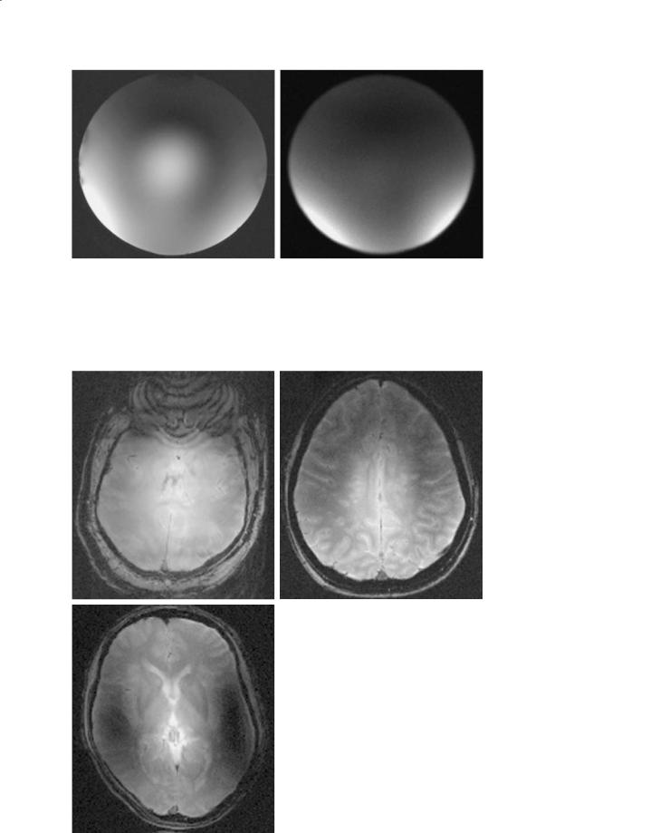
Fig. 10.5a,b. Image of a spherical phantom filled with saline. Both images are gradient echo axial slices with TR=700 ms, TE=11 ms with the same RF power level. Image (a) was acquired with a standard quadrature mode while image (b) was acquired with two-port transmit/receive with variable amplitude and phase difference between the two ports. Notice the vastly improved homogeneity in the image on the right. The bright center of (a) is completely eliminated in (b) as a result of the phase variation on the two ports independently
a |
b |
Fig. 10.6a–c. Axial 1-mm slices of a set of gradient echo images taken at 8 T from a patient with known MS. In these images the development of MS plaques along venous structures are well documented (b). Some of these plaques will appear and disappear at a frequency and in a fashion which diagnostic imaging is yet to correlate to disability onsets. It is important to note that there is a long-stand- ing expectation, supported by pathology, that lesion activity and microvascular inflammation and breakdown (MVIB) are intimately related. This expectation is reasonable since MVIB does influence microcirculation and, as a result, cerebral hemodynamics. Ability to monitor microvasculature changes and inflammation, and thus also hemodynamic changes in vivo,is possible using such HF MRI in MS studies. The images demonstrate the susceptibility artifact in the form of concentric dark bands with the prefrontal lobe (a,b). Susceptibility is much less dramatic in the slices in superior direction (b) and in slices above the semicircular canal (c). The residual effect of susceptibility, in addition to other high-field artifacts such as penetration depth and transmit-receive asymmetry, are still observable as darkening of the image in the frontal lobe (b) and slices above the semicircular canal (c). The reduction of inhomogeneity has enabled better depiction of the
c numerous plaques with central dark spots representing central vessels of each lesion. (a) GRE, BW=50 KHz, FOV=20×20 cm, ST=1 mm, TR=500 ms, TE=7ms

Molecular Imaging and High-Field MRI in Multiple Sclerosis |
139 |
MRI, but only a small fraction of such exacerbations become manifest clinically, the role of: (a) cortical gray matter (CGM) lesions, and (b) comparison of the microscopic structure of white matter plaques, with and without microvasculature involvement, with clinical disability could help reveal the immunological origin of MS. Drugs under investigation for their efficacy could be examined for their ability to modify the MRM profile, and a favorable effect on MRM imaging CGM and microvasculature of white matter plaques could become an important measure of efficacy. In this regard, the effect of interferon, for instance, would be more objectively quantified with MRM.
10.2.1
State of the Art in MR Research in MS
Insofar as the MR assessment of MS is concerned, it has become evident that loss of myelin does not always equal loss of function. Sensitivity of MRI to loss of myelin and its relative insensitivity to changes in axonal function present a challenge to this imaging modality. This is particularly notable since it has become evident that lesions as seen on conventional MRI do not correlate well with the presence or absence of neurological function. This is in spite of the fact that MR imaging and spectroscopy at 1.5 T has permitted some examination of the evolution of the plaques of MS and provided some understanding of the pathogenesis of this disorder that was previously impossible (Filippi et al. 2000). But, although significant advances have been made in this regard, it is becoming increasingly clear that MRI at 1.5 T has limitations in defining all of the pathological events in the brain. Specifically, the total disease burden does not seem to correlate with clinical disability. Even though MR spectroscopy (MRS) (Helms et al. 1999) has identified decreased N-acetyl aspartate (NAA) in NAWM in patients with early relapsing-remitting MS, it has not been able to offer a consistent predictor of disability and quality of life for MS patients (de Stefano et al. 1999; Davie et al. 1997). Decreased levels of NAA, a marker for axons, however, implicate axonal dysfunction in MS. Furthermore, it has been known that MS patients suffer severe disability during which time MR detects no new lesions (Leary et al. 1999). While BBB disruption is related to MS disease activity, it can only be assessed with contrast-enhanced MRI. Even then, direct observation of the internal structure
of the enhanced plaques is difficult. Various techniques, such as Gd-DTPA enhanced MRI, MRS and magnetization transfer (MT) have demonstrated (Leary et al. 1999) promising potential to provide evidence for activity of MS lesions through correlation with enhancement (Horsfield et al. 2000). In many studies, indications such as decreased magnetization transfer ratio (MTR) in NAWM of MS patients support the suggestion that there are inherent changes in NAWM. These observations may point to the possibility that internal changes in NAWM beyond the reach of conventional MRI could cause the disability. Conventional imaging, as such, is incapable of detecting any changes in the affected areas of impaired axonal function. A non-invasive measure of disease activity in MS has not yet been established.
10.2.2 High Field
In search of novel MRI techniques with the ability to probe the internal machinery of brain tissues, a number of basic MR parameters could be considered. Magnetic field (B0) is one parameter known to set a fundamental limit on the SNR, which in turn determines the resolution (Chen et al. 1986; Ugurbil et al. 1993). The gradient strength and novel RF coil designs also play an important role in this regard. We will highlight the role of high magnetic field in MRI in improving MS diagnosis and treatment. Both advantages and challenges involved in applying HF MRI for human subjects will be analyzed.
The nature of the tissues and the etiology of MS are the two most important factors to be considered for the assessment of the opportunities that HF MR will offer. The work employing 7-T and 8-T wholebody human scanners has shown that this technology is indeed feasible (see Fig. 10.6) and offers many advantages in imaging (Burgess et al. 1999; Ugurbil et al. 2003). It has also been shown that neither RF energy of common sequences nor the gradient slew rate poses a safety hazard at field strengths up to 8 T (Kangarlu and Robitaille 2000). It is known that gradient echo (GRE) is the most sensitive sequence for the detection of paramagnetic molecules such as iron and this effect is amplified at higher fields (see Fig.10.2). GRE at HF will provide a simple and sensitive tool that is available for the detection of BBB implications within the lesions. The ultra highresolution images (30–50 µm in-plane resolution) of brain samples of newly deceased MS patients
140 |
|
A. Kangarlu |
a |
b |
c |
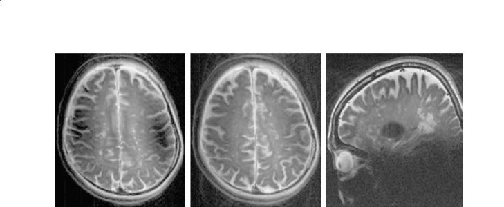
Fig. 10.7a–c. Rapid acquisition with relaxation enhancement (RARE) images in axial plane (a,b) and sagittal plane (c) from a patient with MS. All images are acquired with reduced RF inhomogeneity. The image reveals the ability of spin-echo based images to suppress susceptibility artifacts. These images demonstrate some reminiscence of the susceptibility (a) and coil RF profile (c). The susceptibility of the semicircular canals is much reduced in (a) in a superior direction but still visible. The residual effect of the susceptibility, in addition to other high-field artifacts, is less prominent. In these spin-echo based images the susceptibility and RF inhomogeneity are reduced to the point of allowing detection of the many plaques with central dark spots representing central vessels of each lesion. Acquisition parameters are as follows; RARE, BW=70 KHz, FOV=18×18 cm, ST=2 mm, TR=3 ms, TE=22 ms, matrix=512×512, PW=6 ms
using GRE, SE (see Figs. 10.7 and 10.8) and DWI at 8 T have also proven to be promising sequences in visualizing cortical microanatomy (Kangarlu et al. 2004). For the first time, multilaminar structures have been identified in the CGM and CGM plaques are clearly seen (Fig. 10.8). These CGM plaques are not observed in routine clinical imaging at 1.5 T. The work, to date, has demonstrated that MR scanners at such high fields are capable of developing MRM techniques for in vivo application for human neuroimaging. MRM could produce results that would correlate the clinical deficit with presence or absence of different types of lesions. Development of in vivo human MRM with a 10–100 µm resolution will amount to the next quantum leap in medical imaging. Our images with a 30–50 µm resolution (see Fig. 10.8), have revealed the power of detecting CGM lesions with remarkable contrast. In vivo human MRM will be the precursor to in vivo human cellular imaging. The technical issues in achieving in vivo MRM are presented below.
10.2.3 SNR
Is SNR relevant to the enhancement of MR capabilities in achieving specificity in MS studies? With the advent of whole body HF MR up to 8 T,unprecedented
improvement in SNR was achieved in human subjects (Figs. 10.6 and 10.7). Histopathologic findings have implicated axonal injury in the early stages of the disease (Trapp et al. 1999). This fact is confirmed by MRS in a qualitative way. The pathogenesis of the axonal changes in NAWM will be better defined using HF MRI. In our work, we have shown, using HF MRI, that abnormalities can be detected in NAWM of brains of patients with early relapsing-remitting disease that previously were only seen by pathologic techniques (Kangarlu et al. 2004). Further, experiments with techniques such as MTR at lower fields indicate that such abnormalities can be correlated with future occurrence of progressive disease. The primary MRI techniques used in visualization of the neuroinflammation associated with MS and acute EAE are T2 weighting, T1 weighting, MT, and DWI. The integrity of the BBB has also been examined using gadolinium(III)-diethyltriaminepentaacetic acid (Gd-DTPA). In addition, MRS has been utilized to demonstrate a decrease in NAA and choline levels in NAWM of MS patients and has offered a way of probing possible non-focal magnetically hidden lesion activity. Determination of activities in NAWM would be significant for the detection of microdemyelination (Pan et al. 1996). Decreased levels of NAA, a marker for axons, implicates axonal loss or dysfunction in MS (Arnold 1999). MRI has been able to detect changes in these areas of impaired
Molecular Imaging and High-Field MRI in Multiple Sclerosis |
141 |
|
a |
b |
c |
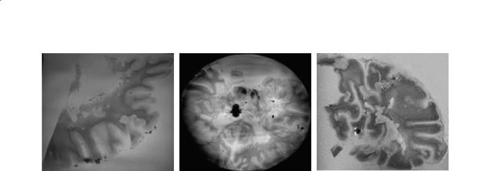
Fig. 10.8a–c. Brain samples imaged with gradient echo (GRE) or diffusion weighted imaging (DWI) in pieces (a,c) and whole (b) within a container filled with distilled water. Slices are taken from a patient with known MS. The in-plane resolutions are 150 µm (a,b) and 300 µm (c) and demonstrate susceptibility artifact in the form of concentric dark bands with prefrontal lobe (a,b). In these images many artifacts, including dielectric resonance, bulk susceptibility, and inductive coupling with the head have been reduced. Reduction of artifacts has also improved visualization of the many plaques. Acquisition parameters are as follows: a GRE, BW=70 KHz, FOV=15.0×15.0 cm, TR=700 ms, TE=11.0 ms, matrix=1024×1024, PW=4 ms; b GRE, BW=70 KHz, FOV=15.0×15.0 cm, ST=5.0 mm, TR=700 ms, TE=11.0 ms, matrix=1024×1024, PW=4 ms; c DWI, BW=50 K, FOV=15.0×15.0 cm, TR=1088 ms, TE=67.8 s, matrix=512×512, PW=4 ms
axonal function. At higher fields, MR SNR increases, making it more sensitive to changes in axonal function through spectroscopy and other techniques such as MT and diffusion. Accordingly, through SNR it may be possible to establish a correlation between axonal loss in NAWM and lesions with presence or absence of neurological function. MR imaging and spectroscopy at lower field strengths have permitted examination of the evolution of the plaques of multiple sclerosis and provided some clues regarding the pathogenesis of this disorder (Arnold 1999). Although significant advances have been made in this regard, it is becoming increasingly clear that HF MRI is needed to define such parameters as the total disease burden and its correlation with clinical disability. In particular, some inherently low SNR sequences such as EPI and its derivatives like DWI and DTI will become possible to implement at higher resolutions. Considering the nature of diffusion (Cercignani et al. 2001) and its sensitivity to sub-voxel events, more basic information on the pathology of inflammation and neurodegeneration becomes available. DTI imaging has shown potential for offering correlation with disability in patients with RR and SP MS (Cercignani et al. 2001). This further reinforces the expectation that HF MRI could make diffusion-based techniques such as DWI and DTI a potent candidate for probing structural information at the microscopic level (Kangarlu et al. 2002; Basser and Pierpaoli
1996).
In addition, HF MRI is capable of examining hypothesis driven experiments in murine model EAE
and directly applying the developed technique to humans within the same system. The SNR at high field is affected by the inherent inhomogeneity of RF distribution (see Fig. 10.6). The characteristic central brightness of these images creates a higher SNR within the deep tissues compared to peripheral tissues (Fig. 10.3). In fact, this effect, also know as dielectric resonance phenomenon (Fig. 10.1), presents a challenge to generating a uniform image. The relatively lower peripheral SNR will particularly hinder cerebral cortex studies (Figs. 10.6 and 10.7).
Depiction of microvascular veins and their position with respect to the lesions (see Figs. 10.6–10.8), new and old, is another way that high SNR and susceptibility based contrast could be utilized for MS studies. BOLD contrast has long been recognized as a mechanism for visualization of veins (Cho 1992), and field-dependent BOLD venography has been demonstrated (Reichenbach et al. 1998, 2001). We have exploited this promising mechanism at 8 T (Kangarlu et al. 2002; Burgess et al. 1999) as well as the utility of paramagnetic deoxyhemoglobin in blood for BBB studies in visualizing microvasculatures within MS plaques (Kangarlu et al. 2004). Even though relevance of blood vessels and formation of plaques around them had been addressed previously (Schelling), it had never been observed in vivo. The clear central vein depiction on some plaques by 8-T MRI (Kangarlu et al. 2002) suggests possible inflammation and breakdown of vessel walls that could be used in distinguishing active plaques from dormant ones.

142 |
A. Kangarlu |
10.2.4 Susceptibility
It has become clear that various MR parameters by themselves will not match the challenges of in vivo microscopy.SNR,relaxation times,chemical exchange mechanisms and diffusion play an important role in defining the image contrast,specificity,and sensitivity to microscopic events. But the short history of MR has shown that interdependence of these parameters will, in spite of initial challenges, inevitably point in the direction that MR evolution has taken, namely a steady increase in magnetic field.As the field increases, there is a proportional increase in the magnetic susceptibility that would enhance MR sensitivity to paramagnetic elements within the body. The susceptibility increase causes a loss of signal on the microscopic and macroscopic scale, the former enhancing MR capabilities and the latter presenting a fundamental challenge for imaging of areas in the near proximity of large cavities such as brain tissues near air interfaces such as the sinuses and mastoids (see Fig. 10.3c). In these areas of the head, magnetic susceptibility discontinuity calls for innovative techniques to suppress the local field inhomogeneities. There have been sequences proposed including GESEPI and z-shim with various degrees of success (Yang et al. 1999; Glover 1999). The impact of susceptibility induced relaxation accelerations to time scales of 1 ms makes it quite challenging to recover the signal losses without a combination of hardware, RF coil, and pulse sequence improvements as human in vivo imaging marches towards 10 T (Gelman 1999; Vymazal et al. 1996; Duewell et al. 1996). The ultimate barrier to achieving microscopic resolution for in vivo imaging is being unable to harness SNR lost to susceptibility and diffusion. In addition, the Boltzmann distribution governing the size of signal from the voxel compounded by the blurring effect of motion convinced the MR community that without resort to simultaneous detection of signal by a number of detectors in parallel valuable signal will be lost to the slow imaging acquisition governed by the serial Fourier phase encoding scheme. HF MRI should be more sensitive to some contrast agents (Tanimoto 2001). T2* contrast agents should be very effective at high field.
10.2.5
RF Penetration
The RF frequency (ω0) used for spin excitation increases with field strength. The electrodynamic
properties of the tissues will vary proportional to ω0. This includes an increase in tissue conductivity which enhances the RF absorption, requiring higher power for implementation of the same pulse sequences. Considering the heterogeneous nature of biological tissues the RF power will be absorbed nonuniformly (Gandhi et al. 1979). A number of other factors, i.e., head position and size, affect the extent of nonuniform distribution of RF (Durney et al. 1986). Furthermore, the image quality, SNR and penetration depth are intimately related to the coil design and dimensions. For volume head coils optimization of image quality is largely a function of the coil design, and the filling factor effect is maximized by constructing the smallest coil for any particular application. Smaller coils and phase array coils with their surfacelike characteristic offer higher SNR. TEM designs are relatively larger and prone to inductive coupling between struts, which creates deep field focusing. This compensates for RF penetration effects, but at the same time causes safety concerns due to the high RF requirements compared to a 1.5-T MR system. Consequently, HF MRI causes more deep RF focal heating than at lower field strengths (Leussler 1999; Singerman 1997; Ibrahim 1999). As such, better understanding of the RF power deposition, penetration depth and innovative coil designs are required for better quality and safe human imaging. In order to better manage RF requirements of HF MRI, new techniques such as parallel excitation and detection of induced EMF within each coil element of a multielement RF coil with ability to independently control the amplitude and phase of each port are necessary (Ibrahim 1999). This will allow RF-intense pulse sequences such as fast spin echo, RARE, and MDEFT (modified driven equilibrium Fourier transform) to be used for clinical HF applications. Moreover, this will allow many beneficial sequences for MS studies such as inverse recovery and magnetization transfer contrast (MTC) techniques to be used.
10.2.6
RF Coil Design
For scanners with magnetic fields above 7 T the imaging in MRI studies are difficult to perform without a new coil design called transverse electromagnetic (TEM) resonators (Roschman 1988; Vaughan 1994). Imaging at 7 and 8 T was made possible due to the construction of such resonators for use in humans and animals for fields ≥7.0 T (Baertline 1999). In spite of the inherent artifacts at high field (Figs. 10.1–10.3), the

Molecular Imaging and High-Field MRI in Multiple Sclerosis |
143 |
TEM resonator has provided an alternative to birdcage design for HF MRI.With TEM coils, however, due to the increasing wavelike behavior of the RF within the subjects of a high dielectric constant, incorporation of a parallel transmit and receive (pT/R) function at high field offers an opportunity to reduce acquisition time and enhance homogeneity.
Forlow-γ nuclei,i.e.,31Pand13C,withfieldstrengths up to 10 T, the existing birdcage coil design will suffice (Hayes et al. 1985). It has been shown that driving this coil in quadrature for low-γ nuclei frequencies provides improved SNR and improved homogeneity compared to similar designs such as the Alderman-Grant coil (Hayes et al. 1985). Unfortunately, at high fields the head-size birdcage resonators with their lumpedelement structure do not lend themselves to operations beyond 300 MHz frequency range even in the presence of radiation shields. Furthermore, the B1 field of the RF wave generated by birdcage coils is inherently inhomogeneous. The need for the ability to evaluate large volumes of the brain without loss of image quality drives the intense research in RF coil design.As was pointed out earlier, high-field coils operating at B0>3T produce head images with bright centers and lower SNR at the peripheries. This makes study of the cerebral cortical pathology difficult (see Fig. 10.2). In our work on the CGM in MS brain samples, since the study was conducted in the whole brain sample (Fig. 10.4), variation of image quality was addressed through the design of a special TEM coil and acquisition of images using optimized sequences for enhanced homogeneity (Kangarlu et al. 2004). Such provisions are difficult to apply for in vivo studies, making high B1 field inhomogeneity within the head a severe challenge (Ibrahim 2001; Collins et al. 2001). It is conceivable that for the studies of deep tissues the dielectric resonance effect provides higher SNR within these tissues enabling depiction of finer structures. Application of independently phase adjusted parallel imaging and phased array techniques (Ibrahim 1999; Griswold et al. 2000) will alleviate this condition and could lead to improved SNR distribution across the entire body parts at high field.
10.2.7
Gradients and Receivers
Gradient coils for high-field systems have already been successfully constructed and many improvements in their design and drive systems have been achieved (Chronik et al. 2000). Availability of slew rates up to 1000 T/m/s is the long-term goal, safety
issues permitting. Already, manufactures are delivering head scanners with 600 T/m/s slew rates. The integration of 3 T within clinical settings has been greatly accelerated following the demonstration of safe exposure of human subjects to fields of up to 8 T (Kangarlu et al. 1999; Schenck 2000). These systems are equipped with the hardware and software to perform basic and clinical scans as well as research quality MRS, fMRI, and DTI fiber tracking. A typical head-only scanner appropriate for MS studies would have gradient hardware of a 36-cm I.D. asymmetric gradient coil capable of imaging at 50–80 mT/m with slew rates of 500–700 T/m/s at a duty cycle of about 70%. Such system will allow a 1- mm slice thickness considering the SNR available at 3 T and will also be capable of executing single shot EPI at a sustained rate of 10–20 images/second. The RF power in state of the art systems should be within a 15to 30-kW range. Considering the new developments in parallel imaging, independent RF preamp channels will enable lower RF power deposition and will provide more homogeneous and faster acquisition. Consequently an 8-RF channel technology for transmitting and receiving would greatly enhance a high resolution MR scanner for MS studies. Such systems must contain software that is designed for PC platforms to enable researchers to benefit from the Unix/Linux open environment and ancillary software development, particularly in image post processing.
Using gradient capabilities of 30–50 mT/m, a slew rate of 150 T/m/s and RF power of about 2.5 kW we have acquired high quality images from the human head at 8 T using TEM resonators driven in single drive, quadrature and 4-port drive mode. In Fig. 10.3, such images are shown indicating improvement in image homogeneity. It is also possible to use the TEM resonator for parallel imaging using different struts as an independent transmitter/receiver. Nonetheless, it is inevitable that new coil designs such as phase array and microstrip design will be added to the RF coil collection of HF MR researchers. We have investigated the possibility of multiport drive on TEM and others have done similar work at 7 T (Ibrahim 1999; Collins 2003).This is expected to further improve the homogeneity of RF distribution as has already been demonstrated using FDTD calculations.
10.2.8 Relaxation E ects
The relaxation mechanisms based on which contrast is generated in MRI are field-dependent.Prolongation

144 |
A. Kangarlu |
and convergence of T1 with B0 creates a situation that makes acquisition of T1-weighted images at very high field more challenging. T1W images have been acquired at 4 T, 7 T and 8 T using MDEFT (Ugurbil et al. 1999; Norris et al. 1999). A simple comparison was made between 3 T and 8 T T1-weighted MDEFT images indicating an eight-fold increase in CNR at 8 T (Norris et al. 1999). This indicates that SNR combined with relaxation effects are responsible for such high CNR. Such CNR will play an important role in characterizing gray matter (GM)/white matter (WM) contrast and consequently rendering the exploration of WM and GM at microscopic level more readily possible. These results are significant in the context of MS imaging. An at least threefold increase in SNR and eightfold increase in CNR at 8 T compared to 3 T point to the necessity for making HF MRI work if the microstructures of WM and GM are to become accessible by MR. By making microscopic MRI more viable, all aspects of the technique such as SNR, relaxation and paramagnetic susceptibility properties of biological tissues appear to have arrived at a critical stage as human MRI marches on towards 400 MHz. Ability, for instance, to acquire high resolution T1weighted images in a short time is an important area of using the high SNR of HF MRI for microstructure visualization in MS. Relaxation effects have also been used through exogenous agents with relaxation modifying properties to generate enhanced contrast in regions of interest such as areas where BBB are compromised. As discussed earlier, capabilities of MRI combined with the conspicuity of new contrast agents such as SPIO have raised hopes of making targeted molecular imaging possible (Frank et al. 2002). A number of developments in novel MR signal amplifiers such as SPIOs have taken place in the past few years which have facilitated their delivery and tolerance. In this regard, approaches based on enzyme-mediated polymerization of paramagnetic substrates into oligomers of higher magnetic relaxivity (Bogdanov et al. 2002) have proved to be particularly promising.
10.2.9 Ultra-High-Resolution MRI
The advent of 8-T whole body MRI made acquisition of high resolution images of the human head with an in-plane resolution of 100 µm possible. A TEM resonator was used to acquire the GRE images from the human head of normal subjects. Serial acquisitions of such images require an average of 15 min.
Though at the limit of clinically accepted time, its implementation triggered an intense HF MR activity within the community. Various technological innovations such as parallel imaging (Pruessmann et al. 1998; Sodickson 1999) have been reported since the first 8-T human head images were acquired that present a realistic opportunity for reducing the acquisition times to 2–4 min. Images acquired at this resolution and within 2 min will produce up to half a gigabyte of unprocessed information from a human head. Acquisition of a 2000×2000 matrix image with a field of view of 20 cm, slice thickness of 1 mm, and flip angle=45º could be TR=750 ms, TE=17 ms, receiver bandwidth=69.4 kHz. Such techniques will allow MRI signal to be acquired from a 0.02-mm3 or 20-nl voxel. The capability of in-vivo acquisition from such a minute volume from the human head begins a new era for high resolution study of pathology such as MS. Combined with susceptibility contrast and parallel imaging, there is the possibility of a ten-fold increase in in-plane resolution relative to the conventional 256×256 images obtained with a 20-cm field of view and a 5-mm slice thickness within a clinically acceptable time. Furthermore, the higher resolution images could be acquired with adequate image quality using new technologies including phased array coil design and parallel imaging. Human head images with a 1k×1k matrix at 8 T reveal numerous small venous structures throughout the image plane and provide reasonable delineation between gray and white matter (see Fig. 10.6). The elevated SNR observed in HF MRI could be utilized to acquire images with a level of resolution approaching the histological level under in vivo conditions. These images represent a significant advance in our ability to examine small anatomical features with noninvasive imaging methods.
10.2.10
New Technologies in MS Imaging
Plaques in MS patients are mostly seen in the white matter. Involvement of gray matter does not lend itself to in vivo detection. The white matter lesions appear in an unpredictable shape, size, and distribution (Trapp et al. 1999). Ability to distinguish the plaques into new and old or active and inactive is important. The newer plaques are identified with inflammation and are producing fat debris due to lipid breakdown. The chronic plaques are mostly characterized with gliosis. The MRI manifestation of these two mechanisms has been difficult.

Molecular Imaging and High-Field MRI in Multiple Sclerosis |
145 |
Histological studies (Lumsden 1970) have found that newer plaques are associated with veins but routine MRI has just been unable to demonstrate this pathogenesis of the plaque in vivo. HF MRI seems to be able to study this vasocentral characteristic of MRI-visible plaques. In order to enhance MRI capability toward in vivo histology, plaques must be distinguished in terms of age, activity, and possibly response to therapeutic techniques based on pathogenic mechanisms. These capabilities would require that MRI features sensitive to MS plaques be probed at the microscopic level. Events at this level are macrophage infiltration, edema, myelin swelling, lymphocyte infiltration and endothelial cell activation (Lassmann et al. 1998). It is difficult for any MR-based technique to target any one or more of these mechanisms. The relaxation effects associated with edema and diffusion manifestation of myelin swelling have been and are subject to imaging exploitation.
10.2.11
HF MRI of MS
The major challenge in achieving the goals summarized above is to enhance MRI specificity for characteristics of individual lesions. The ability to visualize the internal structure of lesions would put MRI on the path to producing distinct contrast for those lesions sharing a particular histopathologic type. The 8-T MRI depiction of WM plaques with their central microvessel was the first step in that direction (see Fig. 10.7). As plaques in WM have been mostly observed for diagnostic and therapeutic purposes in MS, the involvement of GM has received little attention in MRI. Our depiction of CGM lesions in brain samples (Figs. 10.2–10.4), has provided an opportunity for their detection and monitoring in vivo. Through the use of HF MRI it could be feasible to interrogate each plaque based on its structural characteristics and association with CGM lesions as classified by Kidd et al. (1999). Assuming that the technological issues listed above are met then HF MRI could proceed in further expanding its capabilities for classification and probing of MS plaques by developing fMRI, DTI, and quantitative MRS. Imaging at 8 T has detected multiple cortical lesions that are not evident by conventional imaging at 1.5 T (Fig. 10.6– 10.8). Lesions are visualized using gradient echo, spin echo, and diffusion-weighted images (Figs. 10.7 and 10.8). The high quality images at 8 T allow for the identification of the different types of cortical lesions previously described at histology (Kidd et al.). Such
imaging tools, once applied to in vivo detection of CGM pathology, would allow development of techniques to study whether CGM is a primary event or alternately a consequence of the overwhelming white matter pathology. Furthermore, determination of the contribution and sequence of CGM and WM events could be correlated to disabilities. Such imaging capabilities have not been available in multiple sclerosis to date.
10.2.12
Other MRI Methods
One other approach would be the use of dynamic contrast enhanced (DCE) MRI using SPIO contrast media to quantitatively assess the microvessels within the plaques. These contrast agents will help susceptibility enhanced contrast between the microvessels and the brain tissues, which will make these vessels more visible in anatomical images. In addition, perfusion fMRI studies based on the DCE techniques will demonstrate the activity and the extent of BBB breakdown within the plaques. Using computer processing of the imaging data and a relatively simple two-compartment kinetic model, it is possible to non-invasively assay the relative blood volume, microvascular endothelial leakiness, and the interstitial volume of any solid tumor. DCE MRI and its capability in quantifying the permeability of microvessels in the brain at high field will offer an additional tool in characterizing plaque activity and classification. Considering these combined capabilities, HF MRI could offer a powerful predictor of disability and measure of drug efficacy. Using HF MRI and SPIO contrast agents, therapeutic responses could be more accurately detected. Optimization of techniques such as DTI and DCE MRI would complement the high resolution anatomical depiction of MS abnormalities and should lead to an even greater future benefit from diagnostic imaging in MS.
10.3 Conclusion
Observation of the evolution of inflammation will be greatly assisted by targeted MI. Considering that ultra-high-field MRI is producing images whose quality is comparable to histology, and the ability to image regions comparable to cell size, combined with biochemical techniques of attaching molecules with