

Книги по МРТ КТ на английском языке / MR Imaging in White Matter Diseases of the Brain and Spinal Cord - K Sartor Massimo Filippi Nicola De Stefano Vincent Dou
.pdfMR Imaging of Brain Development |
167 |
a 
 b
b
c |
d |
Fig. 11.10a–d. A 4–month-old infant. a T1 axial centrum semi-ovale. The signal is now practically even between the gray and the white matter. There is clearly myelination in process, however, in the white matter subjacent to the central sensory-motor area, and probably also along a longitudinal tract that could be the superior longitudinal fasciculus. b T1 axial, basal ganglia. Loss of contrast between the gray and white matter, except for the internal capsules (anterior and posterior limbs) and portions of the corona radiata such as the thalamo-frontal fibers, or the optic radiations. The internal capsule is now brighter than the adjacent lentiform nuclei and thalami. c T2 axial, centrum semi-ovale. Pattern similar to that of the neonate. The contrast is still strong between the cortex and the white matter, except in the area subjacent to the central cortex. d T2 axial, basal ganglia. Still similar to the neonatal pattern but the PLICs are now clearly and globally myelinated, and also the anterior limbs, the optic radiations and faintly, the thalamo-frontal fibers
Within the cortex, the areas of previously higher contrast are now even. Over this ground of increased signal, the contrast of the basal ganglia is also lost. But the capsules form a striking pattern of high signal corresponding to the whole internal capsules (anterior and posterior limbs) with its radiations. In the white matter, they extend toward the fronto-cen- tral cortex as the cortico-spinal tract, which is now
brighter than the cortex; the optic radiations from the lateral geniculate bodies to the inner occipital cortex; and anteriorly, the thalamo-frontal segment of the corona radiata. The posterior corpus callosum also appears brighter.
In the posterior fossa, the brainstem is diffusely brighter with “hotspots” corresponding to the main fascicles. The whole cerebellar white matter is bright.

168
11.4.2.2 T2 Imaging
In contrast to T1 imaging, little change appears on T2 between birth and four months. The cortex still shows a lower signal in the primary areas. In the white matter, the corticospinal tract is still faint, but on the contrary, the optic radiations are clearly demarcated. This is an important landmark: poorly defined optic radiations at this stage indicate that oligodendrocytes have been damaged, even if a fully fledged picture of periventricular leukomalacia is not observed.
The myelination of the PLICs is more advanced, but still well circumscribed. In the basal ganglia some difference appears between the striatum and the thalamus (more mature), and the globus pallidus (less mature). Little change is apparent in the brainstem and cerebellum.
11.4.3
The Brain from 8 to 12 Months
11.4.3.1 T1 Imaging
At this stage, an obvious maturation gradient is observed between the posterior and the anterior part of the hemispheres. The posterior part looks like fully mature: bright signal of the white matter against the lower signal of the cortex, but the cortical-subcortical junction is still blurred. The thalami are now brighter than the basal ganglia. The more anterior, frontotemporal part of the hemispheres by contrast is still similar to what it was at 4 months.
In the posterior fossa, the fully mature appearance is now attained.
The adult appearance is reached at about 12 months, with a diffusely bright signal of the white matter, brighter than the cortex and the central grey nuclei; the adult pattern of signal gradient between the striatum, globus pallidus and thalamic nuclei is present.
11.4.3.2 T2 Imaging
From 6 to 8 months, the cortex is still darker than the white matter, but this becomes attenuated. The basal ganglia and thalami also appear darker, but not as much as the internal capsule with its radiations toward the frontal lobes, the fronto-central areas, and
C. Raybaud
the visual cortices. The posterior corpus callosum appears clearly darker than the centrum semi-ovale. The posterior fossa is still incompletely myelinated.
But from 8 to 10 months, and later, the signal of the white matter globally equates the signal of the gray matter, in the cortex or in the central grey nuclei, giving the brain a blurry, contrastless appearance. Surprisingly, the corpus callosum and the internal capsules are much darker than the adjacent white matter. The posterior fossa looks almost mature.
11.4.4
The Brain at 18–24 Months
At 18 months, the mature appearance is completed on T1, and almost reached on T2. The white matter is darker than the gray matter, but the centrum semiovale is still diffusely brighter in its central portion, the subcortical arcuate fibers being clearly darker. The final “adult” appearance is reached at 2 years, with the exception of the terminal area, which is the deep white matter lateral and at a distance from the ventricular atria, which remains slightly brighter than the surrounding white matter until adulthood.
11.5
MR Morphometry, Di usion, and Spectroscopy
11.5.1 Morphometry
As the normal developing brain grows, and as any injury will affect this growth in a perhaps unapparent fashion, quantitative volumetry has been developed in healthy and diseased infants, and can be used as a predictor of outcome. Volumetry is achieved by segmentation methods (Hüppi et al. 1998a). It has been shown in presumably normal premature babies (Hüppi et al. 1998a) that at between 29 and 41 weeks, the total volume of the brain is increased 2.7-fold; and that most of this increase is due to the increase of volume of the folding cortical white matter, of which the part in the brain volume increases from 35% at 29 weeks, to 50% at term, in relative terms, and is multiplied four-fold in absolute terms.
MR Imaging of Brain Development |
169 |
a |
b |
c |
d |
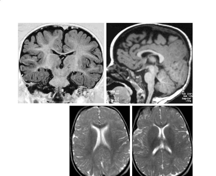
Fig. 11.11a–d. An 18-month-old infant. a T1 coronal, thalami (IR). The signal of the white matter now is approaching the adult pattern, globally brighter than the cortex except in the peripheral white matter close to the cortex (arcuate fibers mostly). b T1 sagittal, midline. The anterior portions of the brainstem now appear brighter than the tegmentum. The corpus callosum looks fully myelinated. c T2 axial, ventricular bodies. Loss of the previous contrast between the white and the grey matter, with a blurry appearance related to the even signals. The corpus callosum, however, appears darker, more myelinated. d T2 axial, basal ganglia. Poor grey/white contrast, diffusely, except for a stronger myelination of the PLICs and of the corpus callosum
11.5.2
Di usion Imaging
Beside the usual T1 and T2 parameters, other methods have been developed to approach brain maturation. Diffusion imaging (DWI) has been shown to be promising (LeBihan et al. 1986). In contrast to conventional MRI, it is not based on relaxation times, but capitalizes on the influence of the structure on the brownian motion of the free water protons.When these protons are in a free space, that is free to move in any direction, there is a loss of the resonant signal (proton dephasing), while this resonant signal
is better preserved when the motion is restricted to smaller spaces (less proton dephasing).This apparent motion can be quantified by calculating the apparent diffusion coefficient (ADC), restituted as a map. The intracranial water is located in the CSF spaces, where it moves freely; within the cells, where its motion is hampered by the numerous cytoplasmic organelles; and in the extracellular spaces, where motion is dependent on the space allowed by the volume of the cells. It is this last water sector that is studied in DWI, together with some of the water crossing the cellular membranes. In the normal mature brain, the extracellular sector represents about 20% of cerebral
170 |
C. Raybaud |
a |
b |
c |
d |
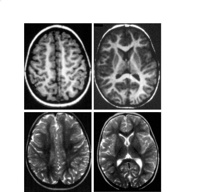
Fig. 11.12a–d. An 18-month-old infant. a,b T1, axials; c,d T2, axials. The adult pattern is practically reached at this stage, on T1 and T2 imaging. Some faintly higher T2-signal areas are acceptable after 2 years in the centrum semi-ovale, and in the deep white matter lateral to the atria (zona terminalis) up to adolescence
volume. Because of the particular organization of the brain, another factor can also be evaluated, which is the anisotropy: the brain contains fibers organized in bundles, so that the motion of the water protons is more restricted in the directions perpendicular to the fibers than in the direction parallel to them. The higher the anisotropy coefficient, the more packed the fibers. It is also possible to identify the direction of the less restricted motion in a given voxel, giving the direction of the fibers, and therefore to reconstruct the organization of the fibers; this is the basis of tractography by diffusion tensor imaging (DTI) (Pierpaoli et al. 1996).
Studies have shown that ADC is high in the immature white matter, and decreases progressively until the age of 6 months when adult values are reached. This is explained in part by the development of the myelination but as the reduction begins before myelination starts, it is likely that other factors also interact, probably the glial multiplication of astrocytes and oligodendrocytes (myelination gliosis) that would limit the extracellular spaces (Wimberger et al. 1995). The anisotropy also increases early, partly explained by the myelination, but also by the multiplication in a parallel fashion of the axons and the dendrites.Therefore this increase expresses the organization of the bundles of white matter.

MR Imaging of Brain Development |
171 |
The ADC decreases also in the gray matter during maturation (multiplication of fibers), but the anisotropy does not change (no fascicular organization within the cortex).
11.5.3 Spectroscopy
Finally, as the maturation of the brain is accompanied by tremendous metabolic changes, MR spectroscopy is also a means to approach it. In clinical settings, proton MRS can bring useful information about the relative amounts of N-acetyl aspartate (NAA) (marker of the neuronal function, but also oligodendroglial marker in the immature brain), choline (cell membrane turnover, including the myelin which is a stack of oligodendrocytic membrane), creatine (high energy products), lactate (anaerobic metabolism), myo-inositol (glial marker), and glutamate–gluta- mine (neurotransmitters, and intermediate form of the metabolism of the amino-acids). Typically, two types of spectra are used, with long and short echo times, either in a single-voxel technique (restricted to a portion of the tissue), or according to the chemical shift imaging (CSI) technique exploring a whole slice of brain.
Using the single-voxel technique, it has been shown that in the fetus, from 30 to 40 weeks, NAA increases significantly, together with myo-inositol and creatine (Heershap et al. 2003). This trend continues after birth, as the concentration of NAA in the newborn is 50% of that in the adult. The evaluation of the metabolites, however, can only be relative: increased NAA in relation to choline and creatine. Because of myelination, the choline peak is high in the developing brain, and then decreases in relation to creatine, together with myo-inositol. Actually, this pattern varies from place to place in the brain and at a given place, at its own pace as the brain matures. The use of spectroscopy is also hampered by the difficulty to get absolute values.
11.6
Imaging, Development, and Brain Disease
In the first half of gestation, there is no clear-cut separation between the inborn and the acquired disorders, part of the morphologic consequences depending on the timing of the insult rather than on its mechanism. For instance, polymicrogyria, which is a
disorder of the cortical development characterized by the accumulation of too many overly small gyri with a (typically) four-layered cortex, and fused molecular layer, may be observed in families (then of genetic origin), after infections by the cytomegalovirus in the early stages of pregnancy, and seemingly as well, as a consequence of fetal anoxia. On the contrary, as the brain is considered as constitutionally achieved after 23 weeks, any later interference is considered a scarring process more than a malformation, even if the difference, in individual cases, may sometimes be academic.
11.6.1
The Early Gross Brain Malformations
From the time of the development of the neural plate, the malformations may be classified according the developmental stage that failed:
Failure of neurulation: the neural tube stays open (myelomeningocele), and mechanically, the development of the rhombencephalon (hindbrain) and of the prosencephalon (forebrain) are altered, resulting in a Chiari 2 deformity (McLone and Knepper 1989). This is both a genetic and an environmental (folic acid deficiency) disorder.
Failure of development of the anterior neural plate. This primordium of the forebrain also gives rise to the face, to the vault and to the anterior cephalic soft tissues.When the process fails,the brain (holoprosencephaly) as well as the face (cyclopia, hypotelorism) are affected (Couly and Le Douarin 1987). Several genes have been invoked, as well as cholesterol and toxic factors such as veratrin, an alkaloid from Veratrum californicum.
Failure of development of the roof of the rhombencephalon: it normally produces the vermis superiorly, and the foramen of Magendie inferiorly. An arrested development produces the typical Dandy-Walker malformation, or one of its minor forms (Raybaud 1982).
Failure of commissuration: the crossing of the commissural fibers results from the complex involvement of tissular (guiding/repulsing molecules, fasciculating factors), cellular (glial sling, glial wedge, indusium griseum, meninges) and genetic influence to go through the inner wall of the hemispheres, cross the midline and enter the opposite side. Disorders of these processes result in the various types of complete, partial and dissociated commissural anomalies (Raybaud and
Girard 1998).

172 |
C. Raybaud |
11.6.2
The Malformations of Cortical Development
The development of the cortical plate goes through the stages of proliferation, differentiation, migration, and organization. Each of these steps may be defective, leading to specific malformations (Barkovich et al. 2001):
Proliferation: if one cycle of division fails, 50% of cells are missing. Microcephaly (three standard deviations or more below normal, commonly with simplified gyral pattern) is usually due to a genetic defect.
Differentiation: in the process of migration from the mantle germinal matrix to the cortical plate, pyramidal neurons differentiate from the guiding radial glia. If the process fails, abnormally differentiated cells are produced, with abnormal migration and poor segregation between the cortex and the white matter. This constitutes the group of the focal cortical dysplasias, the hemimegalencephalies and the tuberous sclerosis complex.
Normal neurons may fail to migrate properly, forming masses or layers of abnormally located normal grey matter: they are called heterotopias, either nodular (periventricular, subcortical) or laminar, with a normal ("band heterotopia", "double-cortex") or a simplified (agyria-pachy- gyria) cortical pattern. Laminar heterotopias are clearly genetic as some of the nodular heterotopias.
The neurons may migrate normally to the cortex but may not get organized properly: this results in the so-called polymicrogyria, which may be due to genetic, infectious, metabolic, and probably anoxic disorders.
The schizencephaly ("cleft brain") is a unior bilateral focal failure of development of the cortical mantle, resulting in clefts lined with a polymicrogyric cortex that is continuous, inwards, with the ependyma. This is rarely familial, more often acquired (cytomegalovirus, toxoplasmosis), or idiopathic.
11.6.3
Destructive Lesions and the Developing Brain
Specific fetal or neonatal pathologies may interfere with development. In fetuses and prematures, hemorrhages in the germinal matrix may not only induce hydrocephalus; they also destroy the source of oligodendrocytes, possibly compromising forever myelination of the brain.
Also in prematures, peri-ventricular leukomalacia may destroy the para-ventricular white matter (resulting in posterior enlargement of the lateral ventricles),but also, in milder cases, may only reduce the population of the late astrocytes, and above all that of the more vulnerable oligodendrocytes. It may also reduce connectivity in prematures (Hüppi et al. 1998b). In the term neonate, anoxia produces specific lesions in the most active parts of the brain, the basal ganglia and thalami (status marmoratus), as well as in the cortex, especially in the deepest part of the sulci, often predominantly in the central sensory motor areas.
Other pathologies have specific developmental consequences.Fetal brain viral infections almost constantly produce microcephaly as well as myelination defects. Ischemia produces different effects depending on the degree of development: in the last trimester of pregnancy, it produces a cavitation with complete detersion of the necrosed tissue, without scar formation. In mature brains, it produces a conventional astrocytic scar. In intermediate stages, it produces the intermediate picture of the multi-cystic encephalomalacia. In all cases, as a portion of the cortex is destroyed, the corresponding white matter fibers are missing, reducing the size of the whole hemisphere and restricting its connectivity. The association and commissural fibers that should have connected with the destroyed area tend to connect with the surrounding preserved cortical margins, creating abnormal neuronal circuits that are thought to explain the high rate of epileptogenicity of these lesions. The ipsilateral half of the brainstem may also be atrophic, as well as the contra-lateral cerebellar hemisphere. Even in older children, longstanding thalamic tumors,especially thalamic germinomas,are associated not with an increased, but with a reduced volume of the hemisphere, because of the reduced number of thalamo-cortical and cortico-thalamic fibers of the corona radiata. In the infant, chronic anoxia-hypoxia such as that produced by arteriovenous steal effects (vein of Galen aneurysm, dural fistula of the torcular) commonly produces atrophy, with sub-cortical calcifications of uncertain mechanism.
In contrast, the potential of the infantile brain to massively produce connections has a positive effect on recovery from any focal cerebral injury: the contrast between the size of the destructive lesions and the relative mildness of the symptomatology is often striking, at least for the perinatal and the infantile injuries. Also, the prognosis of hydrocephalus and its response to its treatment varies significantly depending on the maturational stage. Hydrocephalus in utero is usually devastating, perhaps because of the destruction

MR Imaging of Brain Development |
173 |
of the periventricular germinal matrices. By contrast, early post-natal mechanical hydrocephalus (without associated destructive lesions) often present impressive recoveries, the initially excessively thinned brain mantle being able to re-grow to a normal thickness in a matter a few months. This could be explained by the ability of the neurons to re-grow axons, of which the path-finding is not yet impeded by the accumulation of interstitial astrocytes. In older children, massive hydrocephalus never recovers to such a degree.
Globally, the most sensitive cells of the brain seem to be the oligodendrocytes, which are not only vulnerable to anoxia, but also to immune related disorders, and to specific infections. The mechanism allowing the recovery, or not, of the oligodendrocytic pool is to date poorly understood. Non-myelination (or insufficient myelination) is a common result of varied diseases: periventricular leukomalacia, of which the “milder forms” are those in which only the oligodendrocytes are affected, with no cystic necrosis, as well as hydrocephalus, fetal infections. Malnutrition in late pregnancy and early postnatal period has also been shown to compromise myelination. Leukoencephalopathies are common in mitochondrial cytopathies (Chabrol and Raybaud 2002). Obviously, the metabolic diseases affecting specifically the enzymes related to the metabolism, maintenance and degradation of the myelin (“dysmyelinating” diseases) are genetic diseases concerning the oligodendrocyte. Immune-related, inflammatory disorders may induce demyelination, either acutely (acute disseminated encephalomyelitis or ADEM) or in a more protracted way (group of the scleroses), the oligodendrocyte itself being better preserved in the former, less in the latter. Finally, the oligodendrocyte may be the specific target of particular viruses, like the papova/JCV in the progressive multifocal leukoencephalopathy (PML). Finally, vasculopathies in the elderly may also lead to a demyelination (Binswanger disease). All taken together, the disorders affecting the oligodendrocytes, and therefore the myelin, are among the most common neurological disorders in the child, the adult, and the elderly, because of the multiple ways pathologies may alter them.
11.7 Conclusions
MR imaging is the first imaging modality to be able to demonstrate the morphological and structural changes in the maturing brain, in the fetus, and in
the term-born infant. The fetal brain cellularity first and later, the mass production on myelin with its effects on T1 and the T2, produce age-specific images that make it possible to evaluate the course of brain maturation. This evaluation has opened new diagnostic avenues, since the sensitivity of the oligodendrocytes to injury, as well as the involvement of myelin in numerous pathologic processes, make the evaluation of maturation an essential element of the imaging approach to the pathology. However, there are a few restrictions to be remembered. T1 and T2 images reflect two different things, not necessarily parallel, and should be used as such. Conventional MR images are composite, non-quantitative images, reflecting only the myelination of diverse areas relative to others: it is therefore a biased picture during the entire course of development. This image is tech- nology-dependent, and pictures are not necessarily exactly comparable from one center to the other.Also, MR imaging is blind to the development, and to slight myelin anomalies,that occur after infancy.Finally,the signal in the images equates to the degree of myelination only in normal subjects, as a “high T2”, or a “low T1” signal are not specific, reflecting only the relative concentration of myelin and water, regardless of the mechanism, be it for example an increase of water (edema), or a loss of axons (hence also of myelin). Nevertheless, bearing these restrictions in mind not to“over-read”the images,MRI is an efficient tool,that has allowed a completely new view of brain pathologies in the fetus and the developing infant.
References
Ashikaga R, Araki Y, Ono Y, Nishimura Y, Ishida O (1999) Appearance of normal brain maturation on fluid attenuated inversion-recovery (FLAIR) MR images. AJNR 20:427431
Barkovich AJ (1998) MR of the normal neonatal brain: assessment of deep structures. AJNR 19:1397-1403
Barkovich AJ, Kjos BO, Jackson DE Jr, Norman D (1988) Normal maturation of the neonatal and infant brain: MR imaging at 1.5T. Radiology 166:173-180
Barkovich AJ, Kuzniecky RI, Jckson GD, Guerrini R, Dobyns WB (2001) Classification system for malformations of cortical development. Update 2001. Neurology 57:2168-2178
Barres BA (1999) A new role for glia: generation of neurons. Cell 97:667-670
Battin M, Rutherford MA (2002) Magnetic resonance imaging of the brain in preterm infants: 24 weeks’ gestation to term. In: Rutherford M (ed) MRI of the neonatal brain. Saunders, London, pp 25-49
Baumann N, Pham-Dinh D (2001) Biology of oligodendrocyte and myelin in the mammalian central nervous system. Physiol Rev 81:871-927

174
Bird C, Hedberg M, Drayer BP et al (1989) MR assessment of myelination in infants and children: usefulness of marker sites. AJNR 10:731-740
Brody BA, Kinney HC, Kloman AS, Gilles FH (1987) Sequence of central nervous system myelination in human infancy. I. An autopsy study of myelination. J Neuropathol Exp Neurol 46:283-301
Caviness VS, Takahashi T, Nowakowski RS (1995) Numbers, time and neocortical neurogenesis: a general developmental and evotionary model. Trends Neurosci 18:379-383
Chabrol B, Raybaud C (2002) Inborn and acquired mitochondrial leucodystrophies. In: Desnuelles C, di Mauro S (eds) Mitochondrial disorders. Springer, Berlin Heidelberg New York, pp 221-229
Childs AM, Ramenghi LA, Evans DJ, Ridgeway J, Saysell M, Martinez D, Arthur R, Tanner S, Levine MI (1998) MR features of developing periventricular white matter in preterm infants: evidence of glial cell migration. AJNR 19:971-976
Christophe C, Muller MF, Balériaux D et al (1990) Mapping of normal brain maturation in infants on phasesensitive inversion-recovery images. Neuroradiology 32:173-178
Couly GF, Le Douarin NM (1987) Mapping of the early neural primordium in Quail-chick chimeras. II. The prosencephalic neural plate and neural folds: implications for the genesis of cephalic human congenital abnormalities. Dev Biol 120:198-214
Fees-Higgins A,Larroche JC (1987) Development of the human fetal brain. An anatomical atlas. INSERM/Masson, Paris Fralix TA, Ceckler TL, Wolff SD et al (1991) Lipid bilayer and
water proton magnetization transfer: effect of cholesterol. Magn Reson Med 18:214-223
Girard N, Raybaud C, du Lac P (1991) MRI study of brain myelination. J Neuroradiol 18:320-332
Girard N, Raybaud C, Poncet M (1995) In vivo MR study of brain mayuration in normal fetuses. AJNR 16:407-413 Gressens P, Richelme C, Kadhim HJ, Gadisseux JF, Evrard P
(1992) The germinative zone produces the most cortical astrocytes after neuronal migration in the developing mammalian brain. Biol Neonate 61:4-24
Heershap A, Kok RD, van den Berg PP (2003) Antenatal proton MR spectroscopy of the human brain in vivo. Childs Nerv Syst 19:418-421
Holland BA, Haas DK, Norman D, Brant-Zawadski M, Newton TH (1986) MRI of normal brain maturation. AJNR 7:201208
Holms GL (1986) Morphological and physiological maturation of the brain in the neonate and young child. J Clin Neurophysiol 3:209-238
Hüppi PS, Warfield S, Kikinis R, Barnes PD, Zientara GP, Jolesz FA, Tsuji MK, Volpe JJ (1998a) Quantitative magnetic resonance imaging of brain development in premature and mature newborns. Ann Neurol 43:224-235
Hüppi PS, Maier SE, Peled S, Zientara GP, Barnes PD, Jolesz FA, Volpe JJ (1998b) Microstructural development of human newborn cerebral white matter assessed in vivo by diffusion tensor magnetic resonance imaging. Pediatr Res44:584-590
Huttenlocher PR (1990) Morphometric study of human cebral cortex development. Neuropsychologia 28:517-527
Kastler B, Vetter D, Patay Z, Germain P (2001) Comprendre l’IRM, 4th edn. Masson, Paris
Kinney HC, Brody BA, Kloman AS, Gilles FH (1988) Sequence
C. Raybaud
of central nervous system myelination in human infancy. II. Patterns of myelination in autopsied infants. J Neuropathol Exp Neurol 47:217-234
Koenig SH (1991) Cholesterol of myelin is the determinant of gray-white contrast in MRI of the brain. Magn Reson Med 20:285-291
Kostovic I, Rakic P (1990) Developmental history of the transient subplate zone in the visual and somatosensory cortex of the macaque monkey and human brain. J Comp Neurol 297:441-470
Kostocic I, Judas M, Rados M, Hrabac P (2002) Laminar organization of the human fetal cerebrum revealed by histochemical markers and magnetic resonance imaging. Cerebral Cortex12:536-544
Kucharczyk W, MacDonald P, Stanisz G, Henkelman RM (1994) Relaxivity and magnetization transfer of white matter lipids at MR imaging: importance of cerebrosides and pH. Radiology 192:521-529
Lavdas AA, Grigoriou M, Pachnis V, Parnavelas JG (1999) The medial ganglionic eminence gives rise to a population of early neurons in the developing cerebral cortex. J Neurosci 99:7881-7888
Le Bihan, D, Breton E, Lallemand D, Grenier P, Cabanis E, Laval-Jeantet M (1986) MR imaging of intravoxel incoherent motion: applications to diffusion and perfusion in neurologic disorders. Radiology 161:401-407
Letinic K, Rakic P (2001) Telencephalic origin of human thalamic GABAergic neurons. Nature Neurosci 4:931-936 Marin-Padilla M (1998) Cajal-Retzius cells and the develop-
ment of the neocortex. Trends Neurosci 21:64-71
Martin E, Krassnitzer S, Kaelin P, Boesch C (1991) MR imaging of the brainstem: normal postnatal development. Neuroradiology 33:391-395
McConnell SK (1995) Constructing the cerebral cortex: neurogenesis and fate determination. Neuron 15:761-768
McLone DG, Knepper PA (1989) The cause of Chiari II malformation. A unified theory. Pediatr Neurosci 15:1-12
Meyer G, Schaaps JP, Moreau L, Goffinet AM (2000) Embryonic and early fetal development of the human neocortex J Neurosci 20:1858-1868
Murakami JW, Weinberger E, Shaw DWW (1999) Normal myelination of the pediatric brain imaged with fluid attenuated inversion-recovery (FLAIR) MR imaging. AJNR 20:1406-1411
Nadarajh B, Parnavelas JG (2002) Modes of neuronal migration in the developing cerebral cortex. Nature Rev Neurosci 3:423-432
Pierpaoli C, Jezzard P, Basser P, Barnett A, di Chiro G (1996) Diffusion tensor MR imaging of the human brain. Radiology 201:637-648
Rakic P (1995) A small step for the cell, a giant leap for mankind: a hypothesis of neocortical expansion during evolutiobn. Trends Neurosci 18:383-338
Raybaud C (1982) Cystic malformations of the posterior fossa. Abnormalities of the development of the roof of the 4th ventricle and adjacent meningeal structures. J Neuroradiol 9:103-133
Raybaud C (1990) Development of the arterial supply to the brain tissue. In: Lasjaunias P, Berenstein A (eds) Surgical neuro-angiography. 3. Functional vascular anatomy of brain, spinal cord and spine. Springer, Berlin Heidelberg New York, pp 1-13
Raybaud C, Girard N (1998) Etude anatomique par IRM des

MR Imaging of Brain Development
agénésies commissurales télencéphaliques. Corrélations cliniques et interpétation morphogénétique. Neurochirurgie (Paris) 44 [Suppl 1]:38-60
Romer AS, Parsons TS (1977) The vertebrate body, 5th edn. Saunders, Philadelphia
Sidman RL, Rakic P (1973) Neuronal migration, with special reference to developing human brain: a review. Brain Res 62:1-35
Sie LTL, van der Knaap MS, van Wezl-Meijler G, Valk J (1997) MRI assessment of myelination of motor and sensory pathways in the brain of preterm and term-born infants. Neuropediatrics 28:97-105
Steen RG, Ogg RJ, Reddick WE, Kingsley PB (1997) Age-related changes in the pediatric brain: quantitative MR evidence of maturational changes during adolescence. AJNR 18:819828
Super H, Soriano E, Uylings HB (1998) The functions of the preplate in development and evolution of the neocortex and hippocampus. Brain Res Rev 27:40-64
Tamamaki N, Nakamura K, Okamoto K, Kaneko T (2001) Radial glia is a progenitor of neocortical neurons in the developing cerebral cortex. Neurosci Res 41:51-60
Van der Knaap MS, Valk J (1990) MR imaging of the various stages of normal myelination during the first year of life. Neuroradiology 31:459-470
175
Van der Knaap MS, Valk J, de Neeling N, Nauta JJP (1991) Pattern recognition in magnetic resonance imaging of white matter disorders in children and young adults. Neuroradiology 33:478-493
Van der Knaap MS, van Wezel-Meijler G, Barth PG, Barkhof F, Ader HJ, Valk J (1996) Normal gyration and sulcation in preterm and term neonates: appearance on MR images. Radiology 200:389-396
Van Essen D (1997) A tension-based theory of morphogenesis and compact wiring in the central nervous system. Nature 385:313-318
Wimberger DM, Roberts TP, Barkovich AJ et al (1995) Identification of “premyelination” by diffusion weighted MRI. J Comput Assist Tomogr 19:28-33
Xie Y, Skinner, Landry C, Handley V, Schonmann V, Jacobs E, Fisher R, Campagnoni A (2002) Influence of the embryonic preplate on the organization of the cerebral cortex: a targeted ablation model. J Neurosci 22:8981-8991
Yakovlev PI, Lecours AR (1966) The myelinogenic cycles of regional maturation of the brain. In: Minkovski A (ed) Regional development of the brain in early life. Blackwell, Oxford, UK, pp 3-70
Zhu Y, Li HS, Zhou L, Wu JY, Rao Y (1999) Cellular and molecular guidance of GABAergic neuronal migration from an extracortical origin to the neocortex. Neuron 23:473-485
Imaging of Inherited and Acquired Metabolic Brain Disorders |
177 |
12Imaging of Inherited and
Acquired Metabolic Brain Disorders
Mauricio Castillo
CONTENTS
12.1Predominantly White Matter Disorders with
Increased Head Size 177
12.1.1Alexander Disease 177
12.1.2Canavan Disease 178
12.1.3Krabbe Disease 178
12.1.4Mucopolysaccharidoses 180
12.2Predominantly White Matter Disorders
with Normal Head Size 181
12.2.1Adrenoleukodystrophy 181
12.2.2Metachromatic Leukodystrophy 181
12.3Predominantly White Matter Disorders
with Small Head Size 183
12.3.1 Pelizaeus-Merzbacher Disease 183
12.4Osmotic Myelinolysis 183
12.5 |
Predominantly Gray Matter Disorders 185 |
12.5.1Effects of Liver Failure and Parenteral Nutrition 185
12.5.2Bilirubin Encephalopathy 185
12.5.3Wilson Disease 186
12.5.4Disorders of Iron Metabolism
|
That May Involve the Brain 187 |
12.5.5 |
Hypoglycemia and Hyperglycemia 188 |
12.6 |
Disorders Affecting Gray and White Matter 189 |
12.6.1Mitochondrial Disorders 189
12.6.2Amino Acidurias 191 References 192
12.1
Predominantly White Matter Disorders with Increased Head Size
12.1.1 Alexander Disease
This is a very rare disorder also called fibrinoid leukodystrophy (van der Knaap et al. 2001). Recently, a heterozygous dominant mutation in the glial fibrillary acidic protein has been described. There is no identifiable biochemical defect. Histologic examina-
M. Castillo, MD, FACR
Professor and Chief of Neuroradiology, University of North Carolina School of Medicine, Chapel Hill, NC 27599, USA
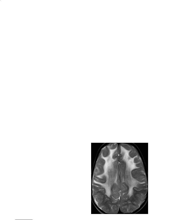
tion shows Rosenthal fibers in the brain, ependyma and pia. It is thought that intracellular deposition of these fibers lead to abnormal functioning of the oligodendrocytes.Arbitrarily it has been divided into infantile, juvenile and adult forms. Macrocephaly is typical and the manifestations include progressive spastic quadriparesis and intellectual failure leading to death (generally all patients die by their teenage years). It generally presents in infancy or adolescence and death occurs as early as 3–10 years after diagnosis. Treatment is only palliative. The disease begins in the frontal regions and then extends posteriorly, and is seen as areas of high T2 signal intensity (Fig. 12.1). The subcortical u-fibers are initially spared but rapidly become affected.The basal ganglia may also appear swollen initially. Although contrast enhancement was, in the past, thought to be rare, it is now known that it occurs in most patients early in the course of the disease. This contrast enhancement
Fig. 12.1. Alexander disease. Axial T2-weighted image shows abnormal hyperintensity in the deep and superficial white matter of the frontal lobes in a symmetrical distribution. (Case courtesy of A. Rossi, Genoa, Italy)