Добавил:
Опубликованный материал нарушает ваши авторские права? Сообщите нам.
Вуз: Предмет: Файл:

Книги по МРТ КТ на английском языке / MR Imaging in White Matter Diseases of the Brain and Spinal Cord - K Sartor Massimo Filippi Nicola De Stefano Vincent Dou

.pdf
Скачиваний:
3
Добавлен:
05.10.2023
Размер:
18.68 Mб
Скачать

232

M. Filippi and M. A. Rocca

Davie et al. 1994; De Stefano et al. 1995a). Recovery of NAA may be related to resolution of edema, increases in the diameter of previously shrunk axons secondary to remyelination and clearance of inflammatory factors and reversible metabolic changes in neurons.

2.Chronic MS lesions are characterized by markedly reduced NAA/Cr peaks. These changes are more pronounced in severely hypointense MS lesions than in isoor mildly hypointense lesions (van Walderveen et al. 1999) and in chronic lesions from patients with SPMS than in those from patients with benign MS (Falini et al. 1998).

3.Cho increase, probably reflecting an altered myelin chemistry or the presence of inflammation, and a decrease in NAA have been also shown in prelesional NAWM (Narayana et al.1998;Sarchielli et al. 1999; Tartaglia et al. 2002).

of the fact that T2-visible lesions represent just a small component of overall brain damage and calls for an accurate assessment of NABT pathology for a better understanding of MS pathophysiology.

3.Metabolite abnormalities, including decrease of NAA and Cho and increase of mI, have also been shown in the cortical GM of MS patients (Kapeller et al. 2001; Sarchielli et al. 2002; Sharma et al. 2001; Chard et al. 2002), since the early phases of the disease (Chard et al. 2002), but not in CIS patients (Kapeller et al. 2002). These changes are more pronounced in patients with SPMS than in those with RRMS (Adalsteinsson et al. 2003). More recently, NAA reduction has also been demonstrated in the thalamus of SPMS (Cifelli et al. 2002) and RRMS patients (Wylezinska et al. 2003).


15.4.3

15.4.2

Correlations with Clinical Manifestations

1H-MRS Findings in NABT

and Disability

1H-MRS also has the potential to provide information on MS pathological changes outside T2-visible lesions. The following are the main findings obtained from the study of the NABT in MS patients using 1H-MRS:

1.Studies of limited and/or selected portions of the brain (Fu et al. 1998; Sarchielli et al. 1999; De Stefano et al. 2002) have shown that NAA reduction is not restricted to MS lesions, but also occurs in the NAWM. These changes are more severe in SPMS and PPMS patients than in those with RRMS (Fu et al. 1998; Suhy et al. 2000); however, they can be detected even in patients with no overt clinical disability (De Stefano et al. 2002) and in those in the early phase of the disease (De Stefano et al. 2001). Diffusely elevated Cho and Cr concentrations have also been described in the NAWM of RRMS (Inglese et al. 2003b) and PPMS (Suhy et al. 2000) patients.

2.The recent development of an unlocalized 1H-MRS sequence for measuring NAA levels in the whole brain (WBNAA) (Gonen et al. 1998) has shown the presence of marked axonal pathology in clinically definite MS (Gonen et al. 2000; Bonneville et al. 2002) and in patients at the earliest clinical phases of MS (Filippi et al. 2003). Interestingly, no correlation has been found between WBNAA concentrations and T2-weighted lesion volumes in patients with RRMS (Bonneville et al. 2002) and in those at presentation with CIS (Filippi et al. 2003a). The lack of such a correlation is likely to be the result

As already mentioned, axonal loss and/or dysfunction is likely to have a major role in the development of fixed clinical disability in MS. In agreement with this notion, a post-mortem study (Bjartmar et al. 2000) has shown a strong correlation between neurological impairment and both axonal density and NAA reduction in the spinal cord of patients with MS. The following are the main in vivo findings obtained from the application of 1H-MRS to the understanding of MS-related disability:

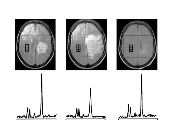
1.Following an acute MS relapse,reversible decreases in NAA have been observed not only in macroscopic lesions responsible for the clinical symptomatology (De Stefano et al. 1995b; Reddy et al. 2000a), but also in the NAWM of the hemisphere contralateral to acute lesions (De Stefano et al. 1999) (Fig. 15.5) and have been correlated with reversal of functional impairment.

2.In patients with RRMS, a longitudinal decrease over time of NAA/Cr in the NAWM correlates strongly with EDSS worsening (De Stefano et al. 1998; Fu et al. 1998), suggesting that progressive axonal damage or loss may be responsible for functional impairment in MS. More recently, it has been demonstrated that brain axonal damage begins in the early stages of MS, develops more rapidly in the earlier clinical stages of the disease and correlates more strongly with disability in patients with mild than in those with more severe disease (De Stefano et al. 2001).

Multiple Sclerosis: Other MR Techniques

233


NAA


NAA


NAA


Cho


Cho

Cr


Cho

Cr


Cr


4.10


p.p.m.

0.78

4.10


p.p.m.

0.78

4.10

p.p.m.

0.78

Fig. 15.5. Proton brain MRI/MRSI examinations of a multiple sclerosis patient performed during the acute phase of the disease (left), 1 month later (center) and 6 months later (right). Conventional proton MRI examinations show a solitary demyelinating lesion that enlarges to involve most of the hemisphere 1 month later (top center), and then decreases in size on the examination at 6 months (top right). Volumes of interest for spectroscopy are shown by the dotted line in each transverse MRI. Averaged spectra from voxels located in normal-appearing white matter contralateral and homologous to the demyelinating lesion (small squares in top panels) are shown in the bottom panels. Note the decrease in the NAA/Cr resonance intensity 1 month after the acute phase of the disease (bottom center). Complete recovery of NAA/Cr intensity ratios occurred at 6 months (bottom right)

3.NAA levels have also been quantified in specific brain regions, whose damage has been related to the impairment of the corresponding functional systems. Davie et al. (1995) showed a significant reduction of NAA concentration in the cerebellar WM of patients with MS and severe ataxia compared with those having little or no cerebellar deficits. Lee et al. (2000a) demonstrated an association between reduction of NAA in the internal capsule and selective motor impairment. Pan et al. (2001) found a relation between cognitive function and NAA levels in the periventricular WM. More recently, Gadea et al. (2004) found a relationship between attentional dysfunction in early RRMS patients and NAA/Cr values in the locus coeruleus nuclei of the pontine ascending reticular activation system.

15.4.4

1H-MRS to Monitor Treatment E cacy

Only four studies have been conducted to evaluate the effect of disease-modifying MS treatments on 1H-MRS-derived parameters (Sarchielli et al. 1998; Narayanan et al. 2001; Schubert et al. 2002; Khan et al. 2003). Using monthly 1H-MRS scans, Sarchielli et al. (1998) found that treatment with interferon beta-1a is associated with increased Cho peaks in spectra of lesions from RRMS patients, suggesting an increase in lesion membrane turnover during the first period of treatment. Narayanan et al. (2001) found an increase of NAA/Cr in a small group of RRMS patients after 1 year of treatment with interferon beta-1b, suggesting a potential effect of treatment in preventing chronic, sublethal axonal

234

M. Filippi and M. A. Rocca

injury. More recently, Schubert et al. (2002) showed a stability of metabolite concentration over time in patients with RRMS treated with interferon beta1b. Finally, Khan et al. (2003) showed an increase of NAA/Cr levels in lesions and NAWM of RRMS patients after 2 years of treatment with glatiramer acetate.

15.5 Functional MRI

Although the resolution of acute inflammation, remyelination, redistribution of voltage-gated sodiumchannels in persistently demyelinated axons, and recovery from sublethal axonal injury are all factors likely to limit the clinical impact of damaging MS pathology (Waxman and Ritchie 1993; De Stefano et al. 1995b), other mechanisms have been recently recognized as potential contributors to the recovery or to the maintenance of function in the presence of irreversible MS-related axonal damage. Brain plasticity is a well known feature of the human brain which is likely to have several pathologic substrates, including an increased axonal expression of sodium channels (Waxman 1998), synaptic changes, increased recruitment of parallel existing pathways or “latent” connections, and reorganization of distant sites. All these changes might have a major adaptive role in limiting the functional consequences of axonal loss. The application of fMRI to the study of the motor, visual and cognitive systems in patients with MS has provided new insights into the mechanisms contributing to the progressive clinical worsening of these patients.

Functional cortical changes have been demonstrated in all MS phenotypes, using different fMRI paradigms. A study of the visual system (Werring et al. 2000b), in patients who had recovered from a single episode of acute optic neuritis (ON), demonstrated that such patients had an extensive activation of the visual network compared to healthy volunteers. An altered brain pattern of movement-associated cortical activations, characterized by an increased recruitment of the contralateral primary sensorimotor cortex (SMC) during the performance of simple tasks (Rocca et al. 2003b; Filippi et al. 2004a) (Fig. 15.6) and by the recruitment of additional “classical” and “higher-order” sensorimotor areas during the performance of more complex tasks (Filippi et al.2004a) has been demonstrated in patients with CIS. An increased recruitment of several sensorimotor areas, mainly located in the cerebral hemisphere ipsilateral to the limb

a

b

Fig. 15.6a,b. Relative contralateral primary sensory-motor cortex (SMC) activation in patients at presentation with clinically isolated syndromes suggestive of multiple sclerosis during the performance of a simple motor task with the right hand in comparison to healthy volunteers (a). The scatterplot of the correlation between the relative activation of the contralateral primary SMC and whole brain N-acetyl aspartate concentrations is shown in (b)

which performed the task has also been demonstrated in patients with early MS and a previous episode of hemiparesis (Pantano et al. 2002a). Interestingly, in patients with similar characteristics, but who presented with an ON, this increased recruitment involved sensorimotor areas which were mainly located in the contralateral cerebral hemisphere (Pantano et al. 2002b). In patients with established MS and a RR course, functional cortical changes have been shown during the performance of visual (Rombouts et al. 1998), motor (Lee et al. 2000b; Reddy et al. 2000a, 2000b; Filippi et al. 2002b; Rocca et al. 2002a), and

Multiple Sclerosis: Other MR Techniques

235

cognitive (Staffen et al. 2002; Hillary et al. 2003; Parry et al. 2003) tasks. Movement-associated cortical changes, characterized by the activation of highly specialized cortical areas, have also been described in patients with SPMS (Rocca et al. 2003c) during the performance of a simple motor task. Two fMRI studies of the motor system (Filippi et al. 2002c; Rocca et al. 2002b)of patients with PPMS suggested a lack of “classical” adaptive mechanisms as a potential additional factor contributing to the accumulation of disability.

The results of all these studies suggest that there might be a “natural history” of the functional reorganization of the cerebral cortex in MS patients, which might be characterized,at the beginning of the disease, by an increased recruitment of those areas “normally” devoted to the performance of a given task, such as the primary SMC and the supplementary motor area (SMA) in case of a motor task.At a later stage, bilateral activation of these regions is first seen, followed by a widespread recruitment of additional areas, which are usually recruited in normal people to perform novel/ complex tasks. This notion has been supported by the results of a recent study (Filippi et al. 2004b), which has provided a direct demonstration that MS patients, during the performance of a simple motor task,activate some regions, that are part of a fronto-parietal circuit, whose activation occurs typically in healthy subjects during object manipulation (Filippi et al. 2004b).

factor modulating movement-associated cortical reorganization, as shown by studies of patients with various disease phenotypes and different levels of disability (Reddy et al. 2000b), patients at presentation with CIS suggestive of MS (Rocca et al. 2003b) (Fig. 15.6), patients with RRMS and no clinical disability (Rocca et al. 2002a), and PPMS and SPMS patients with different degrees of clinical involvement (Filippi et al. 2002c; Rocca et al. 2003c).

d)Subtle GM damage may play a role in modulating cortical excitability, as demonstrated in patients with SPMS (Rocca et al. 2003c) and in patients with clinically definite MS and non-specific (less than three lesions) cMRI findings (Rocca et al. 2003d).

e)Finally, the demonstration of strong correlations between cortical activations and cervical cord damage, quantified using MT MRI, in patients with PPMS (Filippi et al. 2002c), patients with a previous episode of acute myelitis of probable demyelinating origin (Rocca et al. 2003e), and patients with Devic’s neuromyelitis optica (Rocca et al. 2004) suggests that not only brain, but also spinal cord pathology can induce cortical changes with the potential to limit the functional impact of the disease.


15.5.2

15.5.1

Correlations with Clinical Manifestations and

Correlations with Structural Tissue Damage

Disability

The following are the main findings supporting the adaptive role of functional cortical changes in MS:

a)An increased cortical recruitment with increasing T2 lesion load has been shown in patients with relapsing (Lee et al. 2000a; Pantano et al. 2002a; Rocca et al. 2002a), SP (Rocca et al. 2003c) and PP (Rocca et al. 2002b) MS.

b)The severity of intrinsic T2-visible lesion damage has been found to modulate the activity of some cortical areas. A strongly increased recruitment of the primary SMC has been shown to be correlated with the severity of lesion damage of the corticospinal tracts, measured using T1-weighted images (Pantano et al. 2002b) and with the whole brain average lesion MT ratio and mean diffusivity (Rocca et al. 2002a).

c)The severity of NABT damage,measured using 1H- MRS (Reddy et al. 2000b; Rocca et al. 2003b), MT MRI or DW MRI (Filippi et al. 2002c; Rocca et al. 2002a; Rocca et al. 2003c) is another important

Although the actual role of cortical reorganization on the clinical manifestations of MS remains unclear, the demonstration that MS patients may have a normal level of performance despite the presence of diffuse tissue damage suggests that cortical adaptive changes are likely to contribute in limiting the clinical consequences of MS-related structural damage (Filippi and Rocca 2003). The most compelling evidence that cortical reorganization may have a role in recovery from axonal damage derives from the study by Reddy et al. (2000a), who followed a patient after the onset of an acute hemiparesis and a new, large demyelinating lesion located in the corticospinal tract with serial 1HMRS and fMRI exams.In this patient,clinical recovery preceded complete normalization of NAA and was accompanied by increased recruitment of ipsilateral primary SMC and SMA. In line with these findings, in a group of patients who complained of fatigue when compared to matched non-fatigued MS patients,there was a reduced activation of a complex movement-as-

236

sociated cortical/subcortical network, including the cerebellum,the rolandic operculum,the thalamus and the middle frontal gyrus (Filippi et al.2002b).In these patients, a strong correlation between the reduction of thalamic activity and the clinical severity of fatigue was found, indicating that a less marked cortical recruitment might be associated to the appearance of clinical symptomatology in MS. Preliminary work has shown that the pattern of movement-associated cortical activations in MS is determined by both the extent of brain injury and disability and that these changes are distinct (Reddy et al. 2002).

15.6 Conclusions

The application of modern MRI techniques to the assessment of MS patients has considerably improved our understanding of MS pathophysiology and has provided new objective metrics that might be useful to monitor disease evolution, either in natural history studies or in treatment trials. However, none of the quantitative MR-based techniques considered, taken in isolation, is able to provide a complete picture of the complexity of the MS process and this should call for the definition of aggregates of MR quantities, thought to reflect different aspects of MS pathology, to improve our ability to monitor the disease. At present, longitudinal natural history data collected in large samples of MS patients using structural, metabolic and functional MR techniques are needed to gain additional insight into MS pathobiology and on the actual value of modern MR technologies in the management of MS.

References

Adalsteinsson E, Langer-Gould A, Homer RJ et al (2003) Gray matter N-acetyl aspartate deficits in secondary progressive but not relapsing-remitting multiple sclerosis. AJNR Am J Neuroradiol 24:1941–1945

Allen IV, McKeown SR (1979) A histological, histochemical and biochemical study of the macroscopically normal white matter in multiple sclerosis. J Neurol Sci 41:81–91

Arnold DL, Matthews PM, Francis GS et al (1992) Proton magnetic resonance spectroscopic imaging for metabolic characterization of demyelinating plaques. Ann Neurol 31:235–241

Barkhof F, Bruck W, De Groot CJ et al (2003) Remyelinated lesions in multiple sclerosis: magnetic resonance image appearance. Arch Neurol 60:1073–1081

M. Filippi and M. A. Rocca

Basser PJ, Mattiello J, Le Bihan D (1994) Estimation of the effective self-diffusion tensor from the NMR spin-echo. J Magn Reson B 103: 247–254

Bitsch A, Bruhn H, Vougioukas V et al (1999) Inflammatory CNS demyelination: histopathologic correlation with in vivo quantitative proton MR spectroscopy. AJNR Am J Neuroradiol 20:1619–1627

Bjartmar C, Kidd G, Mork S et al (2000) Neurological disability correlates with spinal cord axonal loss and reduced N- acetyl aspartate in chronic multiple sclerosis patients. Ann Neurol 48:893–901

Bjartmar C, Kinkel RP, Kidd G et al (2001) Axonal loss in normal-appearing white matter in a patient with acute MS. Neurology 57:1248–1252

Bonneville F, Moriarty DM, Li BS et al (2002) Whole-brain N-acetyl aspartate concentration: correlation with T2weighted lesion volume and expanded disability status scale score in cases of relapsing-remitting multiple sclerosis. AJNR Am J Neuroradiol 23:371–375

Bozzali M, Cercignani M, Sormani MP et al (2002) Quantification of brain gray matter damage in different MS phenotypes by use of diffusion tensor MR imaging. AJNR Am J Neuroradiol 23:985–988

Caramia F, Pantano P, Di Legge S et al (2002) A longitudinal study of MR diffusion changes in normal appearing white matter of patients with early multiple sclerosis. Magn Reson Imaging 20:383–388

Castriota Scanderbeg A, Tomaiuolo F, Sabatini U et al (2000) Demyelinating plaques in relapsing-remitting and second- ary-progressive multiple sclerosis: assessment with diffusion MR imaging. AJNR Am J Neuroradiol 21:862–868

Cercignani M, Iannucci G, Rocca MA et al (2000) Pathologic damage in MS assessed by diffusion-weighted and magnetization transfer MRI. Neurology 54:1139–1144

Cercignani M, Bozzali M, Iannucci G et al (2001) Magnetisation transfer ratio and mean diffusivity of normal-appear- ing white and gray matter from patients with multiple sclerosis. J Neurol Neurosurg Psychiatry 70:311–317

Chard DT, Griffin CM, McLean MA et al (2002) Brain metabolite changes in cortical grey and normal-appearing white matter in clinically early relapsing-remitting multiple sclerosis. Brain 125:2342–2352

Ciccarelli O, Werring DJ, Wheeler-Kingshott CA et al (2001) Investigation of MS normal-appearing brain using diffusion tensor MRI with clinical correlations. Neurology 56:926–933

Cifelli A, Arridge M, Jezzard P et al (2002) Thalamic neurodegeneration in multiple sclerosis. Ann Neurol 52:650–653 Davie CA, Hawkins CP, Barker GJ et al (1994) Serial proton

magnetic resonance spectroscopy in acute multiple sclerosis lesions. Brain 117:49–58

Davie CA, Barker GJ, Webb S et al (1995) Persistent functional deficit in multiple sclerosis and autosomal dominant cerebellar ataxia is associated with axon loss. Brain 118:1583– 1592

Dehmeshki J, Ruto AC, Arridge S et al (2001) Analysis of MTR histograms in multiple sclerosis using principal components and multiple discriminant analysis. Magn Reson Med 46:600–609

Dehmeshki J, Chard DT, Leary SM et al (2003) The normal appearing grey matter in primary progressive multiple sclerosis: a magnetisation transfer imaging study. J Neurol 250:67–74

Multiple Sclerosis: Other MR Techniques

De Stefano N, Matthews PM, Antel JP et al (1995a) Chemical pathology of acute demyelinating lesions and its correlation with disability. Ann Neurol 38:901–909

De Stefano N, Matthews PM, Arnold DL (1995b) Reversible decreases in N-acetyl aspartate after acute brain injury. Magn Reson Med 34:721–727

De Stefano N, Matthews PM, Fu L et al (1998) Axonal damage correlates with disability in patients with relapsing-remit- ting multiple sclerosis. Results of a longitudinal magnetic resonance spectroscopy study. Brain 121:1469–1477

De Stefano N, Narayanan S, Matthews PM et al (1999) In vivo evidence for axonal dysfunction remote from focal cerebral demyelination of the type seen in multiple sclerosis. Brain 122:1933–1939

De Stefano N, Narayanan S, Francis GS et al (2001) Evidence of axonal damage in the early stages of multiple sclerosis and its relevance to disability. Arch Neurol 58:65–70

De Stefano N, Narayanan S, Francis SJ et al (2002) Diffuse axonal and tissue injury in patients with multiple sclerosis with low cerebral lesion load and no disability. Arch Neurol 59:1565–1571

Dousset V, Gayou A, Brochet B et al (1998) Early structural changes in acute MS lesions assessed by serial magnetization transfer studies. Neurology 51:1150–1155

Droogan AG, Clark CA, Werring DJ et al (1999) Comparison of multiple sclerosis clinical subgroups using navigated spin echo diffusion-weighted imaging. Magn Reson Imaging 17: 653–661

Evangelou N, Esiri MM, Smith S et al (2000) Quantitative pathological evidence for axonal loss in normal appearing white matter in multiple sclerosis. Ann Neurol 47:391–395 Falini A, Calabrese G, Filippi M et al (1998) Benign versus secondary progressive multiple sclerosis: the potential role of 1H MR spectroscopy in defining the nature of disability.

AJNR Am J Neuroradiol 19:223–229

Fazekas F, Ropele S, Enzinger C et al (2002) Quantitative magnetization transfer imaging of pre-lesional white-matter changes in multiple sclerosis. Mult Scler 8:479–484

Ferguson B,Matyszak MK,Esiri MM et al (1997) Axonal damage in acute multiple sclerosis lesions. Brain 120:393–399

Filippi M, Inglese M (2001) Overview of diffusion-weighted magnetic resonance studies in multiple sclerosis. J Neurol Sci 186 [Suppl 1]:S37–S43

Filippi M, Rocca MA (2003) Disturbed function and plasticity in multiple sclerosis as gleaned from functional magnetic resonance imaging. Curr Opin Neurol 16:275–282 (review)

Filippi M, Campi A, Dousset V et al (1995) A magnetization transfer imaging study of normal-appearing white matter in multiple sclerosis. Neurology 45:478–482

Filippi M, Rocca MA, Comi G (1998a) Magnetization transfer ratios of multiple sclerosis lesions with variable durations of enhancement. J Neurol Sci 159:162–165

Filippi M, Rocca MA, Rizzo G et al (1998b) Magnetization transfer ratios in multiple sclerosis lesions enhancing after different doses of gadolinium. Neurology 50:1289–1293

Filippi M, Rocca MA, Martino G et al (1998c) Magnetization transfer changes in the normal appearing white matter precede the appearance of enhancing lesions in patients with multiple sclerosis. Ann Neurol 43:809–814

Filippi M, Grossman RI, Comi G (eds) (1999a) Magnetization transfer in multiple sclerosis. Neurology 53 [Suppl 3]

Filippi M, Iannucci G, Tortorella C et al (1999b) Comparison of

237

MS clinical phenotypes using conventional and magnetization transfer MRI. Neurology 52:588–594

Filippi M, Inglese M, Rovaris M et al (2000a) Magnetization transfer imaging to monitor the evolution of MS: a 1-year follow-up study. Neurology 55:940–946

Filippi M, Tortorella C, Rovaris M et al (2000b) Changes in the normal appearing brain tissue and cognitive impairment in multiple sclerosis. J Neurol Neurosurg Psychiatry 68:157–161

Filippi M, Iannucci G, Cercignani M et al (2000c) A quantitative study of water diffusion in multiple sclerosis lesions and normal-appearing white matter using echo-planar imaging. Arch Neurol 57:1017–1021

Filippi M, Arnold DL, Comi G (eds) (2001a) Magnetic resonance spectroscopy in multiple sclerosis. Springer, Milan Filippi M, Cercignani M, Inglese M et al (2001b) Diffusion

tensor magnetic resonance imaging in multiple sclerosis. Neurology 56:304–311

Filippi M, Dousset V, McFarland HF et al (2002a) The role of MRI in the diagnosis and monitoring of multiple sclerosis. Consensus report of the “White Matter Study Group” of the International Society for Magnetic Resonance in Medicine. J Magn Reson Imag 15:499–504

Filippi M, Rocca MA, Colombo B et al (2002b) Functional magnetic resonance imaging correlates of fatigue in multiple sclerosis. NeuroImage 15:559–567

Filippi M,Rocca MA,FaliniA et al (2002c) Correlations between structural CNS damage and functional MRI changes in primary progressive MS. NeuroImage 15:537–546

Filippi M, Bozzali M, Rovaris M et al (2003) Evidence for widespread axonal damage at the earliest clinical stage of multiple sclerosis. Brain 126:433–437

Filippi M, Rocca MA, Mezzapesa DM et al (2004a) Simple and complex movement-associated functional MRI changes in patients at presentation with clinically isolated syndromes suggestive of MS. Human Brain Mapping 21:108–117

Filippi M, Rocca MA, Mezzapesa DM et al (2004b) A functional MRI study of cortical activations associated with object manipulation in patients with MS. NeuroImage, in press

Fu L, Matthews PM, De Stefano N et al (1998) Imaging axonal damage of normal-appearing white matter in multiple sclerosis. Brain 121:103–113

Gadea M, Martinez-Bisbal MC, Marti-Bonmati L et al (2004) Spectroscopic axonal damage of the right locus coeruleus relates to selective attention impairment in early stage relapsing-remitting multiple sclerosis. Brain 127:89–98

Ge Y, Grossman RI, Udupa JK et al (2001) Magnetization transfer ratio histogram analysis of gray matter in relaps- ing-remitting multiple sclerosis. AJNR Am J Neuroradiol 22:470–475

Ge Y, Grossman RI, Udupa JK et al (2002) Magnetization transfer ratio histogram analysis of normal-appearing gray matter and normal-appearing white matter in multiple sclerosis. J Comput Assist Tomogr 26:62–68

Ge Y, Grossman RI, Babb JS et al (2003) Dirty-appearing white matter in multiple sclerosis: volumetric MR imaging and magnetization transfer ratio histogram analysis. AJNR Am J Neuroradiol 24:1935–1940

Gonen O, Viswanathan AK, Catalaa I et al (1998) Total brain N-acetyl aspartate concentration in normal, age-grouped females: quantitation with non-echo proton NMR spectroscopy. Magn Reson Med 40:684–689

Gonen O, Catalaa I, Babb JS et al (2000) Total brain N-acetyl

238

aspartate. A new measure of disease load in MS. Neurology 54:15–19

Goodkin DE, Rooney WD, Sloan R et al (1998) A serial study of new MS lesions and the white matter from which they arise. Neurology 51:1689–1697

Griffin CM, Chard DT, Ciccarelli O et al (2001) Diffusion tensor imaging in early relapsing-remitting multiple sclerosis. Mult Scler 7:290–297

Hillary FG, Chiaravalloti ND, Ricker JH et al (2003) An investigation of working memory rehearsal in multiple sclerosis using fMRI. J Clin Exp Neuropsychol 25:965–978

Horsfield MA, Lai M, Webb SL et al (1996) Apparent diffusion coefficients in benign and secondary progressive multiple sclerosis by nuclear magnetic resonance. Magn Reson Med 36:393–400

Iannucci G, Minicucci L, Rodegher M et al (1999) Correlations between clinical and MRI involvement in multiple sclerosis: assessment using T1, T2 and MT histograms. J Neurol Sci 171:121–129

Iannucci G, Tortorella C, Rovaris M et al (2000) Prognostic value of MR and magnetization transfer imaging findings in patients with clinically isolated syndromes suggestive of multiple sclerosis at presentation. AJNR Am J Neuroradiol 21:1034–1038

Inglese M, van Waesberghe JH, Rovaris M et al (2003a) The effect of interferon beta-1b on quantities derived from MT MRI in secondary progressive MS. Neurology 60:853–860 Inglese M, Li BS, Rusinek H et al (2003b) Diffusely elevated cerebral choline and creatine in relapsing-remitting mul-

tiple sclerosis. Magn Reson Med 50:190–195

Kalkers NF, Hintzen RQ, van Waesberghe JH et al (2001) Magnetization transfer histogram parameters reflect all dimensions of MS pathology, including atrophy. J Neurol Sci 184155–162

Kapeller P, McLean MA, Griffin CM et al (2001) Preliminary evidence for neuronal damage in cortical grey matter and normal appearing white matter in short duration relaps- ing-remitting multiple sclerosis: a quantitative MR spectroscopic imaging study. J Neurol 248:131–138

Kapeller P, Brex PA, Chard D et al (2002) Quantitative 1H MRS imaging 14 years after presenting with a clinically isolated syndrome suggestive of multiple sclerosis. Mult Scler 8:207–210

Khan O, Shen Y, Ching W et al (2003) Combining immunomodulation and neuroprotection: cerebral axonal recovery in relapsing-remitting multiple sclerosis patients treated with glatiramer acetate. Multiple Sclerosis 9:S63

Kidd D, Barkhof F, McConnell R et al (1999) Cortical lesions in multiple sclerosis. Brain 122:17–26

Kita M, Goodkin DE, Bacchetti P et al (2000) Magnetization transfer ratio in new MS lesions before and during therapy with IFNß-1a. Neurology 54:1741–1745

Le Bihan D, Breton E, Lallemand D et al (1986) MR imaging of intravoxel incoherent motions: application to diffusion and perfusion in neurologic disorders. Radiology 161:401–407

Le Bihan D, Turner R, Pekar J et al (1991) Diffusion and perfusion imaging by gradient sensitization: design, strategy and significance. J Magn Reson Imaging 1:7–8

Lee MA, Blamire AM, Pendlebury S et al (2000a) Axonal injury or loss in the internal capsule and motor impairment in multiple sclerosis. Arch Neurol 57:65–70

Lee M, Reddy H, Johansen-Berg H et al (2000b) The motor

M. Filippi and M. A. Rocca

cortex shows adaptive functional changes to brain injury from multiple sclerosis. Ann Neurol 47:606–613

Loevner LA, Grossman RI, Cohen JA et al (1995) Microscopic disease in normal-appearing white matter on conventional MR images in patients with multiple sclerosis: assessment with magnetization-transfer measurements. Radiology 196:511–515

Lumsden CE (1970) The neuropathology of multiple sclerosis. In: Vinken PJ, Bruyn JW (eds) Handbook of clinical neurology. North-Holland, Amsterdam Vol 9, pp 217-309

McDonald WI,Compston A,Edan G et al (2001) Recommended diagnostic criteria for multiple sclerosis: guidelines from the International Panel on the diagnosis of multiple sclerosis. Ann Neurol 50:121–127

Molyneux PD, Barker GJ, Barkhof F et al (2001) Clinical-MRI correlations in a European trial of interferon beta-1b in secondary progressive MS. Neurology 57:2191–2197

Narayana PA, Doyle TJ, Lai D et al (1998) Serial proton magnetic resonance spectroscopic imaging, contrast-enhanced magnetic resonance imaging, and quantitative lesion volumetry in multiple sclerosis. Ann Neurol 43:56–71

Narayanan S, De Stefano N, Francis GS et al (2001) Axonal metabolic recovery in multiple sclerosis patients treated with interferon beta-1b. J Neurol 248:979–986

Nusbaum AO, Tang CY, Wei TC et al (2000) Whole-brain diffusion MR histograms differ between MS subtypes. Neurology 54:1421–1426

Oreja-Guevara C, Rovaris M, Caputo D et al (2003) Changes in cortical gray matter in untreated relapsing-remitting MS patients: a follow up study. Neurology 60 [Suppl 1]:A297

Pan JW, Krupp LB, Elkins LE et al (2001) Cognitive dysfunction lateralizes with NAA in multiple sclerosis. Appl Neuropsychol 8:155–160

Pantano P, Iannetti GD, Caramia F et al (2002a) Cortical motor reorganization after a single clinical attack of multiple sclerosis Brain 125:1607–1615

Pantano P, Mainero C, Iannetti GD et al (2002b) Contribution of corticospinal tract damage to cortical motor reorganization after a single clinical attack of multiple sclerosis. NeuroImage 17:1837–1843

Parry AM, Scott RB, Palace J et al (2003) Potentially adaptive functional changes in cognitive processing for patients with multiple sclerosis and their acute modulation by rivastigmine. Brain 126:2750–2760

Peterson JW, Bo L, Mork S et al (2001) Transected neurites, apoptotic neurons, and reduced inflammation in cortical multiple sclerosis lesions. Ann Neurol 50:389–400

Pierpaoli C, Jezzard P, Basser PJ et al (1996) Diffusion tensor MR imaging of the human brain. Radiology 201:637–648 Pike GB, De Stefano N, Narayanan S et al (2000) Multiple scle-

rosis: magnetization transfer MR imaging of white matter before lesion appearance on T2-weighted images. Radiology 215:824–830

Reddy H, Narayanan S, Matthews PM et al (2000a) Relating axonal injury to functional recovery in MS. Neurology 54:236–239

Reddy H, Narayanan S, Arnoutelis R et al (2000b) Evidence for adaptive functional changes in the cerebral cortex with axonal injury from multiple sclerosis. Brain 123:2314–2320 Reddy H, Narayanan S, Woolrich M et al (2002) Functional brain reorganization for hand movement in patients with multiple sclerosis: defining distinct effects of injury and

disability. Brain 125:2646–2657

Multiple Sclerosis: Other MR Techniques

Richert ND, Ostuni JL, Bash CN et al (1998) Serial whole-brain magnetization transfer imaging in patients with relapsingremitting multiple sclerosis at baseline and during treatment with interferon beta-1b. AJNR Am J Neuroradiol 19:1705–1713

Richert ND, Ostuni JL, Bash CN et al (2001) Interferon beta1b and intravenous methylprednisolone promote lesion recovery in multiple sclerosis. Mult Scler 7:49–58

Rocca MA, Mastronardo G, Rodegher M et al (1999) Longterm changes of magnetization transfer-derived measures from patients with relapsing-remitting and secondary progressive multiple sclerosis. AJNR Am J Neuroradiol 20:821–827

Rocca MA, Cercignani M, Iannucci G et al (2000) Weekly diffu- sion-weighted imaging of normal-appearing white matter in MS. Neurology 55:882–884

Rocca MA, Falini A, Colombo B et al (2002a) Adaptive functional changes in the cerebral cortex of patients with nondisabling MS correlate with the extent of brain structural damage. Ann Neurol 51:330–339

Rocca MA, Matthews PM, Caputo D et al (2002b) Evidence for widespread movement-associated functional MRI changes in patients with PPMS. Neurology 58:866–872

Rocca MA, Iannucci G, Rovaris M et al (2003a) Occult tissue damage in patients with primary progressive multiple sclerosis is independent of T2-visible lesions-a diffusion tensor MR study. J Neurol 250:456–460

Rocca MA, Mezzapesa DM, Falini A et al (2003b) Evidence for axonal pathology and adaptive cortical reorganisation in patients at presentation with clinically isolated syndromes suggestive of MS. NeuroImage 18:847–855

Rocca MA, Gavazzi C, Mezzapesa DM et al (2003c) A functional magnetic resonance imaging study of patients with secondary progressive multiple sclerosis. NeuroImage 19:1770–1777

Rocca MA, Pagani E, Ghezzi A et al (2003d) Functional cortical changes in patients with MS and non-specific conventional MRI scans of the brain NeuroImage 19:826–836

Rocca MA, Mezzapesa DM, Ghezzi A et al (2003e) Cord damage elicits brain functional reorganization after a single episode of myelitis. Neurology 61:1078–1085

Rocca MA, Agosta F, Mezzapesa DM et al (2004) A functional MRI study of movement-associated cortical changes in patients with Devic’s neuromyelitis optica. NeuroImage, in press

Rombouts SA, Lazeron RH, Scheltens P et al (1998) Visual activation patterns in patients with optic neuritis: an fMRI pilot study. Neurology 50:1896–1899

Rovaris M, Filippi M (1999) Magnetic resonance techniques to monitor disease evolution and treatment trial outcomes in multiple sclerosis. Curr Opin Neurol 12:337–344

Rovaris M,Filippi M,Falautano M et al (1998) Relation between MR abnormalities and patterns of cognitive impairment in multiple sclerosis. Neurology 50:1601–1608

Rovaris M, Filippi M, Minicucci L et al (2000) Cortical/subcortical disease burden and cognitive impairment in multiple sclerosis. AJNR Am J Neuroradiol 21:402–408

Rovaris M, Bozzali M, Santuccio G et al (2001) In vivo assessment of the brain and cervical cord pathology of patients with primary progressive multiple sclerosis. Brain 124:2540–2549

Rovaris M, Bozzali M, Iannucci G et al (2002a) Assessment of normal-appearing white and gray matter in patients

239

with primary progressive multiple sclerosis. Arch Neurol 59:1406–1412

Rovaris M, Iannucci G, Falautano M et al (2002b) Cognitive dysfunction in patients with mildly disabling relapsingremitting multiple sclerosis: an exploratory study with diffusion tensor MR imaging. J Neurol Sci 195:103–109

Rovaris M, Agosta F, Sormani MP et al (2003) Conventional and magnetization transfer MRI predictors of clinical multiple sclerosis evolution: a medium-term follow-up study. Brain 126:2323–2332

Roychowdhury S, Maldijan JA, Grossman RI (2000) Multiple sclerosis: comparison of trace apparent diffusion coefficients with MR enhancement pattern of lesions. AJNR Am J Neuroradiol 21:869–874

Santos AC, Narayanan S, De Stefano N et al (2002) Magnetization transfer can predict clinical evolution in patients with multiple sclerosis. J Neurol 249:662–668

Sarchielli P, Presciutti O, Tarducci R et al (1998) 1H-MRS in patients with multiple sclerosis undergoing treatment with interferon beta-1a: results of a preliminary study. J Neurol Neurosurg Psychiatry 64:204–212

Sarchielli P, Presciutti O, Pelliccioli GP et al (1999) Absolute quantification of brain metabolites by proton magnetic resonance spectroscopy in normal-appearing white matter of multiple sclerosis patients. Brain 122:513–521

Sarchielli P, Presciutti O, Tarducci R et al (2002) Localized (1) H magnetic resonance spectroscopy in mainly cortical gray matter of patients with multiple sclerosis. J Neurol 249:902–910

Schubert F, Seifert F, Elster C et al (2002) Serial 1H-MRS in relapsing-remitting multiple sclerosis: effects of inter- feron-beta therapy on absolute metabolite concentrations. MAGMA 14:213–222

Sharma R, Narayana PA, Wolinsky JS (2001) Grey matter abnormalities in multiple sclerosis: proton magnetic resonance spectroscopic imaging. Mult Scler 7:221–226

Silver NC, Lai M, Symms MR et al (1998) Serial magnetization transfer imaging to characterize the early evolution of new MS lesions. Neurology 51:758–764

Staffen W, Mair A, Zauner H et al (2002) Cognitive function and fMRI in patients with multiple sclerosis: evidence for compensatory cortical activation during an attention task. Brain 156:1275–1282

Suhy J, Rooney WD, Goodkin DE et al (2000) 1H MRSI comparison of white matter and lesions in primary progressive and relapsing-remitting MS. Mult Scler 6:148–155

Tartaglia MC, Narayanan S, De Stefano N et al (2002) Choline is increased in pre-lesional normal appearing white matter in multiple sclerosis. J Neurol 249:1382–1390

Tortorella C, Viti B, Bozzali M et al (2000) A magnetization transfer histogram study of normal-appearing brain tissue in MS. Neurology 54:186–193

Traboulsee A, Dehmeshki J, Brex PA et al (2002) Normalappearing brain tissue MTR histograms in clinically isolated syndromes suggestive of MS. Neurology 59:126– 128

Traboulsee A, Dehmeshki J, Peters KR et al (2003) Disability in multiple sclerosis is related to normal appearing brain tissue MTR histogram abnormalities. Mult Scler 9:566–573 Trapp BD, Peterson J, Ransohoff RM et al (1998) Axonal transection in the lesions of multiple sclerosis. N Engl J Med

338:278–285

van Buchem MA, Grossman RI, Armstrong C et al (1998)

240

Correlation of volumetric magnetization transfer imaging with clinical data in MS. Neurology 50:1609–1617

van Waesberghe JHTM, van Walderveen MA, Castelijns JA et al (1998) Patterns of lesion development in multiple sclerosis: longitudinal observations with T1-weighted spin-echo and magnetization MR. AJNR Am J Neuroradiol 19:675–683

van Waesberghe JH, Kamphorst W, De Groot CJ et al (1999) Axonal loss in multiple sclerosis lesions: magnetic resonance imaging insights into substrates of disability. Ann Neurol 46:747–754

van Walderveen MA, Barkhof F, Pouwels PJ et al (1999) Neuronal damage in T1-hypointense multiple sclerosis lesions demonstrated in vivo using proton magnetic resonance spectroscopy. Ann Neurol 46:79–87

Waxman SG (1998) Demyelinating diseases: new pathological insights, new therapeutic targets. New Engl J Med 338:323–326

Waxman SG, Ritchie JM (1993) Molecular dissection of the myelinated axon. Ann Neurol 33:121–136

M. Filippi and M. A. Rocca

Werring DJ, Clark CA, Barker GJ et al (1999) Diffusion tensor imaging of lesions and normal-appearing white matter in multiple sclerosis. Neurology 52:1626–1632

Werring DJ, Brassat D, Droogan AG et al (2000a) The pathogenesis of lesions and normal-appearing white matter changes in multiple sclerosis. A serial diffusion MRI study. Brain 123:1667–1676

Werring DJ, Bullmore ET, Toosy AT et al (2000b) Recovery from optic neuritis is associated with a change in the distribution of cerebral response to visual stimulation: a functional magnetic resonance imaging study. J Neurol Neurosurg Psychiatry 68:441–449

Wilson M, Tench CR, Morgan PS et al (2003) Pyramidal tract mapping by diffusion tensor magnetic resonance imaging in multiple sclerosis: improving correlations with disability. J Neurol Neurosurg Psychiatry 74:203–207

Wylezinska M, Cifelli A, Jezzard P et al (2003) Thalamic neurodegeneration in relapsing-remitting multiple sclerosis. Neurology 60:1949–1954

Variants of Multiple Sclerosis

241

16 Variants of Multiple Sclerosis

Jack H. Simon and Bette K. Kleinschmidt-DeMasters

CONTENTS

16.1Introduction 241

16.2

Devic’s Neuromyelitis Optica 242

16.2.1General Features 242

16.2.2Neuropathology 244

16.2.3Imaging 244

16.2.4Differential Diagnosis 246

16.3

Acute MS (Marburg Type) 246

16.3.1General Features 246

16.3.2Neuropathology 246

16.3.3Imaging 247

16.3.4Differential Diagnosis 247

16.4

Balo’s Concentric Sclerosis 247

16.4.1General Features 247

16.4.2Neuropathology 248

16.4.3Imaging 248

16.4.4Differential Diagnosis 250

16.5Schilder’s Disease 250

16.5.1General Features 250

16.5.2Neuropathology 251

16.5.3Imaging 251

16.5.4Differential Diagnosis 251 References 252

16.1 Introduction

Around the turn of the last century, several unusual demyelinating conditions were described, including Devic’s disease (1894, neuromyelitis optica), Marburg disease (1906, acute multiple sclerosis, “encephalitis periaxialis diffusa”), Schilder disease (1912, recognized as a childhood variant of Marburg encephalitis periaxialis diffusa by Schilder) and

J. H. Simon, MD, PhD

Departments of Radiology, Neurology, Neurosurgery, University of Colorado Health Sciences Center, 4200 E Ninth Ave, Denver, CO 80262, USA

B. K. Kleinschmidt-DeMasters, MD

Departments of Pathology, Neurology, Neurosurgery, University of Colorado Health Sciences Center, 4200 E Ninth Ave, Denver, CO 80262, USA

Balo disease (1928, “encephalitis periaxialis concentrica,” concentric sclerosis, recognized by Balo to be similar to Marburg and Schilder diseases). All except Devic’s disease were considered by the original authors to represent rare, acute, and/or severe variants of multiple sclerosis (MS).

Debate raged almost immediately as to whether these represented unique demyelinating disorders or MS variants as the original author had often contended. Since several of these original reports were only single cases it took some time before sufficient numbers of patients were accrued by other workers, with tissues reviewed by neuropathologists at autopsy, to put these cases in proper perspective. Adding to the confusion was the fact that Schilder himself subsequently reported two more children with acute demyelinating disorders which he thought represented acute childhood MS, but which subsequently proved to be adrenoleukodystrophy and subacute sclerosing panencephalitis (Prineas et al. 2002).

There are valid differences of opinion as to how rigidly these terms, especially “Devic” and “Balo”, should be applied. Some workers use these eponymic designations when specific distribution patterns are predominantly, but not exclusively, present. An example of this would be the use of the term “Devic’s disease” for MS patients in whom the initial presentation was severe optic nerve and/or spinal cord disease, but who later in the course of the disease develop pathologically proven demyelinative plaques in other central nervous system (CNS) sites. A second instance is the use of these terms when the key variant feature is only minimally or focally present. An example of this would be the use of “Balo disease” for cases of large acute demyelinating lesions that may contain one or more “rings” by neuroimaging studies only in a single lesion, as opposed to the original use of the term which required widespread multiple concentric ring formation throughout cerebral hemispheres, as documented pathologically at autopsy. A final difference is those experts who favor use of