

Книги по МРТ КТ на английском языке / MR Imaging in White Matter Diseases of the Brain and Spinal Cord - K Sartor Massimo Filippi Nicola De Stefano Vincent Dou
.pdfMR Imaging of Brain Development |
157 |
11.3
MR Imaging of the Brain in Fetuses and Prematures
The main anatomic features of the evolving fetal brain have been relatively well established (Fees-Higgins and Larroche 1987; Girard et al. 1995; van der Knaap et al. 1996; Battin and Rutherford 2002). The brain is essentially complete by mid-gestation (20 weeks), but it is still very simple. The migration of the neurons to the cortex is achieved, the anterior posterior development of the corpus callosum is completed, but the brain is small (about 5 cm) and smooth.
There are two ways of calculating the age of a fetus: from the time of conception, which is often un-
certain, and from the time of the last period, which is more easy to identify. By convention, the mode of dating used by obstetricians, based on the last period, is used here. In theory at least, the gestational age is 2 weeks less.
11.3.1
Gross Morphology of the Fetal and Premature Brain
The sylvian pits are largely open with exposed insula at 20 weeks (Fig. 11.2), then the operculation of the adjacent lobes progressively encloses it. Around it, the gyration is just appearing: parieto-occipital fissure (present at 20 weeks), calcarine fissure (ap-
a 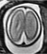
 b
b
c |
d |
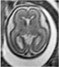
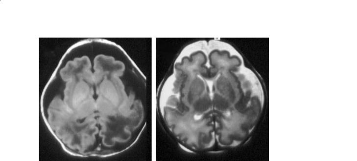
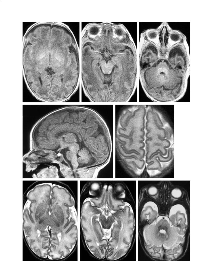
Fig. 11.2a–d. Fetus, 20 weeks. a T2 axial, centrum semi-ovale. Smooth cortex. Within the mantle, from the ventricle to the periphery: germinal matrix of the mantle, adjacent to the ventricle (dark, on the right side); subcortical zone/migrating glia layer (intermediate signal); subplate (brighter); smooth cortical ribbon; wide pericerebral fluid spaces; calvarium. b T2 axial, basal ganglia.Ventricular pseudo-dilatation (colpocephaly, fetal “hydrocephalus”), relative to the still under-developed mantle. Darker cluster of the germinal matrix of the ganglionic eminence over the heads of the caudates. The signal of the basal ganglia is brighter than that of the cortex, and about identical to that of the subventricular zone. c T2 coronal. d T2 sagittal, midline. In the normal posterior fossa (the tentorium facing the inion), the vermis looks small. Wide interparietal subarachnoid spaces
158 |
C. Raybaud |
pearing by |
24 weeks, achieved and Y-shaped by |
28 weeks), |
calloso-marginal sulcus (28 weeks), |
central sulcus (apparent by 24 weeks, achieved by 35 weeks) with the preand post-central sulci (from 26 to 35 weeks), and the superior temporal sulcus (clearly demarcated from its anterior to its posterior end by 28 weeks). These sulci, very shallow at the beginning, become progressively deeper as the gyri develop, until after birth (van de Knaap et al. 1996). Up to the last weeks, these sulci are linear (Fig. 11.4), and devoid of tertiary branching. It is only during the last weeks before term, when the cortex comes to abut against the vault, that the sulcal pattern becomes more complex, and then difficult
to analyze precisely because of the packing against the bone (Fig. 11.7).
The lateral ventricles are less than 10 mm at the atrium at mid-gestation (Fig. 11.2 and 11.3), with a slight reduction toward birth (7.5 mm). The apparent fetal “hydrocephalus”, also called colpocephaly, is only in relation to the brain mantle thickness. The width of the lateral ventricles remains almost constant, whereas the brain is building up around them. But as the brain is growing in every direction, the length of the ventricles also increases. From the beginning, the ventricular segments (frontal, temporal horns, body, atrium, 3rd ventricle, 4th ventricle, even commonly the aqueduct) are clearly rec-
a |
b |
c |
d |
e |
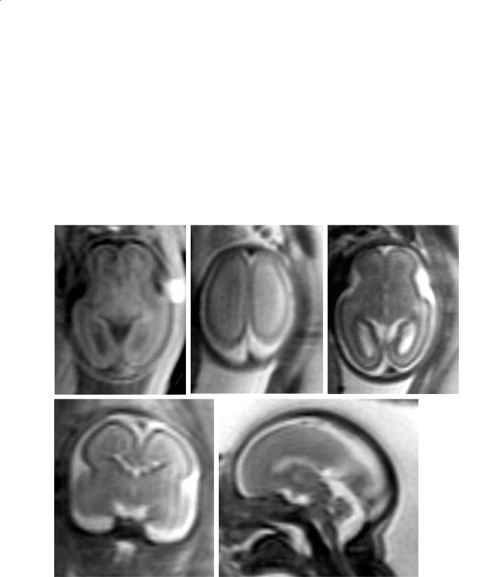
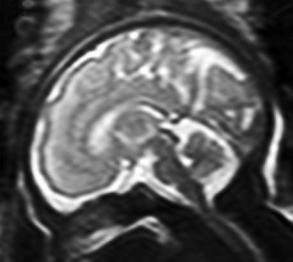
Fig. 11.3a–e. Fetus, 24 weeks. a T1 axial, basal ganglia. Morphology still similar to that of 20 weeks. The grey matter structures (cortex, germinal matrix, basal ganglia, subventricular zone/ migrating glia layer) have a brighter signal than that the subplate subjacent to the cortex. b T2 axial, centrum semi-ovale. Smooth cortex. c T2 axial, basal ganglia. Similar to the 20-week brain. d T2 coronal, 3rd ventricle. Early operculation of the sylvian fissure. e T2 sagittal, midline. Faint appearance of the main vermian sulci
MR Imaging of Brain Development |
159 |
a |
|
b |
|
c |
|
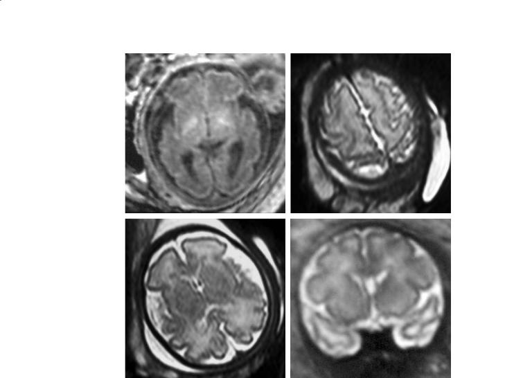
Fig. 11.4a–d. Fetus, 28–29 weeks. a T1 axial, basal ganglia. |
|
|
Early myelination process around the PLICs. The subven- |
|
|
tricular zone/migrating glia layer are not seen anymore. Clear |
|
|
operculation around the Sylvian fissure. Temporal sulcation |
|
|
well seen. b T2 axial, centrum semi-ovale. Clear central sulcus, |
|
|
beginning preand postcentral sulci. c T2 axial, basal ganglia. |
|
|
Seemingly smaller ventricles (in fact the mantle is thicker). |
|
|
The subventricular zone/migrating glia layer has vanished, ex- |
|
|
cept for remnants in front of the frontal horns. The subplate is |
|
|
underlined by a thin denser layer. Temporal sulcation appar- |
|
|
ent. Still large pericerebral fluid spaces. d T2 sagittal, midline. |
|
|
Calloso-marginal sulcus well seen, parieto-occipital and cal- |
|
|
carine fissures prominent. The perivermian fluid spaces are |
|
|
still large; the indentation of the vermian sulci are seen. The |
|
d |
||
tentorium faces the inion |
ognizable. In between the frontal horns, the cavum septi pellucidi is relatively large, sometimes slightly bulging. The corpus callosum (and on occasion the hippocampal commissure as well) is better depicted on the coronal cuts. The peri-cerebral subarachnoid fluid spaces are large and conspicuous in the early pregnancy; they progressively attenuate until 32 weeks, when the cortex comes to abut the vault (Figs. 11.2–11.7).
The cerebellum is disproportionately small in relation to the size of the posterior fossa at about 20 weeks, but grows significantly to nearly fill it at term (Figs. 11.2–11.8), but keeps a clearly demarcated cisterna magna. The limits of the posterior fossa, and especially the insertion of the tentorium facing the inion, are always evident. At 20 weeks (at best) or slightly later, only the main cerebellar fissures are seen on the sagittal cut of the vermis. The other vermian sulci become apparent as the vermis enlarge. It should be noted that not seeing the ver-
mian sulci does not mean that they are not present, but simply that they are blurred by the partial volume effects, since even 2 mm cuts are still exceedingly thick as compared with the structures studied. The brainstem is also well depicted with its three segments, midbrain, pons and medulla (Figs. 11.2– 11.8).
11.3.2
The Intrinsic Anatomy of the Fetal and Premature Brain
In the fetus, or in the premature, the brain is a clearly multilayered structure. This results from the fact that the histogenesis is not yet achieved, even though the neuronal migration is practically complete by 20 weeks. Cellular proliferation goes on until term, in both germinal matrices (mantle and ganglionic eminence), to produce astrocytes and oligodendrocytes.
160 |
C. Raybaud |
a |
b |
Fig. 11.5a,b. Premature baby, 30 weeks. Ex utero imaging with conventional, age-adapted MR sequences: the image definition and contrast are much better than with in utero imaging. a T1 axial, basal ganglia. Clear contrast between the grey and the white matter, even in the PLICs. At least on the right, the subplate is clearly seen as a denser subcortical layer. Residues of the subventricular zone/migrating glia layer in front of the frontal horns. Early myelination process apparent in the posterior lentiform nuclei and anterior-lateral thalami, as well as in the primary acoustic cortices in the posterior insular areas. No myelination seen in the PLICs. b T2 axial, basal ganglia. Clearly established sulcation, globally. Well advanced perisylvian operculation. The subplate is clearly seen in the frontal lobes. A denser appearance of the corpus callosum and fornices reflects a early myelination in respect to the rest of the white matter. Large pericerebral subarachnoid and perhaps, subdural fluid spaces
These cells constitute a transient, highly cellular layer located around the ventricles in the deeper portion of the white matter (Figs. 11.2–11.4). This so-called layer of migrating glial cells (Childs et al. 1998), however, stays away from the cortex, from which it is separated by the (also) transient neuronal layer of the subplate (Figs. 11.2–11.8). All these structures develop and then disappear at different times, giving the cerebral mantle different appearances at different stages of development. In addition, in the last gestational trimester, the myelination is clearly beginning to appear mostly in the deep central structures (Figs. 11.4–11.8).
The germinal matrix lining of the ventricular wall is present until term, mostly from the ganglionic eminence (Fig. 11.7). In relation to the whole brain, it is more prominent at the beginning, then it disappears nearly completely around term. It is highly cellular, and therefore clearly seen as a dark T2, bright T1 periventricular band; on T1 however it tends to blend with the adjacent highly cellular subventricular zone. The germinal matrix appears thicker over the basal ganglia (ganglionic eminence) than along the white matter. Germinal matrix hemorrhages developing in the premature typically affect the ganglionic eminence, but more modern studies in premature babies using MR imaging rather than US or X-CT demon-
strate that the germinal matrix of the mantle, along the ventricles, is also affected. When the germinal matrix of the mantle, lining the white matter, shows discontinuities on fetal MR imaging, a destructive process (infection and/or periventricular leukomalacia) should be suspected.
Until week 28, the dense subventricular zone is seen deep in the white matter, extending to the periphery only to about midway between the germinal matrix and the cortex (Figs. 11.2–11.4). It is generally considered that it contains the differentiating and migrating glial cells, but recent evidence indicates that it is a mixture of a deeper fiber rich zone, of the subventricular cellular zone and migrating glia layer, and of the future white matter (Kostovic et al. 2002). On T1 imaging, it tends to fuse with the image of the germinal matrix. On T2, it is identified by a denser line that separates it from the subplate. The image of this intermediate layer “dissolves” between weeks 28 and 30, but leaves behind a few remnants persisting until term, especially in front of the frontal horns (Figs. 11.4–11.8).
After the disappearance of the subventricular layer, the sub-cortical layer of the transient subplate is better demonstrated against the underlying white matter, as a high T1, low T2 band subjacent to the cortex (Fig. 11.5 to 11.8). Its inner limit is commonly marked
MR Imaging of Brain Development |
161 |
a |
b |
c |
d |
 e
e
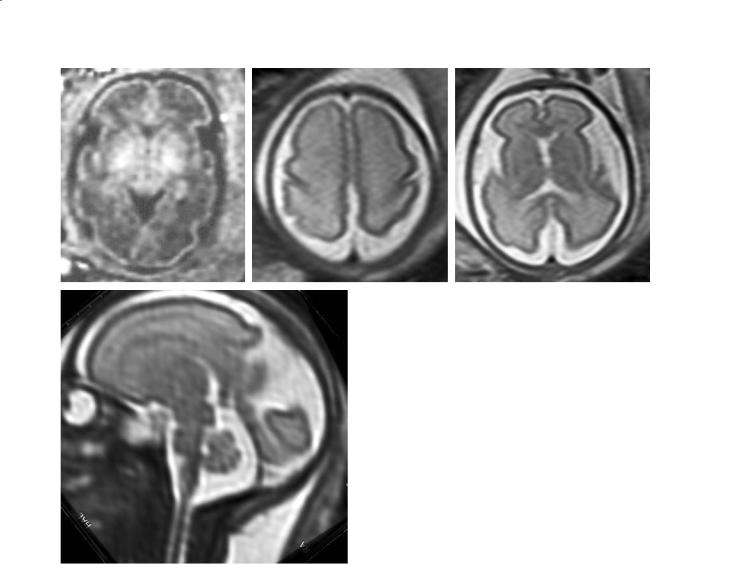
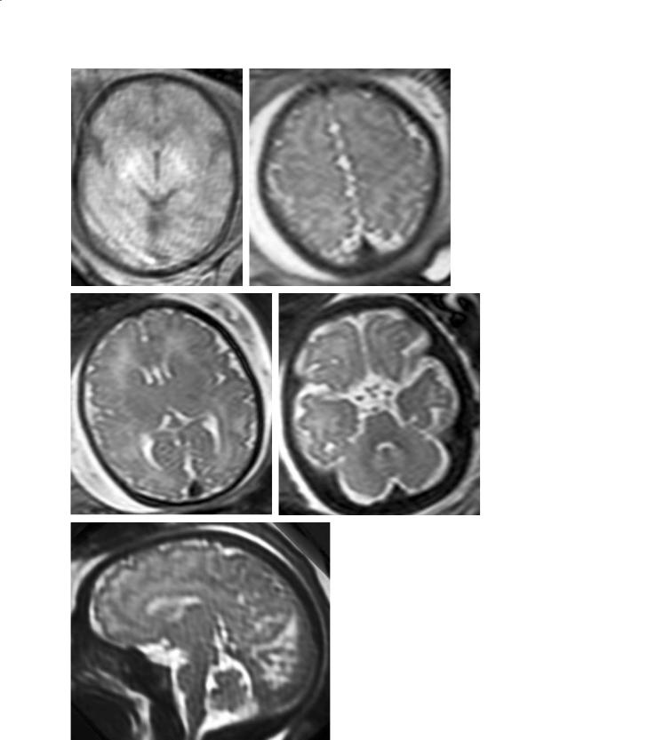
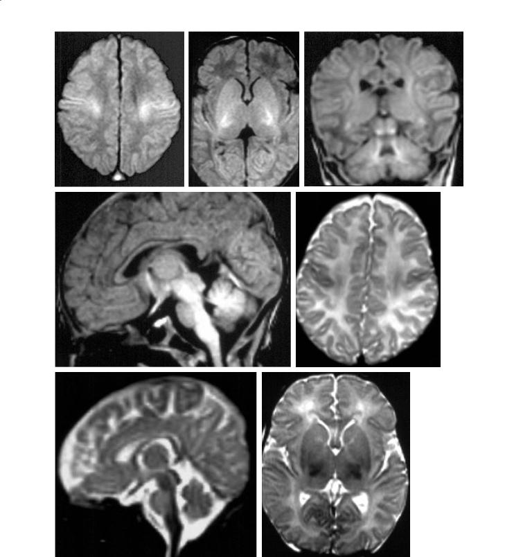
Fig. 11.6a–e. Fetus, 32–33 weeks. a T1 axial, basal ganglia. Clearly seen bright signal of myelination in process in and around the PLICs. The cortex is well convoluted, the pericerebral spaces still large. b T2 axial, centrum semi-ovale. Essentially complete sulcation pattern, even if the sulci are still shallow. c T2 axial, basal ganglia. Clearly delineated subplate. Subventricular residue adjacent to the frontal horns. The thalami are darker, together with the myelinating proximal optic radiations. d T2 coronal. Clearly sulcated cortex. e T2 sagittal, midline. The vermis is larger and now fills the posterior fossa better. Vermian sulci clearly apparent. Sylvian aqueduct well seen. Well developed medial hemispheric sulcal pattern. Dark, already well myelinated brainstem
162 |
C. Raybaud |
a |
b |
c |
d |
Fig. 11.7a–e. Fetus, 37 weeks. a T1 axial, basal ganglia. Clear changes related to the myelination process in the PLICs. b e T2 axial, centrum semi-ovale. The sulcation is well developed,
the cortex is poorly seen because it is now thin in relation to the mantle, and quite convoluted. The pericerebral fluid spaces are not large anymore. c T2 axial, basal ganglia. Focally, the dark cluster of the germinal matrix of the ganglionic eminence persists at the level of the foramen of Monro. The optic radiations are seen lining the ventricular atria, against the non-myelinated, adjacent white matter. The subplate can still be recognized as a subcortical blurring. Discrete areas of myelination in the basal ganglia and thalami adjacent to the unmyelinated PLICs [(compare with (a)]. d T2 axial, posterior fossa. Myelination apparent in the tegmentum pontis and the adjacent brachia pontis. The cerebellum now almost fills the posterior fossa. e T2 sagittal, midline

MR Imaging of Brain Development |
163 |
by a T2 denser line. This layer persists until term. Its signal is related to the composition of its extracellular matrix rather than to the cellular density (Kostovic et al. 2002). It is the subcortical remnant of the primary preplate (or plexiform layer) that has been divided by the developing cortical plate (Kostovic and Rakic 1990). It is an extremely important structure as it serves as a transient relay for the in-coming fi- bers of the corona radiata (thalamo-cortical fibers), of the association paths, and of the commissures (Kostovic and Rakic 1990; Super et al. 1998; Xie et al. 2002). This role in controlling the connectivity of the cortex may be compromised by the occurrence of subcortical injuries.
The cortical ribbon is already close to its final thickness (1–3 mm for most of the cortical areas) at mid-gestation, but because of the developing connections (intracortical fibers, thalamo-cortical fi- bers, association and commissural fibers), it will not be fully organized until birth. It is clearly identified as a bright T1, dark T2 ribbon lining the periphery of the brain.Because it grows tangentially more than the spheric volume of the brain would allow, it progressively develops indentations corresponding to the sulci (Figs. 11.2–11.8).As the brain grows and presses against the vault, it becomes less and less easy to see, and it seems thinner in relation to the globally increased brain size.
The basal ganglia and thalamus form a mass of dense cellularity, brighter on T1 and darker on T2, overlaid by the germinal matrix of the ganglionic eminence, already at 20 weeks (Fig. 11.2). After 30– 32 weeks, the T1 signal increases in and around the PLICs (Figs. 11.4–11.8), reflecting the myelination process that develops early in this area that includes the cortico-spinal tract and the adjoining portions of the basal ganglia and thalami, the optic tracts, as well as in the brainstem. This pre-myelination high signal extends toward the central cortex in the last weeks before term.
11.4
MR Imaging of the Brain in Term-Born Infants
Many publications have been devoted to imaging of the development of the brain in the first months of life (Barkovich et al. 1988; Bird et al. 1989;
Christophe et al. 1990; van der Knaap and Valk
1990; Girard et al. 1991; Martin et al. 1991; Sie et al. 1997; Barkovich 1998). In children born at term, the brain anatomy is essentially complete. Yet the corpus
callosum is thinner than it will be later, not so much due to lack of fibers but because being markedly myelinated, it reaches its final size only after maturation is completed. In the posterior fossa, the neuronal proliferation in the cerebellum continues until the end of the second year, and only then does the cerebellum present its “final” appearance.
From a general point of view, however, most of the changes occurring in the first 2 years are related to myelination. As specified earlier in this chapter, two essential phenomena are responsible for producing the contrast of the MR images. The first is the magnetization transfer effect induced by the precursors of the myelin, which shortens the relaxation time T1 and results in a bright signal on T1w images. The second is the substitution of the achieved myelin to the water, which leads to a general loss of signal and shortens the relaxation time T2, and therefore results in a dark signal on T2w images. The T1 and the T2 images therefore bring different information at this stage. T1 images basically depict the early stages of the myelination processes, while T2 images show the “completed” myelination. Therefore there is an “appearance delay” between the T1 and the T2 images, the changes on T1 being more precocious than on T2, roughly by 4 months. Histologically, this myelination process develops itself according to a precisely determined timetable, both in the cortex, in the white matter, in the central area (gray nuclei and capsules), in the brainstem and in the cerebellum.
11.4.1
The Brain at Term
11.4.1.1 T1 Imaging
At birth, the white matter appears globally darker than the cortex. However, different areas in each structure may show a different appearance. Areas of higher signal are more mature than areas of lower signal.
In the cerebral cortex, four areas have a higher signal than the surrounding structures: the pericentral (peri-rolandic, sensory-motor) cortex, the medial occipital cortex centered on the calcarine fissure (primary visual cortex), the posterior insular temporal area (primary auditory cortex), and the temporo-me- sial structures (hippocampus). These are the primary cortical areas, the oldest phylogenetic cortical struc-
164 |
C. Raybaud |
a |
b |
c |
d |
e |
f |
g |
h |

MR Imaging of Brain Development |
165 |
tures, already present in rudimentary mammals such as the shrew (Romer and Parsons 1977).
In the subcortical white matter, a diffuse higher signal is seen below the central area, more consistent centrally and more diluted peripherally; it corresponds to the cortico-spinal tract. Some higher signal is also seen lining the ventricular atria, fitting the distribution of the optic radiations.
In the central brain, there is a conspicuous high signal centered on the PLICs, and involving the adjacent portions of the lentiform nuclei and the ventrolateral portions of the thalami. In the same area, the anterior commissure and the anterior optic pathways (optic nerves, chiasm, tracts) also show some higher signal.
Fig. 11.8a–i. Premature baby, 37 weeks. Ex utero imaging (compare with Fig. 11.6). a T1 axial, basal ganglia. Myelination in process in the portions of the basal ganglia and thalami adjacent to the PLICs, which themselves appear not yet to myelinate. b T1 axial, midbrain. Clearly brighter signal of the colliculi. c T1 axial, pons. Clear myelination process in the tegmentum pontis. d T1 sagittal midline. Clear myelination process of the midbrain, tegmentum pontis, medulla. Strong high signal of the pituitary gland against the globally lower signal of the (only partially myelinated) brain. e T2 axial, centrum semi-ovale. Clearly well developed sulcation. f T2 axial, basal ganglia. The subplate can still be identified, as well as the remnants of the subventricular zone/migrating glia layer in front of the frontal horns. Clear myelination of the posterior-lateral thalami, and of the rest of the basal ganglia to a lesser degree, but the adjacent PLICs remain apparently not, or only poorly, myelinated. Early myelination of the optic radiations clearly apparent. g T2 axial, midbrain. Global myelination, more advanced in the colliculi. h T2 axial, pons. Fascicular and nuclear myelination in the tegmentum pontis. i T2 sagittal, midline. Well developed sulcation of the medial cortex. Nuclear/fascicular partial myelination of the brainstem and deep vermian white matter. Clear vermian sulcation
In the posterior fossa, the tegmentum (posterior segment) of the brainstem has a higher signal than the anterior pons, as well as the adjacent white matter of the cerebellum. The colliculi are also brighter.
It is worthwhile noting that at this age, the pituitary gland appears exceedingly bright on cerebral imaging. In fact, it has the normal signal of the soft tissue, but it is the brain that has an unusually low signal, to which the imaging windows are adjusted. This contrast between the pituitary gland and the brain eventually disappears as the brain becomes myelinated.
11.4.1.2 T2 Imaging
The images at this stage are nearly,but not exactly,the reverse of the T1 images. Globally, the white matter is brighter than the cortex.
Within the cortex, areas of lower signal (more mature) are found again in the sensory-motor area, the primary motor area, the primary auditory area, and the hippocampus.
In the white matter, areas of faint, decreased signal are seen in the location of the cortico-spinal tracts, but typically not at the level of the optic radiations. An area of lower signal may be observed at some distance in front of the lower part of the frontal horns. This does not reflect myelination, but rather a remnant of the cellular subventricular band.
In the central forebrain, a minute dark fascicle is inconstantly observed in the postero-lateral portion of the PLICs. Dark spots are also seen in the adjacent thalami.
In the brainstem, the tegmentum is darker than the anterior brainstem. The colliculi also appear more mature, as well as the cerebellar white matter surrounding the cerebellar dentate nuclei.
11.4.2
The Brain at 4 Months
11.4.2.1 T1 Imaging
|
Four months is an important time to evaluate myelina- |
|
tion as typically at this stage the diffuse increase of sig- |
|
nal of the white matter matches the signal of the cortex, |
|
globally with a loss of the contrast between the two.The |
|
primary bundles of white matter have progressively |
i |
taken a more mature appearance,from the center to the |
periphery toward their corresponding cortex. |
a |
b |
c |
d |
e |
f |
g |
Fig. 11.9a–g. Term neonate. a T1 axial, centrum semi-ovale. White matter clearly darker than the cortex. Myelination in process of the peri-central cortices and of the subjacent bundles of cortico-subcortical fibers. b T1 axial, basal ganglia. White matter darker than the cortex and the basal ganglia. Bright signal of the myelination process in the whole area surrounding the PLICs, and to a lesser degree of the optic radiations. c T1 coronal, thalami. Myelination in process of the corticospinal bundles (centrum semi-ovale on each side), the optic radiations (lateral to the temporal horns), the colliculi, and of the deep cerebellar white matter. d T1 sagittal, midline. Myelination in process of the hypothalamic structures and optic chiasm, the midbrain, tegmentum pontis, medulla and cerebellar white matter. e T2 axial, centrum semi-ovale. Darker myelination signal of the cortex in the vicinity of the central sulcus, and more faintly, of the subjacent white matter. f T2 axial, basal ganglia. Myelination of the lateral portions of the thalami, the posterior putamina to a lesser degree, as well as of a clearly demarcated fascicle in the posterior lateral part of the PLICs. g T2 sagittal, midline. Strong myelination signal of the fornix and optic chiasm, the midbrain, the tegmentum pontis (and perhaps the anterior pes pontis), and of the medulla