- •Preface
- •Acknowledgements
- •Contents
- •The Team
- •The Instruments
- •Patient Positioning
- •Setup for Upper Abdominal Surgery
- •Setup for Lower Abdominal Surgery
- •The Working Environment
- •Appraisal of Surgical Instruments
- •Trocars
- •Other Instrumental Requirements
- •Troubleshooting Loss of Pneumoperitoneum
- •Principles of Hemostasis
- •Control of Bleeding of Unnamed Vessels
- •Control of Bleeding of a Main Named Vessel
- •Selected Further Reading
- •2 Cholecystectomy
- •Impacted Stone (Hydrops, Empyema, Early Mirizzi)
- •Adhesions Due to Previous Upper Midline Laparotomy
- •Selected Further Reading
- •Selected Further Reading
- •The Need for Specialized Equipment
- •Access to the Liver
- •Maneuvers Common to All Laparoscopic Liver Surgery
- •Resection of Liver Tumors
- •Limited Resection of Minor Lesions
- •Left Lateral Segmentectomy
- •Right Hepatectomy
- •Patient Selection
- •Principles of Surgical Therapy in the Management of Gastroesophageal Reflux Disease
- •Patient Positioning
- •Technique
- •Postoperative Course
- •Management of Complications
- •Paraesophageal Hernia
- •Esophageal Myotomy for Achalasia
- •Vagotomies
- •Bilateral Truncal Vagotomy
- •Highly Selective Vagotomy
- •Lesser Curvature Seromyotomy and Posterior Truncal Vagotomy
- •Selected Further Reading
- •Pyloroplasty
- •Vagotomy with Antrectomy or any Distal Gastrectomy
- •Port Placement
- •Technique
- •Locating the Perforation
- •Abdominal Washout
- •Closure of the Perforation with an Omental Patch
- •Postoperative Course
- •Selected Further Reading
- •7 Appendectomy
- •OR Setup and Port Placement
- •Technique
- •Gangrenous or Perforated Appendicitis
- •Laparoscopic Assisted Appendectomy
- •Left Hemicolectomy
- •Reversing the Hartmann Procedure
- •Selected Further Reading
- •Selected Further Reading
- •Transabdominal Preperitoneal Repair (TAPP)
- •Patient and Port Positioning
- •Dissection of the Preperitoneal Space
- •Dissection of the Cord Structures and the Vas Deferens
- •Placement of the Mesh and Fixation
- •Closure of the Peritoneum
- •Indications
- •Technique
- •Positioning
- •Pneumoperitoneum
- •Port Placement
- •Adhesiolysis
- •Measurement of the Hernia Defect
- •Placement of Mesh
- •Difficult Ventral or Incisional Hernias
- •Pain Following Laparoscopic Ventral or Incisional Hernia Repair
- •Preoperative Requirements and Workup
- •Patient Positioning
- •Port Placement
- •Surgical Anatomy
- •Surgical Principles
- •Technique
- •Division of the Short Gastric Vessels and Exposure of the Tail of the Pancreas
- •Division of the Hilar Vessels and Phrenic Attachments
- •Extraction of the Spleen in a Bag
- •Final Steps of the Procedure
- •Control of an Unnamed Vessel
- •Control of a Major Vessel
- •Splenic Injury
- •Maneuver of Last Resort During Bleeding of the Hilar Vessels
- •Distal Splenopancreatectomy
- •Selected Further Reading
- •13 Adrenalectomy
- •Principles
- •Patient Positioning
- •Technique
- •Immediate Postoperative Complications
- •Late Postoperative Complications
- •Laparoscopic Adjustable Band
- •Technique
- •Complications
- •Laparoscopic Sleeve Gastrectomy
- •Selected Further Reading
- •Laparoscopic Cholecystectomy
- •Laparoscopic Appendectomy
- •Laparoscopic Inguinal Hernia Repair
- •Selected Further Reading
- •Monitors
- •OR Table
- •Trocar Placement and Triangulation
- •Equipment
- •Needle Holders
- •Graspers
- •Suture Material
- •Intracorporeal Knot-Tying
- •Interrupted Stitch
- •Running Stitch
- •Pirouette
- •Extracorporeal Knot-Tying
- •Roeder’s Knot
- •Endoloop
- •Troubleshooting
- •Lost Needle
- •Short Suture
- •Subject Index
Selected Further Reading |
255 |
Arterburn D, Livingston EH, Schifftner T, Kahwati LC, Henderson WG, Maciejewski ML (2009) Predictors of long-term mortality after bariatric surgery performed in Veterans Affairs medical centers. Arch Surg 144(10):914–20
Collins BJ, Miyashita T, Schweitzer M, Magnuson T, Harmon JW (2007) Gastric bypass: why Roux-en-Y? A review of experimental data. Arch Surg 142(10):1000–1003; discussion 1004
Flum DR, Belle SH, King WC, Wahed AS, Berk P, Chapman W, Pories W, Courcoulas A, McCloskey C, Mitchell J, Patterson E, Pomp A, Staten MA,Yanovski SZ, Thirlby R,Wolfe B (2009) Perioperative safety in the longitudinal assessment of bariatric surgery. Longitudinal Assessment of Bariatric Surgery (LABS) Consortium. N Engl J Med 361(5):445–54
Frezza EE, Mammarappallil JG, Witt C, Wei C, Wachtel MS (2009) Value of routine postoperative gastrographin contrast swallow studies after laparoscopic gastric banding. Arch Surg 144(8):766–9
Higa KD, Boone KB, Ho T, Davies OG (2000) Laparoscopic Roux-en-Y gastric bypass for morbid obesity: technique and preliminary results of our first 400 patients. Arch Surg 135(9):1029–33
Katkhouda N, Moazzez A, Gondek S, Lam B (2008) A new and standardized technique for trocar placement in laparoscopic Gastric Bypass. Surg Endosc 23(3):659–62
Khoueir P, Black MH, Crookes PF, Kaufman H, Katkhouda N (2009) Way MY Prospective assesment of axial backpain symptoms before and after bariatric weightloss surgery. Spine J 9(6):454–63
Nguyen NT,Root J,Zainabadi K, Sabio A,Chalifoux S, Stevens CM, Mavandadi S,Longoria M, Wilson SE (2005) Accelerated growth of bariatric surgery with the introduction of minimally invasive surgery. Arch Surg 140(12):1198–202
Puzziferri N, Austrheim-Smith IT, Wolfe BM, Wilson SE, Nguyen NT (2006) Three-year follow-up of a prospective randomized trial comparing laparoscopic versus open gastric bypass. Ann Surg 243(2):181–8
Smith BR, Hinojosa MW, Reavis KM, Nguyen NT (2008) Remission of diabetes after laparoscopic gastric bypass. Ann Surg 74(10):948–52
Suter M,Calmes JM,Paroz A,Romy S,Giusti V (2009) Results of Roux-en-Y gastric bypass in morbidly obese vs superobese patients: similar body weight loss, correction of comorbidities, and improvement of quality of life. Arch Surg 144(4):312–8
Thodiyil PA, Yenumula P, Rogula T, Gorecki P, Fahoum B, Gourash W, Ramanathan R, Mattar SG, Shinde D, Arena VC, Wise L, Schauer P (2008) Selective nonoperative management of leaks after gastric bypass: lessons learned from 2675 consecutive patients. Ann Surg 248(5):782–92
Selected
Further
Reading

Single-Access 15
Laparoscopic
Surgery
(SALS)
The concept of single-access laparoscopic surgery is to perform the procedure through one skin access (port). Several acronyms have been used to describe this concept, such as our single-access laparoscopic surgery (SALS), single port access (SPA) laparoendoscopic single-site surgery (LESS), and single incision laparoscopic surgery (SILS), and they all refer to the same procedure.
The umbilicus is usually used for basic procedures such as laparoscopic cholecystectomy, |
Basic |
appendectomy, and hernias. The incision can circumscribe the umbilicus or go through |
Transumbilical |
the umbilicus, as depicted in Fig. 15.1. In the latter technique, the umbilicus is carefully |
Procedures |
cleaned, and two kocher clamps invert the umbilicus to expose the skin. An eleven blade |
|
is used to cut the skin, and then the skin incision is enlarged with a Kelly. Two S-retractors |
|
are inserted to retract the edges of the skin incision, and a fine scissor is used to create a |
|
pocket, developing a fascial plane slightly larger than the skin incision. It is then impor- |
|
tant to identify the natural defect in the linea alba, which is then enlarged by inserting a |
|
Kelly and spreading it open. Carefully, a blunt 5-mm trocar, preferably without any protu- |
|
berant plastic edges, is inserted and the abdomen is then insufflated. At this point, a spe- |
|
cial long 40 cm, 5 mm, 30° bronchoscope (Karl Storz, Tuttlingen, Germany) is inserted |
|
between and hooked to the camera, and the exploration of the abdomen is performed.All |
|
the trocars are then inserted in a staggered fashion (Fig. 15.2). This figure also clearly |
|
shows the relationship of the different trocars and the long 5 mm scope. Ideally, they |
|
should be short and stealthy to avoid the “knitting needle” effect of the trocars. We prefer |
|
insertion of the 5 mm camera site at about 7 o’clock for a laparoscopic cholecystectomy, |
|
with one 5 mm trocar at 11 and one at 3 o’clock (Fig. 15.3) to allow the two hands to be |
|
positioned comfortably one on top of the other (Fig. 15.4).This describes the technique |
|
N. Katkhouda, Advanced Laparoscopic Surgery,
DOI: 10.1007/978-3-540-74843-4_15, © Springer-Verlag Berlin Heidelberg 2011

258 |
Chapter 15 Single-Access Laparoscopic Surgery (SALS) |
Fig. 15.1 Schematic view of the umbilicus; all the constituting layers are shown; two Kocher clamps evert the umbilical skin
Fig. 15.2 Staggered insertion of trocars; use of a long 45 cm 5 mm 30° scope

Basic Transumbilical Procedures |
259 |
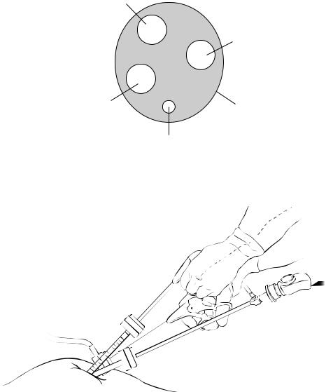
5 mm. left hand
5 mm. right hand
5 mm. |
Umbilicus |
|
camera site |
||
|
Fascial opening for
rigid retracting instrument
Fig. 15.3 Multiple fascial openings for SALS, all located within the umbilicus
Fig. 15.4 Left operating hand supinated and positioned above pronated right hand of surgeon
using multiple small 5 mm facial openings under the divided skin of the umbilicus. One can also use disposable perforated rubber like devices offered by several companies that would require one slightly larger facial incision (around 2 cm).
The fundamental difference between SALS and classic laparoscopy is that the angle of the operation is much wider in the classic technique, therefore allowing a greater degree of freedom outside while the instruments are joining at the tips of the target. In SALS, because of the divergence that occurs early on at the skin level, the two tips reach the target at diverging angles (Fig. 15.5); as such, the only ways to allow for the righthand instrument to reach the target are to cross the hands (Fig. 15.6) or to use the left hand as the dissecting hand, with the right hand as the supporting hand (Fig. 15.7).Again our solution is to avoid unnatural movements of the hands and keep them on top of each other thus allowing for more lateral freedom (Fig. 15.4).

260 |
Chapter 15 Single-Access Laparoscopic Surgery (SALS) |
Crossed hands |
SALS |
Classic triangulation |
|
in SALS |
|||
|
|
Fig. 15.5 Different angulation of instruments; classic triangulation during laparoscopy; SALS
with and without crossing hands
L
R
L R
Fig. 15.6 Dissection of the cystic duct |
Fig. 15.7 Alternative technique: dissec- |
using a hook with the right hand crossed |
tion of the cystic duct using a hook with |
|
the left hand |
