

Adhesions Due to Previous Upper Midline Laparotomy |
35 |
If hemorrhage occurs from the liver bed, the spatula used to dissect the gallbladder can conveniently be used to attempt hemostasis, with an increase in voltage from the cautery unit. If there is severe bleeding in the liver bed, it is possible to introduce a piece of 2×2 radiopaque gauze and apply compression. The steps of managing hemorrhage from the liver bed are:
Avoid obscuring the video laparoscope with blood, pull the camera back, leaving the tip in the port to still provide adequate visualization.
Compress the bleeding with 2 × 2 gauze if available.
Irrigate and clean the area around the bleeding.
Remove the gauze.
Introduce an irrigation/suction device to dry the site of bleeding with the left hand.
Apply high-current electrocautery with the spatula using the right hand. This current creates a crust that halts the bleeding. Care is taken to check that the tip of the cautery does not injure a peripheral bile duct (Duct of Lushka). This can be the cause of postoperative bile leak.
Application of clips is usually a waste of time as it is rarely efficient in controlling oozing in the liver bed.
If these actions do not initially take care of the bleeding the compression should be continued. Hemostatic agents such as Tisseal or Floseal (Baxter Inc, Deerfield, IL) can be used to achieve hemostasis.
If the bleeding is due to a major tear in the liver, and hepatic or portal venous branches are involved, and if all possibilities are exhausted, the only recourse is conversion using a mini-laparotomy. This has very rarely been the case in the author’s experience, but it may occur more frequently in cirrhotic patients. There is no need for a large subcostal incision and usually a 5 cm mini-laparotomy will suffice.
In the case of a supra-umbilical incision with severe midline adhesions that obscure the view, one can place a 5 mm trocar along the left midclavicular line to take those adhesions down using harmonic shears (Fig. 2.14).Another trick is to insert the camera to the right and superior to the umbilicus, closer to the gallbladder. The patient is tilted to the left, possibly on a bean bag; this will allow for a different angle of visualization and a safe cholecystectomy. Trocars for the right and left hand are also placed a little more to the right of the patient (Fig. 2.15).
Controlling
Bleeding in
the Liver Bed
Adhesions Due to Previous Upper Midline Laparotomy

36 |
Chapter 2 Cholecystectomy |
|
A |
D |
B |
E |
|
|
C |
Fig. 2.14 Cholecystectomy in a patient with a history of previous upper midline incision. E additional trocar used to take down adhesions; C insertion of the first camera port using a Hasson technique to the right of the umbilicus; A subxyphoid port; B midclavicular port; D retractor for gallbladder fundus. S surgeon; CA camera assistant; FA first assistant
B
Gallbladder
+
A
D B
C
FA |
S |
CA
Fig. 2.15 Severe adhesions due to prior upper midline incision. Patient tilted after positioning on a bean bag. Camera (c) moved to the right and above the umbilical level. All other trocars (a, b, d) moved to the right
Selected Further Reading |
37 |
Agarwal BB (2009) Patient safety in laparoscopic cholecystectomy. Arch Surg 144(10):979 Baraka A, Jabbour S, Hammoud R et al (1994) End carbon dioxide tension during laparoscopic cholecystectomy, Correlation with the baseline value prior to carbon dioxide
insuffiation. Anaesthesia 49(4):304–306
Bablekos GD, Michaelides SA, Roussou T, Charalabopoulos KA (2006) Changes in breathing control and mechanics after laparoscopic vs open cholecystectomy. Arch Surg 141(1):16–22
Barton JR, Russell RC, Hatfield AR (1995) Management of bile leaks after laparoscopic cholecystectomy. Br J Surg 82(7):980–984
Con P, Tate JJ, Lau WY, Dawson JW, Li AK (1994) Preoperative ultrasound to predict technical difficulties and complications of laparoscopic cholecystectomy. Am J Surg 168(1):54–56
Cushieri A, Dubois F, Mouiel J et al (1991) The European experience with laparoscopic cholecystectomy. Am J Surg 161:385–387
Davidoff AM, Pappas TN, Murray EA et al (1992) Mechanisms of major biliary injury during laparoscopic cholecystectomy. Ann Surg 215:196–202
Eisenstein S, Greenstein AJ, Kim U, Divino CM (2008) Cystic duct stump leaks: after the learning curve. Arch Surg 143(12):1178–1183
Essen P, Thorell A, McNurlan MA et al (1995) Laparoscopic cholecystectomy does not prevent the postoperative protein catabolic response in muscle. Ann Surg 222(1):36–42 Fabiani P, Iovine L, Katkhouda N, Gugenheim J, Mouiel J (1993) Dissection of the triangle
of Calot during laparoscopic cholecystectomy. La Presse Medicale 22:535–537 Fenster LF, Lonborg R, Thirlby RC, Traverso LW (1995) What symptoms does cholecys-
tect omy cure? Insights from an outcomes measurement project and review of the literature. Am J Surg 169(5):533–538
Fredman B, Jedeikin R, Olsfanger D, Flor P, Gruzman A (1994) Residual pneumoperitoneum: a cause of postoperative pain after laparoscopic cholecystecomy. Anesth Analg 79(1):152–154
Freeman JA, Armstrong IR (1994) Pulmonary function tests before and after laparoscopic cholecystectomy. Anaesthesia 49(7):579–582
Fried GM, Barkun JS, Sigman HH et al (1994) Factors determining conversion to laparotomy in patients undergoing laparoscopic cholecystectomy. Am J Surg 167(1):35–41
Fullarton GM, Darling K, Williams J, MacMillan R, Bell G (1994) Evaluation of the cost of laparoscopic and open cholecystectomy. Br J Surg 81(1):124–126
Glaser F, Sannwald GA, Buhr HJ et al (1995) General stress response to conventional and lapafoscopic cholecystectomy. Ann Surg 221(4):372–380
Glerup H, Heindorff H, Flyvbjerg A, Jensen SL, Vilstrup H (1995) Elective laparoscopic cholecystectomy nearly abolishes the postoperative hepatic catabolic stress response. Ann Surg 221(3):214–219
Gold-Deutch R, Mashiach R, Boldur I et al (1996) How does infected bile affect the post operative course of patients undergoing laparoscopic cholecystectomy? Am J Surg 172(3):272–274
Greig JD, John TG, Mahadaven M, Garden OJ (1994) Laparoscopic ultrasonography in the evaluation of the biliary tree during laparoscopic cholecystectomy. Br J Surg 81(8): 1202–1206
Halevy A, Gold-Deutch R, Negri M et al (1994) Are elevated liver enzymes and bilirubin levels significant after laparoscopic cholecystectomy in the absence of bile duct injury? Ann Surg 219(4):362–364
Jatzko GR, Lisborg PH, Perti AM, Stettner HM (1995) Multivariate comparison of complica tions after laparoscopic cholecystectomy and open cholecystectomy. Ann Surg 221(4):381–386
Selected
Further
Reading

38 Chapter 2 Cholecystectomy
Jones DB, Dunnegan DL, Soper NJ (1995) Results of a change to routine fluorocholangiography during laparoscopic cholecystectomy. Surgery 118(4):693–701
Katkhouda N, Mavor E, Mason RJ (2000) Visual identification of the cystic duct–CBD junction during laparoscopic cholecystectomy (visual cholangiography). Surg Endosc 14:88–89
Kendall AP, Bhatt S, Oh TE (1995) Pulmonary consequences of carbon dioxide insuffiation for laparoscopic cholecystectomies. Anaesthesia 50(4):286–289
Koo KP, Thirlby RC (1996) Laparoscopic cholecystectomy in acute cholecystitis. What is the optimal timing for operation? Arch Surg 131(5):540–544
Korman J, Cosgrove I, Furman M, Nathan I, Cohen J (1996) The role of endoscopic retrograde cholangiopancreatography and cholangiography in the laparoscopic era. Ann Surg 223(2):212–216
Kubota K, Bandai Y, Sano K, Teruya M, Ishizaki Y, Makuuchi M (1995) Appraisal of intraoperative ultrasonography during laparoscopic cholecystectomy. Surgery 118(3):555–561 Kuy S, Roman SA, Desai R, Sosa JA (2009) Outcomes following cholecystectomy in preg-
nant and nonpregnant women. Surgery 146(2):358–366
Lanzafame RI (1995) Laparoscopic cholecystectomy during pregnancy. Surgery 118(4): 627–631
Liu CL, Fan ST, Lai EC, Lo CM, Chu KM (1996) Factors affecting conversion of laparoscopic cholecystectomy to open surgery. Arch Surg 131(1):98–101
Lorimer JW, Fairfull-Smith RJ (1995) Intraoperative cholangiography is not essential to avoid duct injuries during laparoscopic cholecystectomy. Am J Surg 169(3):344–347 McLean TR (2006) Risk management observations from litigation involving laparoscopic
cholecystectomy. Arch Surg 141(7):643–648
McMahon AS, Baxter JN, Murray W, Imrie CW, Kenny G, ODwyer PJ (1994) Helium pneumoperitoneum for laparoscopic cholecystectomy: ventilatory and blood gas changes. Br J Surg 81(7):1033–1036
Madariaga JR, Dodson SF, Selby R, Todo S, Iwatsuki S, Starzl TE (1994) Corrective treatment and anatomic considerations for laparoscopic cholecystectomy injuries.Am Coll Surg 179(3):321–325
Morgan RA, van Sonnenberg E,Wittich GR, Nealon WH,Walser EM (1995) Percutaneous management of bile duct injury after laparoscopic cholecystectomy. Am J Roentgenol 165(4):985–990
Nebiker CA, Frey DM, Hamel CT, Oertli D, Kettelhack C (2009) Early versus delayed cholecystectomy in patients with biliary acute pancreatitis. Surgery 145(3):260–264
Ortega AE, Peters JH, Incarbone R et al (1996) A prospective randomized comparison of the metabolic and stress hormonal responses of laparoscopic and open cholecystectomy. J Am Coll Surg 183(3):249–256
Perissat S, Huibregtse K, Keane PB, Russell RC, Neoptolemos JP (1994) Management of bile duct stones in the era of laparoscopic cholecystectomy. Br J Surg 81(6):799–810 Pertsemlidis D (2009) Fluorescent indocyanine green for imaging of bile ducts during
laparoscopic cholecystectomy. Arch Surg 144(10):978
Peters JH, Krailadsiri W, Incarbone R et al (1994) Reasons for conversion from laparoscopic to open cholecystectomy in an urban teaching hospital. Am J Surg 168(6):555–559
Pietrabissa A, Di Candio G, Giulianotti PC, Shimi SM, Cuschieri A, Mosca F (1995) Comparative evaluation of contact ultrasonography and transcystic cholangiography during laparo scopic cholecystectomy: a prospective study. Arch Surg 130(10): 1110–1114
Ponce J, Cutshall KE, Hodge MJ, Browder W (1995) The lost laparoscopic stone. Potential for long-term complications. Arch Surg 130(6):666–668
Redmond HP, Watson RW, Houghton T et al (1994) Immune function in patients undergoing open vs laparoscopic cholecystectomy. Arch Surg 129(12):1240–1246
Selected Further Reading |
39 |
Robinson BL, Donohue JH, Gunes S et al (1995) Selective operative cholangiography. Appropriate management for laparoscopic cholecystectomy. Arch Surg 130(6):625–631 Roush TS, Traverso LW (1995) Management and long-term follow-up of patients with positive cholangiograrns during laparoscopic cholecystectomy. Am J Surg 169(5):484–487 Sanabria JR, Gallinger S, Croxford R, Strasberg SM (1994) Risk factors in elective laparoscopic cholecystectomy for conversion to open cholecystectomy. Am Coll Surg
179(6):696–704
Savader SI, Lillemoe KD, Prescott CA et al (1997) Laparoscopic cholecystectomy-related bile duct injuries: a health and financial disaster. Ann Surg 225(3):268–273
Singh S, Agarwal AK (2009) Gallbladder cancer: the role of laparoscopy and radical resection. Ann Surg 250(3):494–495
Soper NJ, Brunt LM, Callery MP, Edmundowicz SA,Aliperti G (1994) Role of laparoscopic cholecystectomy in the management of acute gallstone pancreatitis. Am J Surg 167(1):42–50
The Southern Surgeons Club (1991) A prospective analysis of 1518 laparoscopic cholecystectomies. N Engl J Med 324:1073–1078
Strasberg SM (2008) Acute calculous cholecystitis. N Engl J Med 358(26):2804–2811 Steiner CA, Bass EB, Talamini MA, Pitt HA, Steinberg EP (1994) Surgical rates and opera-
tive mortality for open and laparoscopic cholecystectomy in Maryland. N Engl J Med 330(6):403–408
Stewart L,Way LW (1995) Bile duct injuries during laparoscopic cholecystectomy. Factors that influence the results of treatment. Arch Surg 130(10):1123–1129
Tate JJ, Lau WY, Li AK (1994) Laparoscopic cholecystectomy for biliary pancreatitis. Br J Surg 81(5):720–722
Teefy SA, Soper NJ, Middleton WD et al (1995) Imaging of the common bile duct during laparoscopic cholecystectomy: sonography versus videofluoroscopic cholangiography. Am J Roentgenol 165(4):847–851
Voyles CR, Sanders DL, Hogan R (1994) Common bile duct evaluation in the era of laparoscopic cholecystectomy. 1050 cases later. Ann Surg 219(6):744–752
Willekes CL, Edoga IK, Castronuovo JJ Jr, Widmann WD, McLean ER Jr, Chevinsky AH (1995) Technical elements of successful laparoscopic cholangiography as defined by radiographic criteria. Arch Surg 130(4):398–400
Woods MS, Traverso LW, Kozarek RA et al (1994a) Characteristics of biliary tract complications during laparoscopic cholecystectomy: a multi-institutional study. Am J Surg 167(1):27–34
Woods MS, Shellito JL, Santoscoy GS et al (1994b) Cystic duct leaks in laparoscopic cholecystectomy. Am J Surg 168(6):56–65
Yaghoubian A, Saltmarsh G, Rosing DK, Lewis RJ, Stabile BE, de Virgilio C (2008) Decreased bile duct injury rate during laparoscopic cholecystectomy in the era of the 80-hour resident workweek. Arch Surg 143(9):847–851
Yamaguchi K, Chijiiwa K, Ichimiya H et al (1996) Gallbladder carcinoma in the era of laparoscopic cholecystectomy. Arch Surg 131(9):981–985

Common Bile 3
Duct Explorations
and Bilioenteric
Anastomosis
If a stone is visualized during an intraoperative cholangiogram, a few measures should be attempted before proceeding to common bile duct (CBD) exploration. First, place the patient in steep reverse Trandelenberg and flush the CBD with saline. Intravenous Glucagon at a dose of 1 mg can also be given to relax the sphincter of Oddi and help the stone pass. If this is not successful, then one can proceed to a CBD exploration.
The transcystic approach is usually accepted as the initial approach in a CBD explora- |
The Transcystic |
tion. In this author’s experience, the transcystic approach is a difficult maneuver that has |
Approach |
not had the 95% success rate that some other authors have reported, and requires |
|
training. |
|
The first step is to place an additional trocar directly above the cystic duct. This |
|
trocar will facilitate access to the cystic duct in a linear fashion. Next the cystic duct |
|
should be dilated by one of the following methods: inserting Maryland forceps into the |
|
duct, or using a dilating balloon, biliary Fogarty, or even stents of different calibers. |
|
The next step is the insertion of a Dormia wire basket to retrieve the stones. This may |
|
be attempted without fluoroscopic guidance because the stones have already been located |
|
by intraoperative cholangiography. However, if retrieval of the stones proves difficult, then |
|
fluoroscopic guidance can be called upon before resorting to a choledochoscope. If the |
|
choledochoscope is needed to locate the stones, they can be removed using a wire basket |
|
introduced through the choledochoscope operating channel. If all these techniques are |
|
not successful, it will be necessary to revert to a laparoscopic choledochotomy. |
|
N. Katkhouda, Advanced Laparoscopic Surgery,
DOI: 10.1007/978-3-540-74843-4_3, © Springer-Verlag Berlin Heidelberg 2011

42 |
Chapter 3 Common Bile Duct Explorations and Bilioenteric Anastomosis |
The Common
Bile Duct
Approach
The first step is dissection of the CBD, as in open surgery, and this is done using very fine instruments in the right hand and an atraumatic grasper in the left hand. The peritoneal adhesions above the CBD are grasped with the left hand and the scissors are then used to peel these adhesions from the CBD. Peelingof the entire CBD is not indicated, as there is a risk of devascularizing the duct. Only an area appropriate for the choledochotomy is dissected out. Sharp micro-scissors are inserted and a choledochotomy is performed longitudinally. The size of the choledochotomy should be appropriate for the size of the CBD. It should also match the size of the stone. The same technique as in open surgery then applies, with the introduction of a choledochoscope with a 2.8 French channel into the midclavicular port to locate the stones. Introduction of the wire basket, either directly into the choledochotomy or via the operating channel of the laparoscope, will usually allow extraction of the stones from the CBD under direct vision. If a flexible choledochoscope is unavailable or stone removal by this method proves difficult, a traditional rigid choledochoscope can be introduced into the subxyphoid skin incision after removal of the 10 mm trocar. A suture can be tightened around the choledochoscope to keep the abdomen airtight. This can also be done directly through a separate skin incision. When the CBD has been cleared of all stones, it should be closed around a T-tube (Fig. 3.1). This T-tube is introduced through a separate small skin incision, and adequate tube length should be left in the abdomen in order to avoid its inadvertent dislodgement if an ileus is encountered postoperatively. The T-tube is cut to the appropriate size and inserted in the choledochotomy (Fig. 3.2). As in open surgery, it should be moved up and down to make sure that it is properly in place in the CBD. 4–0 PDS suture is used to close the choledochotomy in either an interrupted or continuous fashion, depending on the size of the CBD. Interrupted sutures tied intracorporeally are best for thin CBDs, while continuous
Fig. 3.1 Insertion of the T-tube in CBD exploration
The Common Bile Duct Approach |
43 |
Fig. 3.2 Closure of the choledochotomy using 4–0 PDS
Fig. 3.3 Side-to-side choledocoduodenostomy using interrupted 3–0 or 4–0 PDS (full thick- ness on the posterior duodenal wall, extramucosal on the anterior duodenal wall)

44 |
Chapter 3 Common Bile Duct Explorations and Bilioenteric Anastomosis |
|
sutures can be used on larger, dilated CBDs. Extracorporeal knot-tying is not useful in |
|
this setting. The T-tube is then tested for leaks by injecting some saline, before being |
|
properly fixed to the skin. A completion cholangiogram is performed. A drain is always |
|
inserted adjacent to the choledochotomy to monitor bile leakage. |
Choledochodu |
This procedure is similar to open surgery except that the choledochotomy should be |
odenostomy |
performed transversely. The duodenum is then approximated and incised. Interrupted |
|
sutures are placed and knotted intracorporeally to avoid tension that might occur if they |
|
were to be knotted extracorporeally. Running sutures are possible if the CBD is very |
|
dilated. |
|
The ideal suture is 3–0 PDS and square knots are tied using standard techniques |
|
(Fig.3.3).The key to success is proper mobilization of the duodenum.This can be achieved |
|
easily by a partial Kocher maneuver of the duodenum. A drain should be left in place. |
Hepaticoje junostomy with a RouxenY
This is a very difficult operation that should be attempted only by highly skilled laparoscopic surgeons. The operative details are provided here for the reader’s interest, rather than for absolute guidance. Six ports are used, as shown in Fig. 3.4. The operation starts with dissection of the CBD and separation from the important structures of the porta
Fig. 3.4 Trocar port placement for laparoscopic hepaticojejunostomy. A umbilical laparoscope; B surgeon’s right hand (operating port); C irrigation suction probe; D liver fan retractor; E surgeon’s left hand (grasper); F grasper (first assistant)
Hepaticojejunostomy with a Roux-en-Y |
45 |
Fig. 3.5 End-to-side hepaticojejunostomy
hepatis, such as the hepatic artery and portal vein. It is important to use an atraumatic dissector very gently and to stay close to the CBD, using the magnification of the laparoscope as much as possible.
The CBD is encircled and umbilical tape is passed around it to allow gentle retraction of the duct. It is then transected. The distal end is ligated using an Endoloop with 2–0 PDS. The proximal end of the CBD is trimmed conservatively, care being taken to avoid devascularization. One then proceeds to prepare the Roux-en-Y loop (Fig. 3.5).
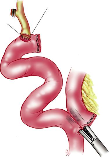
First the angle of Treitz is identified. Following the small bowel, the second jejunal loop is picked up and transection of the loop is performed using an EndoLinear Cutter-45 with white loads (Ethicon EndoSurgery Inc, Cincinatti, OH). One and half firings of the GIA-45 whites divide the mesentery.One should never fire more than two loads of 45-mm cutters in the mesentery to avoid injuring the superior mesenteric artery. The next step is coagulation of any bleeding along the cut edges of the mesentery. It is extremely

46 Chapter 3 Common Bile Duct Explorations and Bilioenteric Anastomosis
important to perform this coagulation either by using electrocautery, or clips to avoid any postoperative bleeding. The harmonic shears are utilized to open up the crotch of this division to further enhance the length of the alimentary loop. Next a 40–60 cm Roux limb is selected and a jejunojejunostomy is performed after opening the proximal and distal limbs with the harmonic scalpel and then firing the 45 mm stapler with white loads. The enterotomy is closed with a running 3–0 Prolene in an extramucosal fashion. The mesentery defect is closed with nonabsorbable running suture. The Roux limb is then brought up and approximated to the end of the CBD. An enterotomy is performed, followed by creation of an anastomosis using a continuous suture on the posterior aspect of the jejunal wall. An interrupted 3–0 PDS is used on the anterior jejunal wall, ensuring the avoidance of the stricture of the hepatic duct.