

Книги по МРТ КТ на английском языке / Normal Findings in CT and MRI
.pdfCervical Spine 75
3Width of spinal canal:
!Transverse diameter at level of pedicles > 20−21 mm
4Width of spinal cord:
!> 6−7 mm in sagittal plane
4
Moeller, Normal Findings in CT and MRI © 2000 Thieme
All rights reserved. Usage subject to terms and conditions of license.

76 CT: Spinal Column
Thoracic Spine
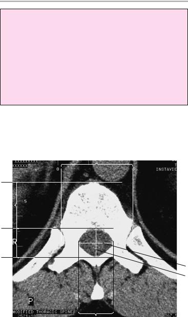
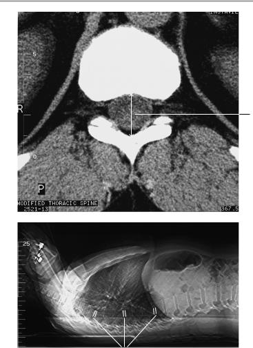
The thoracic spine shows a normal degree of kyphosis in the lateral survey scan, with no segmental malalignment.
The vertebral bodies show normal shape and trabecular structure. The cortical margins are of normal thickness and are free of osteophytes. The bony spinal canal has normal sagittal diameter.
The intervertebral disks show normal CT density and normal posterior concavity. The disks do not project past the posterior surface of the vertebral bodies. The spinal cord is centrally placed and of normal width. It has homogeneous density and shows no circumscribed narrowing or expansion.
The nerve roots show a normal course and passage through the neuroforamina, which are of normal size and structure. The costovertebral and costotransverse joints are unremarkable.
The prevertebral and paravertebral soft tissues show no abnormalities.
Interpretation
The examined segments of the thoracic spine appear normal.
Checklist
Position |
! |
Thoracic kyphosis |
|
! No segmental malalignment (lateral survey |
|
Bony spinal |
! |
scan) |
Width (see below) |
||
canal |
! |
Shape |
Vertebral bodies |
! |
Shape |
|
! Cortex (thickness, margins: smooth, sharp) |
|
|
! |
No marginal osteophytes |
|
! Trabeculae (uniform honeycomb arrangement, |
|
|
|
no rarefaction or circumscribed voids, no nar- |
Intervertebral |
! |
rowing or expansion) |
Width (see below) |
||
disk space |
! |
Margins (smooth, sharp) |
|
! Straight posterior disk contour |
|
|
! No disk protrusion past posterior surface of |
|
Spinal cord |
! |
vertebral bodies |
Position (central) |
||
|
! |
Width |
|
! No circumscribed narrowing or expansion |
|
|
! |
Density (homogeneous) |
Moeller, Normal Findings in CT and MRI © 2000 Thieme
All rights reserved. Usage subject to terms and conditions of license.

Thoracic Spine 77
!Perimedullary thecal space clear: no encroachment from the anterior side (e.g., by an intervertebral disk or osteophyte) or posterior side (e.g., by a hypertrophic ligamentum flavum)
Neuroforamina ! Configuration
!Width
!No encroachment from the anterior side (e.g., by an intervertebral disk or osteophyte) or posterior
side (e.g., by hypertrophic spondylarthrosis) Nerve roots ! Course and passage through the neuroforamina
! No circumscribed expansion Facet joints ! Shape, symmetry
!Pars interarticularis
!Vertebral arches intact
!Spinous processes (shape, length, bony structure)
!Costotransverse joints
!Costovertebral joints (no hypertrophy)
!Ribs
Soft tissues ! Symmetrical arrangement on both sides of the vertebral bodies and spinous processes
!No masses
!Prevertebral soft-tissue structures (especially the lungs, heart, and aorta)
Moeller, Normal Findings in CT and MRI © 2000 Thieme
All rights reserved. Usage subject to terms and conditions of license.
78 CT: Spinal Column
Important Data
1Width of spinal canal:
!Transverse diameter at level of pedicles > 20−21 mm
2Sagittal diameter:
!T1−T11 = 13−14 mm, T12 = 15 mm
3Jones-Thomson ratio (= A × B/C × D):
!Between 0.5 and 0.22 = normal (< 0.22 = spinal stenosis)
4Width of intervertebral disk spaces:
!Smallest at T1
!T6−T11: ca. 4−5 mm
!Largest at T11−T12
3C
3D
3B
1
2
Moeller, Normal Findings in CT and MRI © 2000 Thieme
All rights reserved. Usage subject to terms and conditions of license.
Thoracic Spine 79
2
4
Moeller, Normal Findings in CT and MRI © 2000 Thieme
All rights reserved. Usage subject to terms and conditions of license.

80 CT: Spinal Column
Lumbar Spine
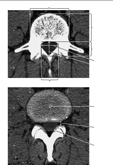
The lumbar spine shows normal lordotic curvature in the lateral survey scan, with no segmental malalignment.
The vertebral bodies have normal shape and trabecular structure. The cortical margins are of normal thickness and are free of osteophytes. The bony spinal canal has a normal sagittal diameter.
The intervertebral disks show normal density and normal posterior concavity. The disks do not project past the posterior surface of the vertebral bodies.
The conus medullaris shows a normal position at L1 with normal subdivision into filaments. The dural sac is of normal width.
The nerve roots show a normal course and passage through the neuroforamina, which are of normal size and structure. The facet joints are unremarkable.
The prevertebral and paravertebral soft tissues show no abnormalities.
Interpretation
The examined segments of the lumbar spine appear normal.
Checklist
Position |
! |
Lumbar lordosis |
|
! Lumbosacral angle (see below) |
|
|
! No segmental malalignment (lateral survey |
|
Bony spinal |
! |
scan) |
Shape |
||
canal |
! |
Width (see below) |
Vertebral bodies |
! |
Shape |
|
! Cortex (thickness, margins: smooth, sharp) |
|
|
! |
No marginal osteophytes |
|
! Trabeculae (uniform honeycomb arrangement, |
|
|
|
no rarefaction or circumscribed voids, no nar- |
Intervertebral |
! |
rowing or expansion) |
Width (see below) |
||
disk space |
! |
Margins (smooth, sharp) |
|
! No disk protrusion past posterior surface of |
|
|
|
vertebral bodies (posterior disk contour is con- |
|
|
cave at L1−L4, straight at L4/5, and slightly con- |
Dural tube |
! |
vex at L5/S1) |
Normal width |
||
Moeller, Normal Findings in CT and MRI © 2000 Thieme
All rights reserved. Usage subject to terms and conditions of license.

Lumbar Spine 81
!No circumscribed narrowing or expansion
!Contents of fluid attenuation
!Conus medullaris (at L1 level, configuration)
!Filaments show normal width and arrangement with no posterior adhesion and no circumscribed anterior encroachment (e.g., by an intervertebral disk or osteophyte) or posterior encroachment (e.g., by a hypertrophic ligamentum
flavum)
Neuroforamina ! Configuration
!Width
!No anterior encroachment (e.g., by an intervertebral disk, osteophyte, or uncovertebral arthrosis)
or posterior encroachment (e.g., by hypertrophic spondylarthrosis)
Nerve roots ! Course and passage through the neuroforamina ! No circumscribed expansion
Facet joints ! Shape, symmetry
!Pars interarticularis
!Vertebral arches intact
!Spinous processes (shape, length, bony struc-
ture)
Soft tissues ! Symmetrical arrangement on both sides of the vertebral bodies and spinous processes
!No masses
!Prevertebral soft-tissue structures (aorta, vena cava), no masses
Moeller, Normal Findings in CT and MRI © 2000 Thieme
All rights reserved. Usage subject to terms and conditions of license.
82 CT: Spinal Column
Important Data
1Lumbosacral angle (S1/horizontal plane):
!26−57°
2Width of intervertebral disk space or height of lumbar intervertebral disks:
!Approx. 8−12 mm, increasing from L1 to L4/5, decreasing again at L5/S1
3Width of spinal canal:
! |
Transverse diameter at level of pedicles: |
L1−L4 > 20− |
|
21 mm, L5 > 24 mm |
|
4 Sagittal diameter: |
|
|
! |
16−18 mm (simple formula: not less than |
15 mm; 11− |
|
15 mm = relative stenosis, less than 10 mm = absolute ste- |
|
|
nosis) |
|
5Jones-Thomson ratio (= A × B/C × D):
!Between 0.5 and 0.22 = normal (<0.22 = spinal stenosis)
6Lateral recess (sagittal diameter):
!> 4−5 mm
7Ligamenta flava:
!Width < 6 mm
8CT density of intervertebral disks:
!70 ± 5 HU
1
2
Moeller, Normal Findings in CT and MRI © 2000 Thieme
All rights reserved. Usage subject to terms and conditions of license.
Lumbar Spine 83
5C
5D
5B
3
 4
4
5A
8
6
7
Moeller, Normal Findings in CT and MRI © 2000 Thieme
All rights reserved. Usage subject to terms and conditions of license.

84
Moeller, Normal Findings in CT and MRI © 2000 Thieme
All rights reserved. Usage subject to terms and conditions of license.