

Книги по МРТ КТ на английском языке / Normal Findings in CT and MRI
.pdfOrbit 105
Optic nerve |
! |
Caliber (see below) |
Eye muscles |
! |
Course |
! |
Position |
|
|
! |
Width (see below) |
Ophthalmic vein |
! |
Course |
! |
Course |
|
Lacrimal gland |
! |
Caliber (see below) |
! |
Size |
|
|
! |
Symmetry |
|
! |
No unilateral or bilateral enlargement |
|
! |
Position (see below) |
|
! |
No excavation or destruction of adjacent bone |
|
! |
Homogeneous internal structure |
|
! |
No hypointense or hyperintense changes |
Neurocranium |
! |
Smooth borders |
! |
Temporal lobes |
|
|
! |
Frontal lobes |
Paranasal sinuses ! |
Maxillary sinuses |
|
|
! |
Ethmoid cells |
|
|
|
1a 
2 |
|
3a |
|
||
|
|
3b
4a
4b
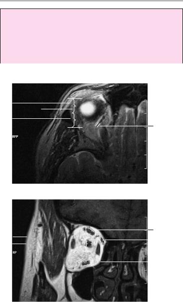
Axial image through center of orbits
Moeller, Normal Findings in CT and MRI © 2000 Thieme
All rights reserved. Usage subject to terms and conditions of license.
106 MRI: Head and Neck
Important Data
1Diameter of globe:
a Axial image plane: right 28.6 ± 1.2 mm
left 29.4 ± 1.4 mm
b Sagittal image plane: right 27.8 ± 1.2 mm left 28.2 ± 1.2 mm
6 |
|
|
|
1/2 |
|||
|
|||
6 |
|
1/2 |
|
|
|||
5a
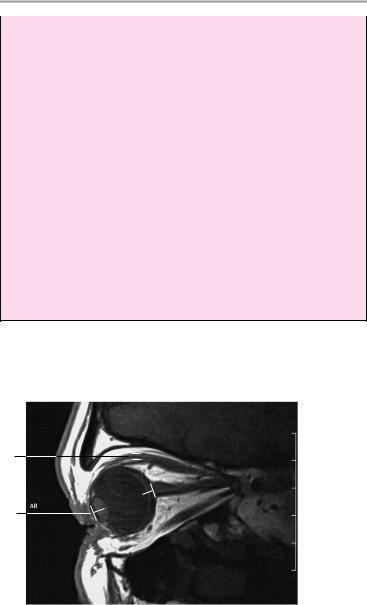
Axial image through roof of orbit
4c
5b |
|
|
|
|
|
|
|
|
|
|
|
|
4d |
|
|
|
|
|
|
|
|
|
|
|
|||||
3a |
|
|
|
|
|
|
|
|
|
|
|
|
|
|
|
|
|
|
|
|
|
|
|
|
4e |
||||
|
|
|
|
|
|
|
|
|
|
|
|
|
|
|
|
|
|
|
|
|
|
|
|
|
|
||||
Coronal image
Moeller, Normal Findings in CT and MRI © 2000 Thieme
All rights reserved. Usage subject to terms and conditions of license.
Orbit 107
2Position of globe:
!Posterior margin of globe is 9.9 mm ± 1.7 mm behind interzygomatic line
3Optic nerve (axial image plane):
a Retrobulbar segment: 5.5 mm ± 0.8 mm
b Narrowest point (at approximately mid-orbit): 4.2 mm ± 0.6 mm
4Eye muscles:
a Lateral rectus: 2.9 mm ± 0.6 mm b Medial rectus: 4.1 mm ± 0.5 mm c Superior rectus: 3.8 mm ± 0.7 mm d Oblique: 2.4 mm ± 0.4 mm
e Inferior rectus: 4.9 mm ± 0.8 mm
f Levator palpebrae superioris: 1.75 mm ± 0.25 mm
5Ophthalmic vein:
a 1.8 mm ± 0.5 mm (axial image, 4 mm slice thickness) b 2.7 mm ± 1 mm (coronal image)
6Lacrimal gland:
!Less than one-half of the gland is anterior to the frontozygomatic process
4f
1b
Sagittal image
Moeller, Normal Findings in CT and MRI © 2000 Thieme
All rights reserved. Usage subject to terms and conditions of license.

108 MRI: Head and Neck
Paranasal Sinuses
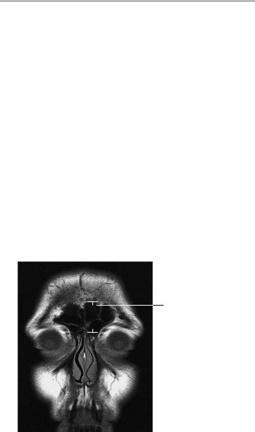
The frontal sinuses are normally developed, clear, and pneumatized with smooth wall contours.
The ethmoid cells show normal development and intact bony walls, with no defects on the orbital side. There are no areas of wall erosion or mucosal thickening.
The sphenoid sinus is normally developed and presents a coarse honeycomb structure. There are no fluid collections or mucosal swelling. The maxillary sinuses are bilaterally symmetrical and have smooth walls of normal thickness. The sinuses are clear and aerated with no foci of bone erosion or destruction. The nasal septum is centered on the midline. The nasal turbinates show a normal arrangement and normal signal intensity.
The nasal cavity, pharynx, and imaged parapharyngeal structures show no abnormalities.
Interpretation
The paranasal sinuses appear normal.
Checklist
Frontal sinuses |
! |
Anatomy |
|
! |
Wall contours (smooth) |
Ethmoid cells |
! |
Pneumatization |
! |
Anatomy |
|
|
! |
Pneumatization |
|
! Bony structures (especially bordering the orbit: |
|
|
! |
contours are smooth, sharp, and intact) |
|
No wall erosion |
|
|
! |
No mucosal thickening |
Sphenoid sinus ! Anatomy (coarse honeycomb structure) |
||
|
! |
Clear and pneumatized |
|
! |
No fluid collection |
|
! |
No mucosal swelling |
|
! Bony structures (smooth, intact walls, no ero- |
|
sion)
! No extrinsic wall indentations
Maxillary sinuses ! Anatomy
!Size (bilaterally symmetrical)
!Bony structures (smooth, intact contours, normal wall thickness, no bone erosion or destruction)
Moeller, Normal Findings in CT and MRI © 2000 Thieme
All rights reserved. Usage subject to terms and conditions of license.
Paranasal Sinuses 109
|
! |
Pneumatization |
Nasal cavity |
! No tooth roots projecting through sinus floor |
|
! Anatomy (symmetry) |
||
|
! |
Size |
|
! |
Aeration (clear) |
|
! Septum centered on the midline |
|
|
! |
Nasal turbinates (three per side: superior, |
|
! |
middle, inferior) are normally developed |
Pharynx and |
Signal characteristics |
|
! Anatomy (symmetry) |
||
parapharyngeal |
! |
Size |
structures |
! |
Wall thickness |
|
! |
No foreign bodies |
Neurocranium |
! |
No masses |
! |
Cortex |
|
(especially the |
! |
White matter |
temporal and |
! |
Gyration |
frontal lobes) |
! Signal characteristics |
|
Orbit |
! |
Eye muscles (width, signal characteristics) |
|
! Optic nerve (width, course) |
|
|
! Globe (shape, size, signal characteristics) |
|
|
! Retrobulbar fat (no masses) |
|
|
|
|
1
Coronal image
Moeller, Normal Findings in CT and MRI © 2000 Thieme
All rights reserved. Usage subject to terms and conditions of license.
110 MRI: Head and Neck
Important Data
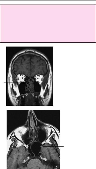
1Frontal sinus:
!Height ca. 1.5−2 cm
2Sphenoid sinus:
!Width 0.9−1.4 cm
3Maxillary sinuses; a Width ca. 2 cm b Height ca. 2 cm
Coronal image
3b
Axial image
2
Moeller, Normal Findings in CT and MRI © 2000 Thieme
All rights reserved. Usage subject to terms and conditions of license.

Paranasal Sinuses 111
3a
Axial image
3b
Sagittal image
Moeller, Normal Findings in CT and MRI © 2000 Thieme
All rights reserved. Usage subject to terms and conditions of license.

112 MRI: Head and Neck
Cervical Soft Tissues
The cervical soft tissues show normal configuration. The position of the cervical spine is normal.
The oral floor muscles are normally developed and bilaterally symmetrical. The spaces of the oral cavity and neck are clear and well defined.
Imaged portions of the parotid and submandibular glands show no abnormalities.
The pharynx and larynx show normal boundaries and normal wall thickness.
The thyroid gland shows reasonable symmetry and normal size. The thyroid lobes display normal internal structure.
Cervical vessels that are evaluable with MRI have normal appearance. The muscular structures of the neck are normal.
There are no signs of cervical lymphadenopathy.
No abnormalities are seen in the cervical spinal cord or cervical plexus.
Interpretation
The cervical soft tissues appear normal.
|
|
|
|
|
|
|
|
|
|
|
|
Visceral space |
|
|
|
|
|
|
|
|
|
|
|
|
|
Retropharyn- |
|
|
|
|
|
Anterior cer- |
||||||
|
|
|
|
|
||||||||
|
|
|
|
|
vical space |
|||||||
geal space |
|
|
|
|
|
|
|
Carotid space |
||||
|
|
|
|
|
|
|||||||
|
|
|
|
|
|
|
|
|
|
|||
|
|
|
|
|
|
|
|
|
|
|
|
|
Posterior |
|
|
|
|
|
|
|
|
|
|
|
Perivertebral |
|
|
|
|
|
|
|
|
|||||
cervical space |
|
|
|
|
|
|||||||
|
|
|
|
|
||||||||
|
|
|
|
|
|
|
|
|
|
|
|
space (pre- |
Perivertebral |
|
|
|
|
|
|
|
|
|
vertebral |
||
|
|
|
|
|
|
|
|
portion) |
||||
|
|
|
|
|
|
|||||||
space |
|
|
|
|
|
|
||||||
(paraspinal |
|
|
|
|
|
Superficial |
||||||
portion) |
|
|
|
|
|
|||||||
|
|
|
|
|
space |
|||||||
|
|
|
|
|
|
|
|
|
|
|
|
|
Spaces of the neck |
|
|
|
|
|
|
||||||
Moeller, Normal Findings in CT and MRI © 2000 Thieme
All rights reserved. Usage subject to terms and conditions of license.

Cervical Soft Tissues 113
Checklist
Cervical soft |
! |
Configuration |
|
tissues |
! |
Normal anatomy |
|
Oral floor |
! Normal position of the cervical spine (see below) |
||
! |
Anatomy |
||
muscles |
! |
Width |
|
|
! |
Bilateral symmetry |
|
|
! |
Delineation |
|
|
! |
Internal structure |
|
Submandibular |
! Spaces of oral floor are clearly defined |
||
! |
Size (symmetry) |
||
and parotid |
! Signal characteristics |
||
glands |
! |
No dilatation of glandular duct |
|
|
! No hypointense or hyperintense areas within the |
||
Pharynx and |
|
glandular tissue |
|
! Shape (symmetrical) |
|||
larynx |
! |
Size |
|
|
! |
Smooth walls |
|
|
! |
Normal wall thickness |
|
|
! |
No masses |
|
Cervical spaces ! Retropharyngeal space |
|||
|
! Parapharyngeal space (visceral space) |
||
|
! |
Carotid space |
|
|
! Anterior and posterior cervical spaces |
||
|
! Perivertebral space (prevertebral and paraspinal |
||
|
|
portions): |
|
|
|
— |
Configuration |
|
|
— Borders |
|
|
|
— Symmetry |
|
|
|
— |
Internal structure |
Esophagus |
! |
— |
Width (see below) |
Position |
|||
|
! Wall thickness (see below) |
||
|
! |
Borders |
|
Thyroid gland |
! |
No masses |
|
! Anatomy (consists of two lobes, reasonably sym- |
|||
|
! |
metrical) |
|
|
Size (see below) |
||
|
! |
Internal structure (homogeneous) |
|
|
! |
No cysts |
|
|
! |
No nodules |
|
Moeller, Normal Findings in CT and MRI © 2000 Thieme
All rights reserved. Usage subject to terms and conditions of license.

114 MRI: Head and Neck
Cervical vessels |
! |
Course |
|
! |
Caliber (see below) |
Neck muscles |
! No abrupt caliber changes |
|
! |
Anatomy |
|
|
! |
Symmetry |
|
! |
Borders |
Lymph node |
! Signal characteristics (internal structure) |
|
! No lymphadenopathy |
||
stations (if |
|
|
evaluable) |
|
|
Cervical spine |
! Vertebral bodies |
|
(if evaluable) |
|
— Number |
|
|
— Shape |
|
|
— Position |
|
! |
— Contours |
|
Bone marrow signal |
|
|
! |
Intervertebral disk spaces |
|
! |
Spinal canal: |
|
|
— Width |
|
|
— No circumscribed narrowing |
|
! Normal width of cervical spinal cord |
|
|
! |
No masses |
|
! |
No narrowing |
Cervical plexus ! No appreciable narrowing
! No masses (including lymphadenopathy)
Important Data
Ch = Chamberlain’s line (line connecting the posterior part of the hard palate with the posterior rim of the foramen magnum):
!Tip of the dens projects no more than 1 ± 6.6 mm past Chamberlain’s line
Prevertebral soft tissues:
1Retropharyngeal:
!Approximately 1.7 ± 0.7 mm
2Retroglottic:
!Approximately 6.0 ± 1.1 mm
3Retrotracheal:
!Approximatley 8.4 ± 2.5 mm
Moeller, Normal Findings in CT and MRI © 2000 Thieme
All rights reserved. Usage subject to terms and conditions of license.