

Книги по МРТ КТ на английском языке / Normal Findings in CT and MRI
.pdfLiver 45
Checklist
Liver |
! |
Position: |
|
! |
— Directly below the right hemidiaphragm |
|
Size (see below) |
|
|
! |
Borders: |
|
|
— Smooth |
|
|
— Sharp |
|
! Attenuation values (see below) |
|
|
! Homogeneous internal parenchymal structure, |
|
|
! |
no focal abnormalities |
|
Intrahepatic bile ducts: |
|
|
|
— Course (centrifugal) |
|
|
— Width (general rule: ducts should no longer |
|
|
be visible after contrast administration) |
|
|
— No calculi |
|
! |
— No air |
|
Extrahepatic bile ducts: |
|
|
|
— Course (from porta hepatis to head of pan- |
|
|
creas) |
|
|
— Width (see below) |
|
|
— Contents of homogeneous fluid density |
|
|
— No calculi |
|
! |
— No air |
|
Gallbladder: |
|
|
|
— Size (see below) |
|
|
— Contours (smooth and sharp) |
|
|
— Wall thickness (see below; no general or cir- |
|
|
cumscribed thickening) |
|
! |
— No pericholecystic fluid |
|
Gallbladder contents: |
|
|
|
— Homogeneous |
— Fluid density (see below)
— No calculi (hypodense or hyperdense)
— No air
— Porta hepatis occupied by the hepatic artery, common bile duct, and portal vein; no masses or lymphadenopathy
— Costophrenic sinus clear and aerated on both sides; no pleural effusion, no infiltrates, no masses
Moeller, Normal Findings in CT and MRI © 2000 Thieme
All rights reserved. Usage subject to terms and conditions of license.

46 CT: Abdomen
Spleen |
! |
Position |
|
! |
Configuration |
|
! |
Size (see below) |
|
! |
Density (homogeneous internal structure) |
Pancreas |
! |
Contours (smooth) |
! |
Position |
|
|
! |
Configuration |
|
! |
Size |
|
! |
Density (homogeneous internal structure) |
|
! |
Contours (smooth, lobulated) |
|
! |
Pancreatic duct |
Adrenal glands, |
! |
Para-aortic region unremarkable |
! |
Position |
|
kidneys (if |
! |
Size (see below) |
visualized) |
! |
Internal structure |
Abdominal cavity ! |
Intestinal structures (if visualized and evaluable: |
|
|
! |
configuration, width, wall thickness) |
|
No free extraintestinal or intra-abdominal air or |
|
Soft tissues |
|
fluid |
|
|
|
|
|
|
Important Data
Dimensions
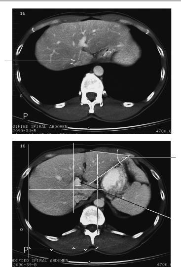
1Liver:
a Angle of left hepatic border: ca. 45°
b Caudate lobe/right lobe (CL/RL) = 0.37 ± 0.16 (e.g., 0.88 ± 0.2 in cirrhosis. Reference lines [from medial side]: line I is tangent to the medial border of the caudate lobe; line II is parallel to I and tangent to the lateral aspect of the portal vein; line III is tangent to the lateral hepatic border and perpendicular to a line midway between the portal vein and inferior vena cava and perpendicular to I and II.
c Left lobe (anteroposterior diameter measured on the paravertebral line): up to 5 cm
2Portal vein:
!Up to 1.5 cm
3Hepatic veins:
!Up to 0.5 cm
Moeller, Normal Findings in CT and MRI © 2000 Thieme
All rights reserved. Usage subject to terms and conditions of license.

Liver 47
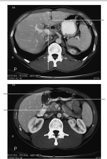
4Gallbladder:
!Horizontal diameter up to 5 cm (> 5 cm is suspicious for hydrops)
5Width of gallbladder wall:
!1−3 mm
6Width of common bile duct:
!! 8 mm (after cholecystectomy: ! 10 mm)
Spleen:
!Depth (D): 4−6 cm
!Width (W): 7−10 cm
!Length (L): 11−15 cm
!Splenic index: D×W×L = 160−440 Kidneys:
!Craniocaudal diameter: 8−13 cm
!Anteroposterior diameter: ca. 4 cm
!Transverse diameter 5−6 cm Position of superior poles:
!Right: superior border of L1
!Left: inferior border of T12
Width of renal cortex: ! 4−5 mm
Gerota fascia (thickness): ! 1−2 mm
Adrenal glands (variable):
!Crural thickness < 10 mm Diameter of abdominal aorta:
!Approximately 18−30 mm
Lymph nodes larger than 1 cm are suspicious for pathology.
Attenuation values
!Liver: 65 ± 10 HU
!Gallbladder contents: 0−25 HU
!Spleen: 45 ± 5 HU
!Pancreas: 40 ± 10 HU
!Fat: -65 to -100 HU
!Kidneys: 35−45 HU without contrast medium
!Adrenal glands: 25−40 HU without contrast medium
!Muscle: 45 ± 5 HU
!Blood vessels: ca. 40−55 HU without contrast medium
Moeller, Normal Findings in CT and MRI © 2000 Thieme
All rights reserved. Usage subject to terms and conditions of license.
48 CT: Abdomen
3
1a
 2c
2c
1b |
RL |
CL |
Moeller, Normal Findings in CT and MRI © 2000 Thieme
All rights reserved. Usage subject to terms and conditions of license.
Liver 49
1c
4
5
6
Moeller, Normal Findings in CT and MRI © 2000 Thieme
All rights reserved. Usage subject to terms and conditions of license.

50 CT: Abdomen
Pancreas
The pancreas is orthotopic and presents a normal size and smooth borders.
Lobulation of the pancreas is normal and appropriate for age. The parenchyma shows normal internal structure and attenuation values with no hypodense or hyperdense intrapancreatic changes. The pancreatic duct shows normal course and caliber.
The duodenal colon is unremarkable. The common bile duct is of normal width and has an unobstructed, fluid-filled lumen.
The visualized intrahepatic and extrahepatic bile ducts appear normal. No abnormalities appear in the peripancreatic fat.
The splenic vein and mesenteric vessels appear normal. The mesenteric root is normal.
Imaged portions of the liver, spleen, kidneys, and adrenals are unremarkable, as are the pararenal and paracolic spaces.
The major vessels appear normal, and there are no signs of lymphadenopathy.
Interpretation
The pancreas appears normal at CT.
Checklist
Pancreas |
! |
Position |
|
|
! |
Configuration |
|
|
! Size appropriate for age (see below) |
||
|
! Normal lobulation (borders are usually straight |
||
|
|
in adolescents and show increased lobulation |
|
|
! |
with aging) |
|
|
Smooth outer contours |
||
|
! Internal parenchymal structure (homogeneous |
||
|
|
in young patients, becomes slightly nonhomo- |
|
|
|
geneous with aging) |
|
|
! No focal abnormalities (e.g., calcifications, cysts, |
||
|
! |
tumors) |
|
|
Pancreatic duct: |
||
|
|
— Position (centered in the pancreas) |
|
|
|
— |
Width (see below) |
|
|
— |
No obstruction |
|
|
— No circumscribed or beaded dilatation or nar- |
|
|
|
|
rowing |
Moeller, Normal Findings in CT and MRI © 2000 Thieme
All rights reserved. Usage subject to terms and conditions of license.
|
|
|
Pancreas |
51 |
|
|
|
— Termination (usually opens into duodenum at |
|
||
|
|
|
Vater papilla with intrapancreatic part of |
||
|
|
|
common bile duct) |
|
|
|
! No peripancreatic fluid (exudate tracks along the |
||||
|
|
left and right pararenal spaces, into the omental |
|||
|
|
bursa, and along the paracolic gutters) |
|
|
|
|
! Para-aortic region |
|
|
||
|
! |
Splenic vein: |
|
|
|
|
|
— Course (lies against posterior surface |
of |
||
|
|
|
pancreas, runs from splenic hilum to portal |
||
|
|
|
vein) |
|
|
|
|
— Width |
|
|
|
|
! Lymph nodes (especially the parapancreatic and |
||||
|
|
pancreaticosplenic stations): |
|
|
|
|
|
— |
Size |
|
|
|
! |
— Number |
|
|
|
|
Peripancreatic fat: |
|
|
||
|
|
— |
Fat attenuation |
|
|
|
|
— |
No infiltration |
|
|
|
|
— |
No fluid |
|
|
|
! Mesenteric artery and vein: |
|
|
||
|
|
— Course |
|
|
|
|
|
— |
Size |
|
|
|
! Duodenum (descending part: directly adjacent |
||||
|
|
to head of pancreas; horizontal part: directly ad- |
|||
|
! |
jacent to uncinate process) |
|
|
|
|
Transverse mesocolon |
|
|
||
|
! |
Stomach: |
|
|
|
|
|
— |
Smooth posterior surface |
|
|
|
|
— |
Normal wall thickness |
|
|
|
! |
— Surrounded by fatty tissue |
|
|
|
|
Extrahepatic bile ducts: |
|
|
||
|
|
— Course (from porta hepatis to head of pan- |
|||
|
|
|
creas) |
|
|
|
|
— |
Width (see below) |
|
|
|
|
— Contents of homogeneous fluid density |
|
|
|
|
|
— |
No calculi |
|
|
Liver |
! |
— No air |
|
|
|
Position |
|
|
|||
|
! |
Size (if evaluable) |
|
|
|
|
! |
Borders |
|
|
|
|
|
— Smooth |
|
|
|
|
|
— Sharp |
|
|
|
Moeller, Normal Findings in CT and MRI © 2000 Thieme
All rights reserved. Usage subject to terms and conditions of license.

52 CT: Abdomen
|
! |
Attenuation values (see below) |
|
! |
Homogeneous internal parenchymal structure |
|
! |
Intrahepatic bile ducts |
|
|
— Course (centrifugal) |
|
|
— Width (general rule: should no longer be vis- |
|
! |
ible after contrast administration) |
|
Gallbladder (if visualized): |
|
|
|
— Size |
|
|
— Contents |
|
! |
— Wall thickness |
|
Porta hepatis occupied by the hepatic artery, |
|
|
|
common bile duct, and portal vein; no masses or |
Spleen |
! |
lymphadenopathy |
Position |
||
|
! |
Size |
|
! |
Borders |
Kidneys |
! |
Density (see below) |
! |
Paired |
|
|
! |
Position |
|
! |
Size |
|
! |
Smooth contours |
|
! |
Width of parenchyma |
Adrenal glands |
! |
Density (see below) |
! |
Shape |
|
|
! |
Size (see below) |
|
! |
Symmetrical crura |
Intestinal |
! |
No circumscribed expansion |
! |
Normal wall thickness |
|
structures (colon ! |
Homogeneous opacification by oral contrast me- |
|
haustrations, |
|
dium |
small bowel) |
! |
No free extraintestinal or intra-abdominal air or |
Blood vessels |
! |
fluid |
Size |
||
(aorta, inferior |
! |
No luminal obstruction |
vena cava) |
|
|
Lymph node |
! |
No lymphadenopathy |
stations (para- |
|
|
aortic, retro- |
|
|
crural) |
|
|
|
|
|
Moeller, Normal Findings in CT and MRI © 2000 Thieme
All rights reserved. Usage subject to terms and conditions of license.

Pancreas 53
Important Data
Dimensions of pancreas (normal ranges for age):
Age (years) |
Head 1 (mm) |
Body 2 (mm) |
Tail 3 |
(mm) |
! 20−30 |
25−32 |
17−21 |
16−20 |
|
! 31−40 |
23−29 |
16−20 |
15−18 |
|
! 41−50 |
22−29 |
16−19 |
14−17 |
|
! 51−60 |
21−27 |
14−18 |
14−17 |
|
! 61−70 |
20−26 |
14−18 |
13−16 |
|
! 71−80 |
17−25 |
12−17 |
11−15 |
|
Rule of thumb: head ! 3.5 cm, body and tail ! 2.5 cm
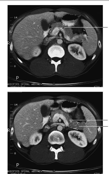
4Pancreatic duct:
!Width 1−3 mm
5Width of common bile duct:
!! 8 mm (! 10 mm after cholecystectomy)
Attenuation values:
!Pancreas: 40 ± 10 HU
!Liver: 65 ± 10 HU
!Spleen: 45 ± 5 HU
!Kidneys: 35−45 HU without contrast medium; renal cortex after contrast medium: ca. 140 HU
!Adrenal glands: 25−40 HU without contrast medium
!Muscle: 45 ± 5 HU
!Blood vessels: ca. 40−55 HU without contrast medium
!Gallbladder contents: 0−25 HU
!Fat: -65 to -100 HU
Dimensions:
!Spleen: width 7−10 cm, height 4−6 cm, length 11−15 cm
!Adrenal glands (variable): crural thickness ! 10 mm
!Gallbladder: horizontal diameter up to 5 cm (> 5 cm is suspicious for hydrops)
!Width of gallbladder wall: 1−3 mm
!Gerota fascia (thickness): 1−2 mm
!Diameter of abdominal aorta: approx. 18−30 mm
!Lymph nodes larger than 1 cm are suspicious for pathology.
Moeller, Normal Findings in CT and MRI © 2000 Thieme
All rights reserved. Usage subject to terms and conditions of license.
54 CT: Abdomen
2
4
3
Moeller, Normal Findings in CT and MRI © 2000 Thieme
All rights reserved. Usage subject to terms and conditions of license.