

Книги по МРТ КТ на английском языке / Normal Findings in CT and MRI
.pdfParanasal Sinuses 25
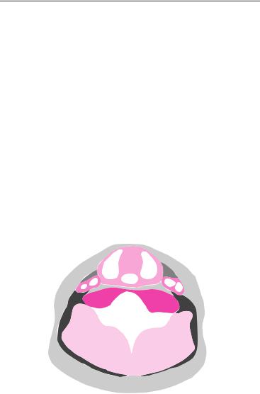
Important Data
1Frontal sinus:
!Height ca. 1.5−2 cm
2Sphenoid sinus:
!Width 0.9−1.4 cm
3Maxillary sinuses: a Width ca. 2 cm b Height ca. 2 cm
 2
2
Axial scan
 3a
3a
Axial scan
Moeller, Normal Findings in CT and MRI © 2000 Thieme
All rights reserved. Usage subject to terms and conditions of license.
26 CT: Head and Neck
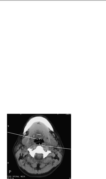
Cervical Soft Tissues
The cervical soft tissues show normal configuration, and the cervical spine is normally positioned.
The oral floor muscles are bilaterally symmetrical and normally developed. The spaces of the oral floor and neck are clear and well defined.
Imaged portions of the parotid and submandibular glands show no abnormalities.
The pharynx and larynx show normal boundaries and normal wall thickness.
The thyroid gland shows reasonable symmetry and normal size. The thyroid lobes have a normal internal structure.
Cervical vessels that can be evaluated with CT have a normal appearance.
The muscular structures of the neck appear normal, and there are no signs of cervical lymphadenopathy.
Interpretation
The cervical soft tissues appear normal.
|
|
|
|
|
|
|
|
|
|
|
|
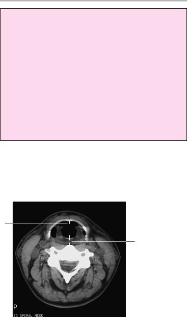
Visceral space |
|
|
|
|
|
|
|
|
|
|
|
|
|
Retropharyn- |
|
|
|
|
|
Anterior cer- |
||||||
|
|
|
|
|
||||||||
|
|
|
|
|
vical space |
|||||||
geal space |
|
|
|
|
|
|
|
Carotid space |
||||
|
|
|
|
|
|
|||||||
|
|
|
|
|
|
|
|
|
|
|||
|
|
|
|
|
|
|
|
|
|
|
|
|
Posterior |
|
|
|
|
|
|
|
|
|
|
|
Perivertebral |
|
|
|
|
|
|
|
|
|||||
cervical space |
|
|
|
|
|
|||||||
|
|
|
|
|
||||||||
|
|
|
|
|
|
|
|
|
|
|
|
space (pre- |
Perivertebral |
|
|
|
|
|
|
|
|
|
vertebral |
||
|
|
|
|
|
|
|
|
portion) |
||||
|
|
|
|
|
|
|||||||
space |
|
|
|
|
|
|
||||||
(paraspinal |
|
|
|
|
|
Superficial |
||||||
portion) |
|
|
|
|
|
|||||||
|
|
|
|
|
space |
|||||||
|
|
|
|
|
|
|
|
|
|
|
|
|
Moeller, Normal Findings in CT and MRI © 2000 Thieme
All rights reserved. Usage subject to terms and conditions of license.

Cervical Soft Tissues 27
Checklist
Cervical soft |
! |
Configuration |
|
tissues |
! |
Normal anatomy |
|
Oral floor |
! Normal position of cervical spine |
||
! |
Anatomy |
||
muscles |
! |
Width |
|
|
! |
Bilateral symmetry |
|
|
! |
Boundary definition |
|
|
! |
Internal structure |
|
Submandibular |
! Spaces of oral floor are clearly defined |
||
! |
Size (symmetry) |
||
gland (and |
! |
Density |
|
parotid gland) |
! No dilatation of glandular duct |
||
|
! No hypodense or hyperdense areas within the |
||
Pharynx and |
|
glandular tissue |
|
! Shape (symmetrical) |
|||
larynx |
! |
Size |
|
|
! |
Smooth walls |
|
|
! |
Normal wall thickness |
|
|
! |
No masses |
|
Cervical spaces ! Retropharyngeal space |
|||
|
! Parapharyngeal space (visceral space) |
||
|
! |
Carotid space |
|
|
! Anterior and posterior cervical spaces |
||
|
! Perivertebral space (prevertebral and paraspinal |
||
|
|
portions): |
|
|
|
— |
Configuration |
|
|
— Boundaries |
|
|
|
— Symmetry |
|
|
|
— |
Internal structure |
Esophagus |
! |
— |
Width (see below) |
Position |
|||
|
! Wall thickness (see below) |
||
|
! |
Boundaries |
|
Thyroid gland |
! |
No masses |
|
! Anatomy (two lobes, largely symmetrical) |
|||
|
! |
Size (see below) |
|
|
! |
Internal structure (homogeneous) |
|
|
! |
No cysts |
|
|
! |
No nodules |
|
|
! |
No calcifications |
|
Moeller, Normal Findings in CT and MRI © 2000 Thieme
All rights reserved. Usage subject to terms and conditions of license.
28 CT: Head and Neck
Cervical vessels |
! |
Course |
|
! |
Caliber (see below) |
|
! No abrupt caliber changes |
|
Neck muscles |
! |
No calcifications |
! |
Anatomy |
|
|
! |
Symmetry |
|
! |
Borders |
Lymph node |
! |
Internal structure |
! No lymphadenopathy |
||
stations |
|
|
Cervical spine |
! Vertebral bodies |
|
(if evaluable) |
|
— Number |
|
|
— Shape |
|
|
— Position |
|
! |
— Contours |
|
Intervertebral disk spaces |
|
|
! |
Spinal canal: |
|
|
— Width |
|
|
— No circumscribed narrowing |
|
! Normal width of cervical spinal cord |
|
|
! |
No masses |
|
! |
No narrowing |
|
|
|
4 
 1
1
Moeller, Normal Findings in CT and MRI © 2000 Thieme
All rights reserved. Usage subject to terms and conditions of license.
Cervical Soft Tissues 29
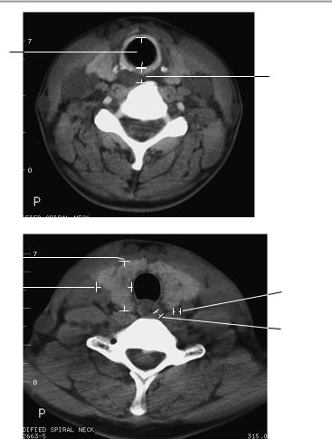
Important Data
Prevertebral soft tissues
1Retropharyngeal: ca. 1.7 ± 0.7 mm
2Retroglottic: ca. 6.0 ± 1.1 mm
3Retrotracheal: ca. 8.4 ± 2.5 mm
Lumina of upper respiratory tract (normal respiration):
4Laryngeal inlet (hyoid level): ca. 19 ± 4 mm
5Glottis: ca. 21 ± 4 mm
6Trachea: ca. 17 ± 3 mm
7Thyroid dimensions:
a Length: 3.6−6 cm (reconstruction) b Width: 1.5−2 cm
c Depth: 1−2 cm
Vascular calibers (at level of thyroid gland)
8Common carotid artery: 6−10 mm
9Esophagus: wall thickness 3 mm
5
2
Moeller, Normal Findings in CT and MRI © 2000 Thieme
All rights reserved. Usage subject to terms and conditions of license.
30 CT: Head and Neck
6
3
7c |
|
|
|
|
|
||
7b |
|
|
|
8 |
|||
|
|||
|
9 |
||
Moeller, Normal Findings in CT and MRI © 2000 Thieme
All rights reserved. Usage subject to terms and conditions of license.

31
CT: Chest
Thoracic Organs
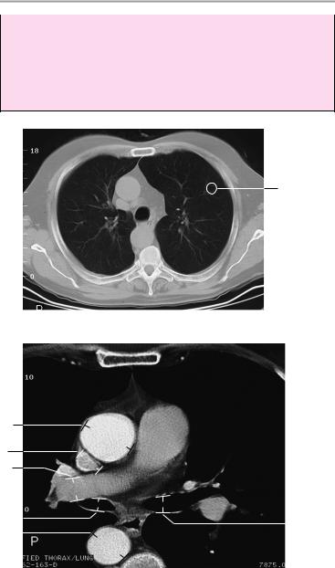
Both lungs are normally aerated and are applied to the chest wall on all sides. There is no sign of circumscribed pleural thickening and no fluid collection.
Pulmonary structure is normal and shows normal vascular markings. There are no intrapulmonary nodules or patchy opacities.
The mediastinum is centered and of normal width. There is no evidence of masses in the anterior, central, or posterior compartments.
The hilar region on each side is unremarkable, and the main bronchi appear normal.
The heart is orthotopic with normal configuration. The cardiac chambers are of normal size.
Major intrathoracic vessels and imaged portions of the supra-aortic vessels are unremarkable.
The thoracic skeleton and thoracic soft tissues show no abnormalities.
Interpretation
The thoracic organs appear normal at CT.
Checklist
Lungs |
! |
Anatomy (paired and symmetrical) |
|
! Fully apposed to the chest wall |
|
|
! |
No pleural thickening |
|
! |
No wall thickening |
|
! No fluid collection (patchy or circumscribed) |
|
|
! |
Complete aeration |
|
! Normal attenuation values of lung parenchyma |
|
|
! |
(see below) |
|
Pulmonary structure |
|
|
! Vascular markings (diminish from center to pe- |
|
|
! |
riphery) |
|
No pulmonary nodules |
|
|
! No patchy opacities (e.g., plaques or infiltrates) |
|
Moeller, Normal Findings in CT and MRI © 2000 Thieme
All rights reserved. Usage subject to terms and conditions of license.

32 CT: Thorax
Mediastinum |
! |
Configuration, position: |
|
|
— Centered |
|
|
— Width (see below) |
|
|
— No masses in the anterior, central, or poste- |
|
! |
rior compartment |
|
Hilar region: |
|
|
! |
— No masses or lymphadenopathy |
|
Main bronchi: |
|
|
|
— Anatomy |
|
|
— Course |
|
! |
— Width (see below) |
|
Heart: |
|
|
|
— Position (centered slightly left of midline) |
|
|
— Configuration |
|
|
— Size (cardiac chambers—see below) |
Vessels |
! |
— Myocardium (width—see below) |
Intrathoracic vessels (ascending aorta, aortic |
||
|
|
arch, descending aorta, vena cava—see below): |
|
|
— Anatomy |
|
|
— Size |
|
! Supra-aortic vessels (subclavian artery, brachio- |
|
|
|
cephalic trunk, left common carotid artery): |
|
|
— Anatomy |
Diaphragm |
|
— Size |
! Shape (no contour abnormalities, costophrenic |
||
|
|
angle is sharp and clear) |
|
! Position (approximately the level of the 10th− |
|
11th posterior rib)
! Width (no circumscribed widening, no defect)
Thoracic skeleton ! Position (ribs, clavicle, ! Structure
sternum, scapula) ! Contours and symmetry
|
! No bony expansion or destruction |
|
|
! |
Thoracic spine: |
|
|
— Position |
Thoracic soft |
! |
— Shape of thoracic vertebrae |
Configuration |
||
tissues |
! |
Width |
|
! |
Symmetry |
|
! |
Density |
|
|
|
Moeller, Normal Findings in CT and MRI © 2000 Thieme
All rights reserved. Usage subject to terms and conditions of license.

Thoracic Organs 33
Important Data
1CT density of lung parenchyma:
!-403 ± 25 HU
2Diameter of aorta:
!< 4 cm
aAscending aorta:
!At level of pulmonary trunk bifurcation: 3.2 cm ± 0.5 cm
!At level of aortic root: 3.7 cm ± 0.3 cm
bDescending aorta:
!2.5 cm ± 0.4 cm
!Aortic arch: 1.5 cm ± 1.2 cm
Ratio of ascending to descending aortic diameters = 1.5:1
3Diameter of superior vena cava:
!At level of aortic arch: 1.4 cm ± 0.4 cm
!At level of pulmonary trunk bifurcation: 2 cm ± 0.4 cm
4Diameter of pulmonary arteries:
!Pulmonary trunk: 2.4 cm ± 0.2 cm
!Proximal right pulmonary artery: 1.9 cm ± 0.3 cm
!Distal right pulmonary artery: 1.5 cm ± 0.3 cm
!Left pulmonary artery: 2.1 cm ± 0.4 cm
5Width of main bronchi:
!Right ca. 15 mm
!Left ca. 13 cm
6Mediastinum:
!Transverse diameter of thymus: 1−2 cm
Heart
Dimensions of cardiac chambers
7Right atrium:
!Maximum transverse diameter: 4.4 cm
—At level of aortic root: 1.9 cm ± 0.8 cm
—At level of mitral valve: 3.2 cm ± 1.2 cm
—At center of ventricles: 2.8 cm ± 0.4 cm
8Left atrium:
a Maximum anteroposterior diameter: 4−5 cm
!At level of aortic root: 2.4 cm ± 4.5 cm
!At level of mitral valve: 2.9 cm ± 4.9 cm b Maximum transverse diameter: 9 cm
!At level of aortic root: 5.5 cm ± 8.4 cm
!At level of mitral valve: 4.9 cm ± 9.1 cm
9Angle between midsagittal plane and septum = 38°
Moeller, Normal Findings in CT and MRI © 2000 Thieme
All rights reserved. Usage subject to terms and conditions of license.
34 CT: Thorax
10Thickness of ventricular septum:
!Approximately 5−10 mm
11Thickness of pericardium:
!1−2 mm
12Thickness of myocardium:
!10−12 mm
1
Lung window
2a
3
4a
5a |
|
|
|
5b |
||
|
|
|||||
2b |
|
|
|
|
||
|
|
|
|
|
||
|
|
|
||||
Contrast bolus scan at level of pulmonary trunk bifurcation
Moeller, Normal Findings in CT and MRI © 2000 Thieme
All rights reserved. Usage subject to terms and conditions of license.