

Книги по МРТ КТ на английском языке / Normal Findings in CT and MRI
.pdf
|
|
|
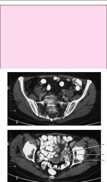
Female Pelvis |
65 |
Uterus |
! |
Position |
|
|
|
! |
Size |
|
|
|
! Borders (smooth outer contours) |
|
||
|
! |
Density (see below) |
|
|
|
! |
Uterine cavity: |
|
|
|
|
— |
Configuration |
|
|
|
— |
Size |
|
|
|
— Density |
|
|
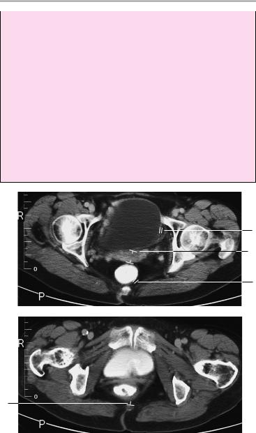
Cervix, vagina |
! |
— Contents |
|
|
Position |
|
|||
|
! |
Size |
|
|
Ovaries |
! |
Borders |
|
|
! |
Position |
|
||
|
! |
Size |
|
|
|
! |
Density |
|
|
|
! |
Symmetry |
|
|
|
! No masses of soft-tissue or fluid density |
|
||
Urinary bladder ! Adequate distention |
|
|||
|
! |
Smooth outer contours |
|
|
Vessels |
! Wall thickness (see below) |
|
||
! |
Caliber |
|
||
|
! |
Course |
|
|
Lymph node |
! No significant intimal calcifications |
|
||
! No nodal enlargement (>1 cm) |
|
|||
stations |
|
|
|
|
Pelvic skeleton |
! |
Configuration |
|
|
|
! Margins (cortex smooth and sharp with no dis- |
|||
|
! |
continuities) |
|
|
|
Bony structures |
|
||
|
! No circumscribed hypoor hyperdense areas |
|
||
|
! Femoral heads are rounded and centered |
in |
||
|
|
acetabula |
|
|
|
! Sacroiliac joints are smooth and of normal width |
|||
Subcutaneous |
! Symphysis pubis (see below) |
|
||
! |
Density |
|
||
tissue and |
! |
Extent |
|
|
muscles |
! |
Borders |
|
|
|
! |
Symmetry |
|
|
|
|
|
|
|
Moeller, Normal Findings in CT and MRI © 2000 Thieme
All rights reserved. Usage subject to terms and conditions of license.
66 CT: Abdomen
Important Data
1Sacroiliac joint spaces:
!Cartilage thickness 2−5 mm (anterior and inferior: 2−3 mm)
2Uterus:
!Size (variable): Prepubescent: length up to 3 cm, transverse diameter ca. 1 cm
!Nullipara: length up to 8 cm, transverse diameter ca. 4 cm
! Multipara: length up to 9.5 cm, transverse diameter ca.
5.5 cm
!Postmenopausal: length up to 6 cm, transverse diameter ca. 2 cm
aTransverse diameter of upright uterus (= well-distended bladder) ! 5 cm
bUterine cervix: transverse diameter ! 3 cm
 1
1
3a
3b
2a
Moeller, Normal Findings in CT and MRI © 2000 Thieme
All rights reserved. Usage subject to terms and conditions of license.
Female Pelvis 67
3Ovaries:
!Prepubescence: a, length up to 2.5 cm; b, transverse diameter ca. 2.5 cm
!Sexual maturity: a, length up to 4 cm; b, transverse diameter ca. 2.5 cm
!Postmenopausal: a, length up to 3 cm; b, transverse diameter ca. 1.5 cm
4Urinary bladder:
!Wall thickness (of well-distended bladder): ca. 3 mm
5Rectum:
!Wall thickness ! 5 mm
6Symphysis pubis:
!Width < 6 mm
7Pelvic dimensions:
!Pelvic outlet: anteroposterior (= coccyx to posterior edge of symphysis): ca. 9 cm
4
2b
5
6
7
Moeller, Normal Findings in CT and MRI © 2000 Thieme
All rights reserved. Usage subject to terms and conditions of license.

68 CT: Abdomen
Male Pelvis
The pelvic inlet appears normal, with normal configuration of the iliac wings and iliopsoas muscles.
Imaged bowel structures, especially the cecum and rectum, show no abnormalities with no evidence of wall thickening or mass lesions. The perirectal fat and ischiorectal fossa are unremarkable. The adequately distended urinary bladder has smooth outer contours and normal wall thickness. The seminal vesicles are of normal size. The angle between the bladder and seminal vesicle is clear and normal on each side.
The prostate shows normal size and configuration and a homogeneous internal structure.
The vessels of the lesser pelvis are normal in course and caliber. There are no signs of lymphadenopathy.
The appearance of the pelvic skeleton, especially the femoral heads and sacroiliac joints, is normal.
There are no soft-tissue abnormalities.
Interpretation
The lesser pelvis appears normal at CT.
Checklist
Pelvic inlet |
! |
Configuration |
|
! |
Width |
|
! |
Symmetry |
|
! |
Iliac wings (shape) |
|
! |
Iliopsoas muscles: |
|
|
— Size |
|
|
— Density |
Intestinal struc- |
! |
— Symmetry |
Position |
||
tures (especially |
! |
Wall thickness (when normally distended, see |
the cecum and |
|
below) |
rectum) |
! |
No circumscribed wall thickening |
Perirectal fat |
! |
Well-opacified lumen with no soft-tissue mass |
! |
Density (fat attenuation) |
|
|
! |
No infiltration |
|
! |
No masses |
Ischiorectal fossa ! |
Bilateral symmetry |
|
|
! |
No masses |
|
! |
No lymphadenopathy |
Moeller, Normal Findings in CT and MRI © 2000 Thieme
All rights reserved. Usage subject to terms and conditions of license.

Male Pelvis 69
Urinary bladder ! Adequate distention
!Smooth outer contours
!Wall thickness (see below)
Seminal vesicles ! Position (behind the bladder)
|
! |
Size (see below) |
|
|
|
! |
Symmetry |
|
|
|
! Angle between bladder and seminal vesicle (see |
|||
Prostate |
! |
below) is clear on each side |
|
|
Position (central at bladder outlet) |
|
|||
|
! |
Configuration (rounded) |
|
|
|
! |
Size (see below) |
|
|
|
! Density (homogeneous, see below) |
|
||
|
! |
No calcifications |
|
|
|
! No unilateral nonhomogeneity after contrast ad- |
|||
Vessels |
! |
ministration |
|
|
Caliber |
|
|
||
|
! |
Course |
|
|
Lymph node |
! No significant intimal calcifications |
|
||
! |
No adenopathy |
|
|
|
stations |
|
|
|
|
Pelvic skeleton |
! |
Configuration |
|
|
|
! Margins (cortex smooth and sharp, no discon- |
|||
|
! |
tinuities) |
|
|
|
Bony structures |
|
|
|
|
! No circumscribed |
hypodense or |
hyperdense |
|
|
|
areas |
|
|
|
! Femoral heads are |
rounded and |
centered in |
|
|
|
acetabula |
|
|
|
! Sacroiliac joints are smooth and of normal width |
|||
|
! |
(see below) |
|
|
Subcutaneous |
Symphysis pubis |
|
|
|
! |
Density |
|
|
|
tissue and |
! |
Extent |
|
|
muscles |
! |
Borders |
|
|
|
! |
Symmetry |
|
|
|
|
|
|
|
Moeller, Normal Findings in CT and MRI © 2000 Thieme
All rights reserved. Usage subject to terms and conditions of license.
70 CT: Abdomen
Important Data
1Sacroiliac joint spaces:
!Width 2−5 mm (anterior and inferior: 2−3 mm)
2Urinary bladder:
!Wall thickness (of well-distended bladder): ca. 3 mm
3Seminal vesicles:
!Size (highly variable):
aLength up to 5 cm
bWidth up to 2 cm, height up to 2.5 cm
cAngle between bladder and seminal vesicle: clear on each side
4Prostate:
!Size (varies with age, 20−70 years): a Anteroposterior diameter 2.5−3 cm
b Lateral (and craniocaudal diameter) 3−5 cm Attenuation value: 40−65 HU
5Rectum:
!Wall thickness ! 5 mm
 1
1
Moeller, Normal Findings in CT and MRI © 2000 Thieme
All rights reserved. Usage subject to terms and conditions of license.

Male Pelvis 71
2
3c
3b
3a
4a
4b
5
Moeller, Normal Findings in CT and MRI © 2000 Thieme
All rights reserved. Usage subject to terms and conditions of license.

72
CT: Spinal Column
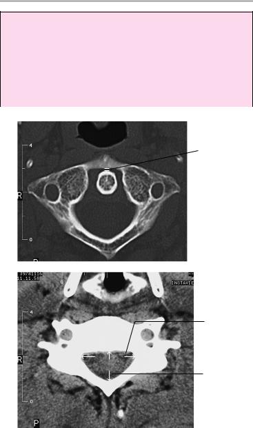
Cervical Spine
The cervical spine shows a normal degree of lordosis in the lateral survey scan, with no segmental malalignment.
The vertebral bodies show normal configuration and trabecular structure. The cortical margins are of normal thickness and are free of osteophytes.
The bony spinal canal shows normal sagittal diameter.
The intervertebral disks show normal CT density and normal posterior concavity. The disks do not project past the posterior surface of the vertebral bodies. The spinal cord is centrally placed and of normal width. It has homogeneous density and shows no circumscribed narrowing or expansion.
The nerve roots show a normal course and passage through the neuroforamina, which are of normal size and structure. The facet joints and uncovertebral joints are unremarkable.
The prevertebral and paravertebral soft tissues show no abnormalities.
Interpretation
The examined segments of the cervical spine appear normal.
Checklist
Position |
! |
Cervical lordosis |
|
! No segmental malalignment (lateral survey |
|
|
|
scan) |
Bony spinal |
! Normal position of dens (see below) |
|
! |
Width (see below) |
|
canal |
! |
Shape |
Vertebral bodies |
! |
Shape |
|
! Cortex (thickness, margins: smooth, sharp) |
|
|
! |
No marginal osteophytes |
! Trabeculae (uniform honeycomb arrangement, no rarefaction or circumscribed voids, no narrowing or expansion)
Moeller, Normal Findings in CT and MRI © 2000 Thieme
All rights reserved. Usage subject to terms and conditions of license.
|
|
Cervical Spine 73 |
|
! |
Bony structure (if evaluable: no lytic defects, |
Intervertebral |
! |
fracture lines, or osteoplastic areas) |
Width |
||
disk space |
! |
Margins (smooth, sharp) |
|
! |
Straight posterior disk contour |
|
! |
No disk protrusion past posterior surface of adja- |
Spinal cord |
|
cent vertebral bodies |
! Position (central) |
||
|
! |
Width (see below) |
|
! No circumscribed narrowing or expansion |
|
|
! |
Density (homogeneous) |
|
! Perimedullary thecal space clear: |
|
|
|
— No encroachment from the anterior side (e.g., |
|
|
by an intervertebral disk or osteophyte) or |
|
|
posterior side (e.g., by a hypertrophic liga- |
Neuroforamina |
! |
mentum flavum) |
Configuration |
||
|
! |
Width |
|
! No encroachment from the anterior side (e.g., by |
|
|
|
an intervertebral disk, osteophyte, or uncoverte- |
|
|
bral arthrosis) or posterior side (e.g., by hyper- |
Nerve roots |
|
trophic spondylarthrosis) |
! Course and passage through the neuroforamina |
||
Facet joints |
! |
No circumscribed expansion |
! Shape, symmetry |
||
|
! |
Pars interarticularis |
|
! |
Vertebral arches intact |
|
! Spinous processes (shape, length, bony struc- |
|
Soft tissues |
|
ture) |
! Symmetrical arrangement on both sides of the |
||
|
! |
vertebral bodies and spinous processes |
|
No masses |
|
|
! Prevertebral soft-tissue structures (especially |
|
|
|
the pharynx and thyroid gland; no masses) |
|
|
|
Moeller, Normal Findings in CT and MRI © 2000 Thieme
All rights reserved. Usage subject to terms and conditions of license.
74 CT: Spinal Column
Important Data
1Anteroposterior diameter of preodontoid space:
!< 2 mm
2Sagittal diameter:
!C1 ! 21 mm
!C2 ! 20 mm
!C3 ! 17 mm
!C4−C7 = 14 mm
1
3
2
Moeller, Normal Findings in CT and MRI © 2000 Thieme
All rights reserved. Usage subject to terms and conditions of license.