

Книги по МРТ КТ на английском языке / Normal Findings in CT and MRI
.pdfMagnetic Resonance Imaging
Moeller, Normal Findings in CT and MRI © 2000 Thieme
All rights reserved. Usage subject to terms and conditions of license.
Moeller, Normal Findings in CT and MRI © 2000 Thieme
All rights reserved. Usage subject to terms and conditions of license.

87
MRI: Head and Neck
Neurocranium
The interhemispheric fissure is centered on the midline. The cerebrum and cerebellum exhibit normal cortical sulcation.
The cerebral ventricles are of normal size and symmetrical with normal circulation of CSF. There are no signs of increased intracranial pressure. The cortex and white matter show normal development and normal signal intensity, especially in the periventricular white matter.
No abnormalities are seen in the basal ganglia, internal capsule, corpus callosum, or thalamus.
The brain stem and cerebellum show no abnormal changes in signal characteristics.
The sella and pituitary are normal, and parasellar structures are unremarkable.
The cerebellopontine angle area appears normal on each side. The internal acoustic meatus has normal width.
The paranasal sinuses and mastoid air cells show normal development and pneumatization. The orbital contents are unremarkable.
Interpretation
Cranial MRI is normal.
Checklist
Interhemispheric ! |
Centered on the midline |
|
fissure |
! |
No displacement |
|
! |
Falx cerebri: |
|
|
— Width |
|
|
— Signal characteristics |
|
|
— Flow in the dural sinuses (if the sequence per- |
Cortical sulcation ! |
mits flow assessment) |
|
Configuration |
||
in the cerebrum |
! |
Number of sulci |
and cerebellum |
! |
Width of sulci |
(arbor vitae) |
! |
No coarsening of sulci |
Moeller, Normal Findings in CT and MRI © 2000 Thieme
All rights reserved. Usage subject to terms and conditions of license.

88MRI: Head and Neck
!No circumscribed widening or narrowing
!Cisterns and cortical markings are well defined
Cerebral cortex |
! |
Width |
|
! Distribution (no ectopic tissue) |
|
|
! Signal characteristics (no hyperintense [demy- |
|
|
|
elination, edema, hemorrhage] or hypointense |
|
|
[calcification, hemorrhage] changes) |
|
! No areas of separation from the calvarium |
|
|
! No abnormal fluid collection (convex or con- |
|
|
|
cave) between the cerebral cortex and calvar- |
Ventricles |
! |
ium |
Shape |
||
|
! Size normal for age (see below) |
|
|
! Symmetry (no unilateral or circumscribed en- |
|
|
|
largement) |
|
! Evidence of flow in the (centrally located) aque- |
|
|
|
duct |
|
! Fourth ventricle is tent-shaped and not dilated |
|
|
! No signs of increased intracranial pressure |
|
|
|
(e.g., effaced sulci, narrowed or widened ven- |
White matter |
|
tricles) |
! Signal characteristics (maturity appropriate for |
||
|
|
age; homogeneous signal intensity, especially at |
|
|
periventricular sites; no patchy or circumscribed |
|
|
hyperintense [demyelination, edema, hemor- |
|
|
rhage] or hypointense [calcification, hemor- |
|
|
rhage] signal changes) |
Basal ganglia, |
! Normal width in relation to cortex |
|
! |
Position |
|
internal and |
! |
Size |
external capsule, |
! |
Delineation |
thalamus |
! |
Signal intensity |
Corpus callosum |
! |
Anatomy |
|
! |
Configuration |
|
! |
Size |
|
! No circumscribed narrowing or expansion |
|
|
! No foci of demyelination |
|
Brain stem |
! |
No masses |
! |
Shape |
|
|
! |
Signal intensity (homogeneous) |
|
! |
No focal abnormalities |
|
! Cranial nerves (presence, course, width, sym- |
|
|
|
metry) |
Moeller, Normal Findings in CT and MRI © 2000 Thieme
All rights reserved. Usage subject to terms and conditions of license.
|
|
Neurocranium 89 |
Cerebellum |
! |
Anatomy (symmetry) |
|
! |
Cortex (width, sulcation) |
Intracranial |
! |
White matter (homogeneous signal intensity) |
! |
Course |
|
vessels |
! |
Width |
|
! |
No abnormal dilatation |
Sella and |
! |
No vascular malformations |
! |
Size (see below) |
|
pituitary |
! |
Configuration (surface flat or slightly concave, |
|
! |
infundibulum centered) |
|
Signal intensity (neurohypophysis and adeno- |
|
|
|
hypophysis, no circumscribed change in signal |
|
|
intensity before or after contrast administra- |
|
! |
tion) |
|
Parasellar structures (optic chiasm, suprasellar |
|
|
|
CSF spaces, carotid siphon, cavernous sinus) are |
Petrous pyramids ! |
unremarkable |
|
Cerebellopontine angle area: |
||
|
|
— Width of internal auditory canals (see |
|
|
below) |
|
|
— CSF spaces (symmetrical, fluid intensity) |
|
|
— No masses |
|
! |
— Vestibulocochlear nerve clearly defined |
|
Mastoid cells, mastoid antrum |
|
|
|
— Anatomy |
|
|
— Pneumatization |
|
|
— Borders (wall thickness, smooth and continu- |
|
|
ous contours) |
|
|
— No masses |
|
! |
— Not fluid-filled |
|
Cochlea and semicircular canals: |
|
|
|
— Anatomy |
|
|
— Configuration |
Paranasal sinuses ! |
— Smooth borders |
|
Anatomy |
||
|
! |
Pneumatization |
|
! |
Borders (wall thickness, smooth and continuous |
|
! |
contours) |
|
Nasal cavity: |
|
|
|
— Pneumatization |
|
|
— Septum on midline |
— Turbinates (presence of superior, middle, and inferior turbinates; width)
Moeller, Normal Findings in CT and MRI © 2000 Thieme
All rights reserved. Usage subject to terms and conditions of license.

90 MRI: Head and Neck
Orbit |
! |
Configuration of orbital cone |
|
! |
Contents: |
|
|
— Globe (position, size, signal intensity, wall |
|
|
thickness) |
|
|
— Eye muscles (position, course, signal inten- |
|
|
sity, width) |
|
|
— Optic nerve (course, width—see below) |
|
|
— Ophthalmic vein (course, width—see below) |
|
|
|
Important Data
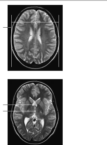
Vetricular dimensions:
1Cella media index:
!B/A > 4 = normal
2Frontal horn of lateral ventricle (at level of foramen of Monro):
!Under 40 years: < 12 mm
!Over 40 years: < 15 mm
3Width of third ventricle:
!< 5 mm in children (slightly more in infants)
!< 7 mm in adults under age 60
!< 9 mm in adults over age 60
Moeller, Normal Findings in CT and MRI © 2000 Thieme
All rights reserved. Usage subject to terms and conditions of license.
Neurocranium 91
B
1
A
Axial image
2
3
Axial image
Moeller, Normal Findings in CT and MRI © 2000 Thieme
All rights reserved. Usage subject to terms and conditions of license.
92 MRI: Head and Neck
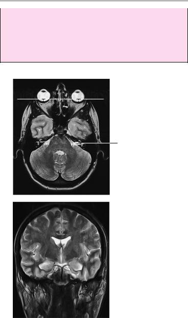
4Width of ophthalmic vein:
!3−4 mm
5Optic nerve (axial image):
a Retrobulbar segment: 5.5 mm ± 0.8 mm
b Narrowest point (at approximately mid-orbit): 4.2 mm ± 0.6 mm
Axial image
4
Axial image
5a
5b
Moeller, Normal Findings in CT and MRI © 2000 Thieme
All rights reserved. Usage subject to terms and conditions of license.
Neurocranium 93
6Position of globe:
!Posterior margin of globe 9.9 mm ± 1.7 mm behind interzygomatic line
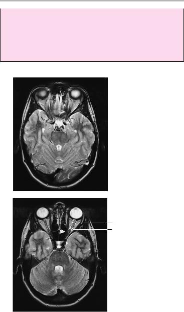
7Internal auditory canal:
!Approximately 5−10 mm, with no more than 1 mm difference between the right and left sides
Axial image
 6
6
7
Coronal image
Moeller, Normal Findings in CT and MRI © 2000 Thieme
All rights reserved. Usage subject to terms and conditions of license.
94 MRI: Head and Neck
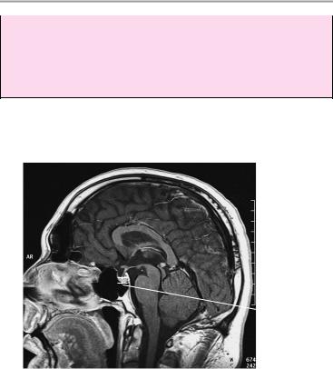
8Pituitary:
!Height of pituitary in sagittal plane: 2−6 mm Caution: normal size variations during
—Pregnancy: up to 12 mm
—Puberty: up to 10 mm in girls, up to 8 mm in boys
 8
8
Sagittal image
Moeller, Normal Findings in CT and MRI © 2000 Thieme
All rights reserved. Usage subject to terms and conditions of license.