

Книги по МРТ КТ на английском языке / Normal Findings in CT and MRI
.pdfLumbar Spine 175
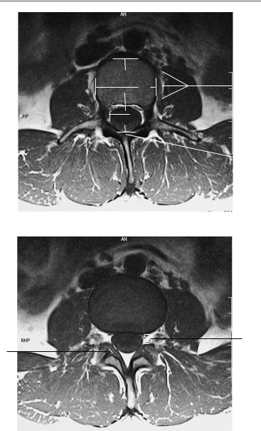
T2-weighted sagittal image at level of lateral recess
1
2
3
T2-weighted midsagittal image
4
Moeller, Normal Findings in CT and MRI © 2000 Thieme
All rights reserved. Usage subject to terms and conditions of license.
176 MRI: Spinal Column
D
C |
|
6 |
|
B A 

 5
5
 4
4
T2-weighted axial image at level of pars interarticularis
7
8
T1-weighted axial image at level of neuroforamina
Moeller, Normal Findings in CT and MRI © 2000 Thieme
All rights reserved. Usage subject to terms and conditions of license.

Lumbar Spine 177
Coronal image
Moeller, Normal Findings in CT and MRI © 2000 Thieme
All rights reserved. Usage subject to terms and conditions of license.

178 MRI: Spinal Column
Sacroiliac Joints
The sacroiliac joints are normally shaped with normal development of the sacrum and iliac wings and a normal-appearing lumbosacral junction. The joint space is of normal width on both sides. The joint contours are smooth and sharply defined.
The subchondral bone marrow appears normal. There are no marginal osteophytes.
The sacrum and iliac wings also contain normal bone marrow and present smooth, intact cortical boundaries. The sacral neuroforamina are of normal width.
The nerve filaments shows a normal course and diameter, and the width of the sacral spinal canal is normal.
The muscles and the imaged organs of the lesser pelvis show no abnormalities.
Interpretation
The sacroiliac joints appear normal.
Checklist
Joint |
! |
Shape: |
|
|
— Articular surfaces converge posteriorly |
|
! |
— Bilateral symmetry |
|
Contours: |
|
|
|
— Margins: smooth, sharp |
|
|
— Cortical thickness (uniform) |
|
|
— No steps or discontinuities |
|
! |
— No marginal osteophytes |
|
Joint space: |
|
|
|
— Uniform normal width (see below) |
|
|
— No circumscribed narrowing or expansion |
|
|
— No obliteration (ankylosis) |
|
|
— No unilateral increase in joint fluid |
|
|
— No signal voids within the joint space (air, cal- |
|
|
cifications) |
|
|
— No marginal osteophytes (caution: the ileum |
|
|
normally contains areas of hyperostosis) |
|
|
— Normal thickness of articular cartilage (see |
|
|
below) |
|
|
— No abnormal contrast enhancement |
|
|
— No thickening of joint capsule |
Moeller, Normal Findings in CT and MRI © 2000 Thieme
All rights reserved. Usage subject to terms and conditions of license.

Sacroiliac Joints 179
|
! |
Subchondral region: |
|
|
— Homogeneous, fat-equivalent signal intensity |
|
|
of bone marrow |
|
|
— No erosive or destructive changes |
|
|
— No increase in T2-weighted signal intensity |
|
|
(e.g., circumscribed due to cysts or patchy due |
|
|
to bone-marrow edema) |
|
|
— No decrease in T1-weighted or T2-weighted |
|
|
signal intensity (e.g., sclerosis on the sacral |
|
|
side or fatty infiltration of the periarticular |
Sacrum |
|
bone marrow) |
! Anatomy (four vertebral bodies, four neuro- |
||
|
! |
foramina) |
|
Shape |
|
|
! Symmetry (lateral sacral mass) |
|
|
! Width and arrangement of neuroforamina |
|
|
! Bone marrow signal (fat-equivalent, no marrow- |
|
|
|
replacing process) |
|
! Bony spinal canal (width) |
|
|
! |
Shape (closed) |
|
! Dural tube (width, no circumscribed narrowing |
|
|
|
or expansion) |
|
! Filaments have normal size and arrangement, |
|
|
|
and are not fused together; no posterior adhe- |
|
|
sions |
Iliac wings |
! Sacral plexus (course, width) |
|
! |
Shape |
|
|
! |
Symmetry |
|
! |
Margins: smooth, sharp |
|
! Cortical thickness (continuous and uniform; no |
|
|
|
steps or discontinuities) |
|
! Bone marrow signal (fat-equivalent, no marrow- |
|
|
|
replacing process) |
Lumbar spine |
! Symphysis and femoral heads |
|
! |
Position: |
|
|
|
— Lumbar lordosis (sagittal survey image) |
|
! Lumbosacral angle (see below) |
|
|
! Bony spinal canal (shape, width—see below) |
|
|
! Vertebral bodies (shape, margins, bone-marrow |
|
|
|
signal) |
|
! Height of intervertebral disk spaces |
|
|
! |
Intervertebral disks |
|
! |
Dural tube |
Moeller, Normal Findings in CT and MRI © 2000 Thieme
All rights reserved. Usage subject to terms and conditions of license.

180MRI: Spinal Column
!Neuroforamina
!Nerve roots:
— Origin and course
!Facet joints
!Vertebral arches intact
!Spinous processes
!Coccyx (shape, structure, position—see below)
Soft tissues |
! Muscles (especially the iliac, psoas, gluteals, and |
|
|
|
intrinsic back muscles) |
|
! Fat and intra-abdominal structures (e.g., sigmoid |
|
|
|
colon and rectum, bladder, prostate or uterus |
|
! |
and ovaries) |
Vessels |
No masses |
|
! |
Aorta |
|
|
! |
Iliac arteries |
|
! |
Vena cava |
Lymph nodes |
! |
Iliac veins |
! |
Lymph node stations (particularly the iliac |
|
|
|
nodes) |
|
|
|
Important Data
1Lumbosacral angle (S1/horizontal plane):
!26−57°
2Angle between sacrum and coccyx:
!Anterior angle ca. 10−30° (sagittal survey image, large range of variation)
3Width of joint space:
!4−5 mm
4Articular cartilage: a Sacral: 3 mm
b Iliac: 1 mm
Moeller, Normal Findings in CT and MRI © 2000 Thieme
All rights reserved. Usage subject to terms and conditions of license.
Sacroiliac Joints 181
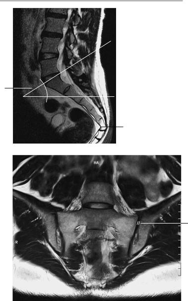
Sagittal image
1
2
3
Paracoronal image parallel to the sacrum
Moeller, Normal Findings in CT and MRI © 2000 Thieme
All rights reserved. Usage subject to terms and conditions of license.
182 MRI: Spinal Column
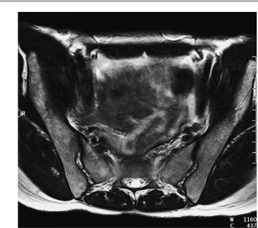
 4a
4a  4b
4b
Para-axial image at right angles to sacrum
Moeller, Normal Findings in CT and MRI © 2000 Thieme
All rights reserved. Usage subject to terms and conditions of license.

183
MRI: Joints
Temporomandibular Joint
The mandibular condyle has normal configuration and articulates with a normally shaped glenoid fossa.
The joint space is of normal width, and the articular surfaces have smooth, sharp borders. Cortical thickness and signal intensity of the bone marrow are normal.
The articular disk presents a hoodlike configuration on paracoronal images. It is dumbbell-shaped on parasagittal images. The posterior ligament is at approximatley the 11 o’clock position relative to the circumference of the mandibular condyle.
When the mouth is opened, the articular disk follows the movement of the mandibular condyle onto the articular tubercle.
Imaged portions of the paranasal sinuses are clear and pneumatized. Imaged portions of the neurocranium show no abnormalities.
Interpretation
The temporomandibular joint appears normal.
Checklist
Mandibular ! Cylindrical shape (coronal plane) condyle ! Spherical shape (sagittal plane)
Glenoid ! Shape (posteriorly convex, largely congruent with the mandibular condyle when the mouth is closed)
!Articular surfaces:
—Margins (smooth, sharp)
!Joint space:
—Width (see below)
—No effusion
!Cortex:
—Thickness
—No subchondral changes
—No osteophytes
Moeller, Normal Findings in CT and MRI © 2000 Thieme
All rights reserved. Usage subject to terms and conditions of license.

184 MRI: Joints
|
! Signal intensity of bone marrow (fat-equivalent) |
||
Articular disk |
! No circumscribed signal changes |
||
! |
Coronal plane: |
||
|
|
— Configuration (tubular or cylindrical) |
|
|
|
— |
Width (approximately uniform, 2−3 mm |
|
|
|
thick) |
|
|
— Position (surmounts mandibular condyle like |
|
|
|
|
a hood, does not project past medial or lateral |
|
! |
|
aspect of condyle) |
|
Sagittal plane: |
||
|
|
— |
Configuration (dumbbell-shaped: anterior |
|
|
|
ligament, intermediate zone, posterior liga- |
|
|
|
ment) |
|
|
— |
Position: |
|
|
— With mandible in resting position, posterior |
|
|
|
|
ligament is at about the 11−12 o’clock posi- |
|
|
|
tion relative to circumference of mandibular |
|
|
|
condyle |
|
|
— When mouth opens, articular disk moves |
|
|
|
|
with condyle (anterior ligament is anterior to |
|
|
|
condyle or at about the 11 o’clock position |
|
|
|
relative to condyle circumference) onto the |
Surrounding |
|
|
articular tubercle |
! Muscles (particularly the masseter and lateral |
|||
soft tissues |
|
pterygoid) |
|
|
! Normal-appearing periarticular fat |
||
|
! |
No masses |
|
Bony boundaries |
! |
No infiltration |
|
! |
Smooth |
||
(skull base supe- |
! |
Sharp |
|
riorly, external |
! |
Intact |
|
auditory canal |
|
|
|
and mastoid |
|
|
|
posteriorly) |
|
|
|
Adjacent struc- |
! |
Unremarkable |
|
tures (temporal |
|
|
|
lobe, mastoid |
|
|
|
process) |
|
|
|
Paranasal sinuses ! Clear and pneumatized
(if imaged)
Moeller, Normal Findings in CT and MRI © 2000 Thieme
All rights reserved. Usage subject to terms and conditions of license.