

Книги по МРТ КТ на английском языке / Normal Findings in CT and MRI
.pdfNeurocranium 5
Petrous pyramids ! Cerebellopontine angle area:
|
|
— Width and symmetry of bony portions of in- |
|
|
ternal auditory canals (see below) |
|
|
— CSF spaces symmetrical and of normal size, |
|
|
no masses |
|
! Mastoid air cells, mastoid antrum |
|
|
|
— Anatomy |
|
|
— Pneumatization |
|
|
— Borders (wall thickness, smooth contours |
|
|
with no discontinuities) |
|
|
— No masses |
|
|
— No fluid-dense opacification |
|
! Cochlea and semicircular canals: |
|
|
|
— Anatomy |
|
|
— Configuration |
Paranasal sinuses ! |
— Smooth borders |
|
Anatomy |
||
|
! |
Pneumatization |
|
! Borders (wall thickness, smooth and continuous |
|
|
! |
contours) |
|
Nasal cavity: |
|
|
|
— Pneumatization |
|
|
— Septum on midline |
|
|
— Turbinates (presence of superior, middle, and |
Orbit |
! |
inferior turbinates; width) |
Configuration of orbital cone |
||
|
! |
Contents: |
|
|
— Globe (position—see below; size, density, |
|
|
wall thickness) |
|
|
— Eye muscles (position, course, density, width) |
|
|
— Optic nerve (course, width—see below) |
Calvarium |
! |
— Ophthalmic vein (course, width—see below) |
Configuration |
||
|
! Contours (smooth, sharp, no expansion or bony |
|
|
|
outgrowths, no osteolytic or osteoplastic areas) |
|
|
|
Moeller, Normal Findings in CT and MRI © 2000 Thieme
All rights reserved. Usage subject to terms and conditions of license.

6 CT: Head and Neck
Important Data
Normal attenuation values: |
White matter |
Cortex |
! Noncontrast: |
39 HU |
32 HU |
! Postcontrast: |
41 HU |
33 HU |
(Each value has a deviation of ± 2 HU [Hounsfield units].) Attenuation difference between cortex and white matter: approximately 7 HU
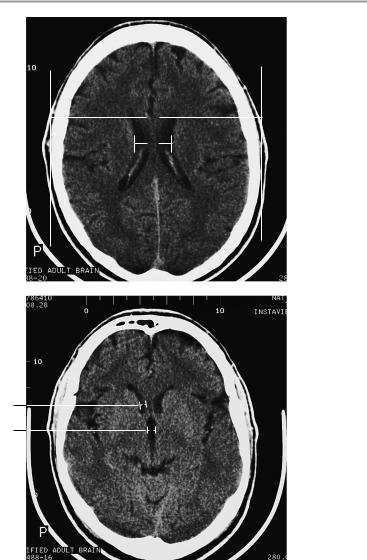
Ventricular dimensions
1Cella media index:
!B/A > 4 = normal
2Frontal horn of lateral ventricle (at level of foramen of Monro):
!Under age 40: < 12 mm
!Over age 40: < 15 mm
3Width of third ventricle:
!< 5 mm in children (slightly more in infants)
!< 7 mm in adults under age 60
!< 9 mm in adults over age 60
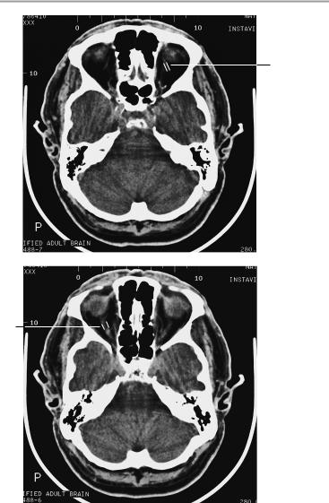
4Width of ophthalmic vein:
!3−4 mm
5Optic nerve (axial plane):
aRetrobulbar segment: 5.5 mm ± 0.8 mm
bNarrowest point (at approximately midorbit): 4.2 mm ± 0.6 mm
6 Position of globe:
!Posterior margin of globe is 9.9 mm ± 1.7 mm behind the interzygomatic line
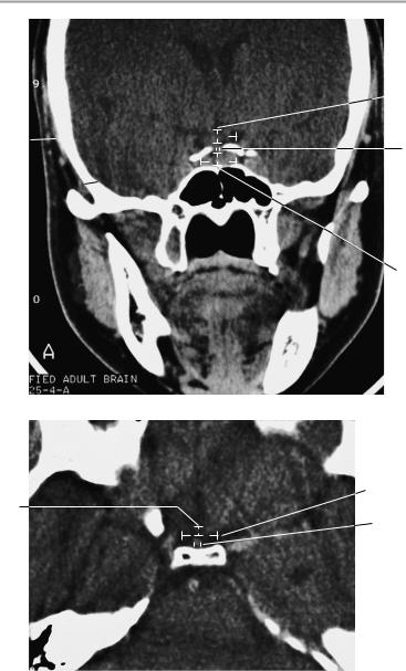
Pituitary: Height of pituitary in sagittal reconstruction: 2−7 mm Caution: normal size variations during:
—Pregnancy: up to 12 mm
—Puberty: up to 10 mm in girls, up to 8 mm in boys Internal auditory canal: 5−10 mm (average 7.6 mm); should be !
1 mm difference between the right and left sides
Moeller, Normal Findings in CT and MRI © 2000 Thieme
All rights reserved. Usage subject to terms and conditions of license.
Neurocranium 7
B
1 |
A |
2
3
Moeller, Normal Findings in CT and MRI © 2000 Thieme
All rights reserved. Usage subject to terms and conditions of license.
8 CT: Head and Neck
4
5b
Moeller, Normal Findings in CT and MRI © 2000 Thieme
All rights reserved. Usage subject to terms and conditions of license.

Neurocranium 9
5a
 6
6
Moeller, Normal Findings in CT and MRI © 2000 Thieme
All rights reserved. Usage subject to terms and conditions of license.

10 CT: Head and Neck
Pituitary
The sella shows normal size, position, and configuration. The borders of its floor and walls are smooth and sharply defined.
The pituitary shows normal position, shape, and size. The pituitary tissue shows normal, homogeneous density both before and after contrast administration. It contains no circumscribed hypodense or hyperdense areas.
The infundibulum is centered and of normal size.
The optic chiasm and suprasellar CSF spaces appear normal. The cavernous sinus and imaged portions of the internal carotid artery and carotid siphon are unremarkable.
Evaluable portions of the neurocranium show no abnormalities. The sphenoid sinus is clear and pneumatized.
Interpretation
The pituitary appears normal.
Important Data
1Pituitary:
a Height (in the midcoronal plane): 2−7 mm Caution: allow for normal size variations during:
!Pregnancy: up to 12 mm
!Puberty: up to 10 mm in girls, up to 8 mm in boys
b Width (transverse extent in coronal plane, women of childbearing age): 12.9 mm ± 1.6 mm
Area of the pituitary in the coronal plane (height x width, women of childbearing age): 93 mm2 ± 1.6 mm2
2Optic chiasm:
!Coronal: a, width 9−18 mm; b, height 3−6 mm
!Axial: c, width 12−27 mm; d, depth 4−9 mm
3Pituitary stalk:
!< 4 mm
Moeller, Normal Findings in CT and MRI © 2000 Thieme
All rights reserved. Usage subject to terms and conditions of license.
Pituitary 11
 2b
2b
2a

3
1b 
1a
Coronal scan
2c
2d
3
Axial scan
Moeller, Normal Findings in CT and MRI © 2000 Thieme
All rights reserved. Usage subject to terms and conditions of license.

12 CT: Head and Neck
Checklist
Sella |
! |
Position |
|
! |
Configuration (U shape) |
|
! Walls steep, not splayed, of normal size |
|
|
! Normal width of floor and walls |
|
Pituitary |
! Borders smooth and sharp |
|
! |
Position: |
|
|
! |
— At the center of the sella |
|
Configuration: |
|
|
|
— Bean-shaped |
|
|
— Superior border straight or slightly concave |
|
! |
(convex only during puberty or pregnancy) |
|
Size (see below) |
|
|
! |
Density: |
|
|
— Pituitary tissue homogeneous on noncontrast |
|
|
scans |
|
|
— Homogeneous contrast enhancement |
|
|
— No circumscribed hypodense or hyperdense |
Infundibulum |
! |
areas within the pituitary |
Position (centered) |
||
Optic chiasm |
! |
Size (see below) |
! |
Position |
|
|
! |
Size (see below) |
|
! |
Symmetry |
Suprasellar CSF |
! Course of optic nerve |
|
! Shape (symmetrical) |
||
spaces (chias- |
! Width (no circumscribed narrowing) |
|
matic cistern) |
|
|
Cavernous sinus ! Shape (symmetrical) |
||
|
! |
Size (see below) |
Internal carotid |
! |
No infiltration |
! |
Size |
|
artery |
! |
Course |
(siphon area) |
! |
Density |
Neurocranium |
! |
Temporal lobe |
|
! |
Hypothalamus |
Sphenoid sinus |
! Floor of third ventricle |
|
! Borders: smooth, normal width (especially of |
||
|
! |
roof), contours |
|
Pneumatization |
|
|
|
|
Moeller, Normal Findings in CT and MRI © 2000 Thieme
All rights reserved. Usage subject to terms and conditions of license.

Petrous Pyramids 13
Petrous Pyramids
The petrous pyramids are normally developed and symmetrical. They have smooth, intact cortical margins and a normal trabecular structure. The internal auditory canal is smooth and sharply defined on each side, with normal shape and diameter. The cochlea and semicircular canals appear normal. The mastoid air cells are normally developed, clear and pneumatized. Their bony walls are of normal thickness. The tympanic cavity is normally developed, and the auditory ossicles have a normal appearance.
Configuration of the cerebellopontine angle area on each side is normal, with clear delineation of the cerebellopontine angle cistern. The brain stem has normal configuration and CT density.
The external auditory canal appears normal on each side.
Other visualized portions of the neurocranium show no abnormalities.
Interpretation
Both petrous pyramids appear normal at CT.
Checklist
Petrous pyramids ! |
Configuration |
|
|
! |
Shape (triangular) |
|
! |
Bilateral symmetry |
|
! |
Delineation (cortical margins smooth and sharp) |
|
! |
Trabecular internal structure (no fracture lines, |
|
|
no bone destruction or circumscribed hy- |
|
! |
podense or hyperdense areas) |
|
Smooth, sharp boundary with the calvarium |
|
Internal auditory ! |
(sinodural angle) |
|
Shape |
||
canals |
! |
Course |
|
! |
Width (see below) |
|
! |
Bony boundaries (smooth, sharp) |
|
! |
Vestibulocochlear nerve (cranial nerve VIII) and |
|
|
facial nerve (cranial nerve VII), if visualized: |
|
|
— Width (uniform, no right-left disparity) |
Cochlea and |
! |
— Enhancement characteristics (nonenhancing) |
Anatomy |
||
semicircular |
! |
Configuration |
canals |
! |
Smooth borders |
Moeller, Normal Findings in CT and MRI © 2000 Thieme
All rights reserved. Usage subject to terms and conditions of license.

14CT: Head and Neck
!Tympanic cavity:
—Anatomy
—Shape
—Borders
—Pneumatization
!Auditory ossicles (malleus, incus, stapes: presence, shape, relative positions in ossicular chain)
Mastoid |
! |
Cellular anatomy (antrum, retrofacial cells, peri- |
|
|
|
tubal cells, peribulbar cells, marginal cells, ter- |
|
|
|
minal cells): |
|
|
|
— Cells small, large, or of mixed sizes; normal = |
|
|
|
|
uniform enlargement of cells from antrum to |
|
|
|
terminal cells) |
|
|
— Pneumatization |
|
|
|
— Borders (septal thickness, smooth contours |
|
|
|
|
with no discontinuities) |
|
|
— No masses |
|
|
|
— Not opacified by abnormal fluid or soft-tissue |
|
Cerebellopontine ! |
|
density |
|
Brain stem |
|||
angle area |
|
— Shape |
|
|
|
— Density (homogeneous) |
|
|
|
— |
No focal abnormalities |
|
! Vestibulocochlear and facial nerve nuclei: |
||
|
|
— No hypodensity |
|
|
|
— No masses |
|
|
! Entry sites of vestibulocochlear nerve (enters |
||
|
|
pons and medulla at lateral extension of medul- |
|
|
|
lopontine sulcus) and facial nerve: |
|
|
! |
— |
Bilaterally symmetrical |
|
CSF spaces: |
||
|
|
— Cerebellopontine angle cistern (symmetrical, |
|
|
|
|
fluid density) |
|
|
— No masses |
|
|
|
— |
Well delineated |
|
! |
— |
No vascular loop |
|
External auditory canals: |
||
|
|
— Anatomy |
|
|
|
— Course |
|
|
|
— Width |
|
|
|
— Borders |
|
Moeller, Normal Findings in CT and MRI © 2000 Thieme
All rights reserved. Usage subject to terms and conditions of license.