

Книги по МРТ КТ на английском языке / Normal Findings in CT and MRI
.pdfMR Angiography of the Renal Arteries 225
1
6
5a |
|
|
|
5b |
|
||||
|
|
4 |
|
|
|
3 |
|||
|
|
||||||
|
|
|
|
|
|
8 |
|
9 |
|
|
|
|
|
||
|
|
|
|
||||
|
|
|
|
|
|
|
|
|
|
|
|
|
|
|
|
2
7
10
Moeller, Normal Findings in CT and MRI © 2000 Thieme
All rights reserved. Usage subject to terms and conditions of license.

226 MRI: Special Investigations
9 Contrast excretion into the pyelocaliceal system:
! 3 minutes
10Width of ureter:
!4−7 mm Inferior vena cava:
!Transverse diameter up to 2.5 cm
4
Moeller, Normal Findings in CT and MRI © 2000 Thieme
All rights reserved. Usage subject to terms and conditions of license.

Pelvic and Lower Limb Vessels 227
Pelvic and Lower Limb Vessels
The abdominal aorta is normal in its course, diameter, and filling. The aortic bifurcation occurs at a normal level, with normal visualization of the common, internal, and external iliac arteries.
Both common femoral arteries have normal calibers, smooth walls, and a homogeneous intraluminal signal. The femoral arteries show a normal course, caliber, and distribution.
The superficial femoral artery appears normal, especially within the adductor canal. Like the popliteal artery, the vessel shows a normal course and no irregularities in its caliber. It divides normally into the three lower leg arteries, which show normal course, caliber, and distribution.
Interpretation
The vascular system of the pelvic and lower limb arteries appears normal.
Checklist
Vascular course ! Abdominal aorta:
and caliber |
|
— |
Position: slightly to left of midline |
(described from |
|
— |
Almost straight course |
center to |
|
— |
Bifurcation (see below) |
periphery) |
! Common iliac artery |
||
|
! |
External iliac artery |
|
|
! |
Internal iliac artery |
|
|
! |
Common femoral artery |
|
|
! Superficial femoral artery (see below) |
||
|
! |
Circumflex femoral artery |
|
|
! |
Profunda femoris artery |
|
|
! Popliteal artery (see below) |
||
|
! |
Anterior tibial artery |
|
|
! |
Posterior tibial artery |
|
|
! |
Peroneal (fibular) artery: |
|
|
|
— |
Position (no displacement) |
|
|
— Course (no excessive tortuosity or coiling) |
|
|
|
— |
Caliber |
— Signal characteristics (homogeneous intraluminal signal, no filling defect)
Moeller, Normal Findings in CT and MRI © 2000 Thieme
All rights reserved. Usage subject to terms and conditions of license.

228MRI: Special Investigations
—Contours (smooth; no circumscribed, segmental or beaded constrictions; particularly note superficial femoral artery in the adductor canal)
—No circumscribed outpouching (e.g., popliteal artery)
Veins |
! |
No arteriovenous communications |
Vessels |
! No pathologic vessels or cutoffs |
|
Soft tissues and |
! |
(If evaluable) |
bony structures |
|
|
|
|
|
2
1
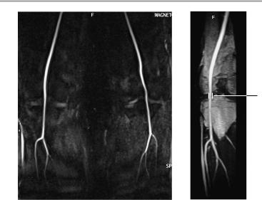
Phase-contrast angiogram of the pelvic vessels
Moeller, Normal Findings in CT and MRI © 2000 Thieme
All rights reserved. Usage subject to terms and conditions of license.

Pelvic and Lower Limb Vessels 229
Important Data
1Bifurcation:
!At approximately the L4-L5 level Vascular calibers:
2Abdominal aorta:
!Approximately 2−4 cm
3Superficial femoral artery:
!Approximately 0.7−1.5 cm
4Popliteal artery:
!Approximately 0.6−1 cm
3
Phase-contrast angiogram of the femoral vessels
Moeller, Normal Findings in CT and MRI © 2000 Thieme
All rights reserved. Usage subject to terms and conditions of license.
230 MRI: Special Investigations
4
Phase-contrast angiogram of the vessels about the knee joints
Moeller, Normal Findings in CT and MRI © 2000 Thieme
All rights reserved. Usage subject to terms and conditions of license.

MR Cholangiopancreatography 231
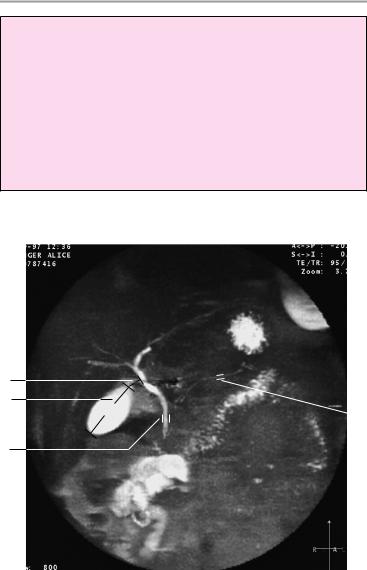
MR Cholangiopancreatography
The common bile duct shows normal position, caliber, and length with a homogeneous, fluid-equivalent intraluminal signal. The cystic duct and imaged portions of the intrahepatic bile ducts also appear normal. The gallbladder is of normal size; it has smooth borders and homogeneous contents.
The pancreatic duct shows normal position, length, and caliber with homogeneous internal structure and smooth contours.
Interpretation
The bile ducts, gallbladder, and pancreatic duct appear normal.
Checklist
Common bile |
! |
Course: |
duct |
! |
— Usually slightly convex toward the left side |
|
Size: |
|
|
|
— Tapers slightly from its origin (the right and |
|
|
left hepatic ducts and common hepatic duct |
|
|
are of equal size) |
|
|
— No circumscribed caliber irregularities, espe- |
|
|
cially in the papillary area (e.g., prestenotic |
|
|
dilatation, discrete or segmental stenosis due |
|
|
to tumor or fibrosis) |
|
|
— No circumscribed narrowing (stricture) or di- |
|
! |
latation |
|
Shape: |
|
|
|
— Contours (smooth, straight) |
|
! |
— Number (one) |
|
Internal structure: |
|
|
|
— Homogeneous fluid-equivalent signal inten- |
|
|
sity |
|
|
— No calculi |
Gallbladder |
! |
— No tumor |
Position |
||
|
! |
Number (one) |
|
! |
Shape |
|
! |
Possible septation |
|
! |
Size (see below) |
Moeller, Normal Findings in CT and MRI © 2000 Thieme
All rights reserved. Usage subject to terms and conditions of license.

232MRI: Special Investigations
!Contours:
—Smooth
—Straight
—No diverticula
!Internal structure:
—Homogeneous fluid-equivalent signal
—No filling defect (sludge, stone, papilloma, carcinoma)
Cystic duct and |
! Position (presence and number) |
|
intrahepatic |
! |
Course |
bile ducts |
! |
Size |
|
! |
Contours |
Pancreatic duct |
! |
Filling |
! |
Position: |
|
|
|
— Horizontal |
|
! |
— Ascends toward left side |
|
Size: |
|
|
|
— Diameter tapers uniformly toward the |
|
|
duodenum |
|
|
— No circumscribed change in diameter (e.g., |
|
|
constriction by a tumor, cyst, or inflamma- |
|
|
tion; prestenotic dilatation due to a tumor; |
|
|
segmental ectasia such as the segmental ir- |
|
! |
regularities in pancreatitis) |
|
Shape: |
|
|
|
— Contours (smooth with straight walls) |
|
|
— No irregular margins |
|
|
— Not sacciform |
|
! |
— Not tortuous or dilated |
|
Internal structure: |
|
|
|
— Homogeneous fluid-equivalent signal |
— No calculi
— No tumor
Moeller, Normal Findings in CT and MRI © 2000 Thieme
All rights reserved. Usage subject to terms and conditions of license.
MR Cholangiopancreatography 233
Important Data
1Gallbladder:
!Horizontal diameter up to 5 cm (> 5 cm is suspicious for hydrops)
2Width of common bile duct:
!! 8 mm (after cholecystectomy: ! 10 mm)
3Cystic duct:
!Length ca. 4 cm
4Pancreatic duct:
!Width: 1−3 mm
3
1
 4
4
2
Moeller, Normal Findings in CT and MRI © 2000 Thieme
All rights reserved. Usage subject to terms and conditions of license.

234 MRI: Special Investigations
Cervical Arteries
The aortic arch presents smooth walls and normal configuration.
The brachiocephalic trunk arises normally and undergoes a normal division into the subclavian artery, common carotid artery, and right vertebral artery. The left common carotid artery arises directly from the aortic arch, has a normal caliber, and shows no luminal narrowing or filling defects. The vertebral artery appears normal.
The carotid bifurcation occurs at a normal level on each side and is normally shaped. The external carotid artery and particularly the internal carotid artery are symmetrical on each side and have normal calibers. There is no circumscribed narrowing or expansion.
The vessels display a homogeneous intraluminal signal. The carotid siphon appears normal, showing no displacement or extrinsic compression.
The vertebral arteries are symmetrically disposed and take a normal course. They show normal luminal diameters with no filling defects or caliber irregularities as far as the basilar artery.
The portions of the neck that are imaged and evaluable show no abnormalities.
Interpretation
The supra-aortic system of arterial cervical vessels appears normal.
Checklist
Aortic arch |
! |
General form |
|
! |
Course |
|
! |
Caliber |
|
! |
Signal characteristics |
|
|
Origins: brachiocephalic trunk, left common |
Brachiocephalic |
! |
carotid artery, left subclavian artery |
Origin |
||
trunk |
! |
Division into right subclavian and right common |
Left and right |
! |
carotid arteries |
Position |
||
subclavian |
! |
Course |
arteries |
! |
Caliber |
|
! |
Signal characteristics |
Moeller, Normal Findings in CT and MRI © 2000 Thieme
All rights reserved. Usage subject to terms and conditions of license.