

Книги по МРТ КТ на английском языке / Normal Findings in CT and MRI
.pdfLiver 135
|
! |
Gallbladder: |
|
|
— Size (see below) |
|
|
— Contours (smooth and sharp) |
|
|
— Wall thickness (see below, no general or cir- |
|
|
cumscribed thickening) |
|
! |
— No pericholecystic fluid |
|
Gallbladder contents: |
|
|
|
— Homogeneous |
|
|
— Fluid-equivalent signal intensity |
|
|
— No filling defects (calculi, polyps) |
|
! |
— No air |
|
Porta hepatis: |
|
|
|
— Occupied by the hepatic artery, common bile |
|
|
duct, and portal vein |
|
|
— No masses |
|
! |
— No lymphadenopathy |
|
Costophrenic sinus is clear and aerated on each |
|
Spleen |
! |
side |
Normal size (see below) |
||
Pancreas |
! |
Homogeneous internal structure |
! |
Normal size (see below) |
|
|
! |
Pancreatic duct unobstructed (see below) |
Para-aorticregion ! Unremarkable |
||
Kidneys |
! |
Position |
(if visualized) |
! |
Size |
Intestinal |
! |
Internal structure |
! |
Normal |
|
structures |
! |
No free extraintestinal or intra-abdominal air or |
|
|
fluid |
|
|
|
Moeller, Normal Findings in CT and MRI © 2000 Thieme
All rights reserved. Usage subject to terms and conditions of license.
136 MRI: Abdomen
Important Data
Dimensions:
1Liver:
a Left lobe (anteroposterior diameter on the left paravertebral line): up to 5 cm
b Right lobe (craniocaudal diameter measured on the midclavicular line): up to ca. 15 cm
Angle of hepatic border:
c Right side: ca. 75° (inferior border, sagittal plane) d Left side: ca. 45° (left lateral and inferior borders)
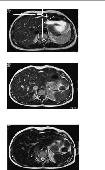
e Caudate lobe/right lobe (CL/RL) = 0.37 ± 0.16 (e.g., 0.88 ± 0.2 in cirrhosis). Reference lines [from medial side]: line I is tangent to the medial border of the caudate lobe; line II is parallel to I and tangent to the lateral aspect of the portal vein; line III is tangent to the lateral hepatic border and perpendicular to a line midway between the portal vein and inferior vena cava and perpendicular to I and II.
2Gallbladder:
a Horizontal diameter up to 5 cm (> 5 cm is suspicious for hydrops)
b Width of gallbladder wall: 1−3 mm
3Width of common bile duct:
! ! 8 mm (after cholecystectomy: ! 10 mm)
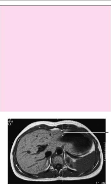
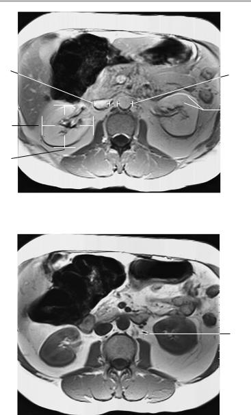
1a
T1-weighted noncontrast axial image
Moeller, Normal Findings in CT and MRI © 2000 Thieme
All rights reserved. Usage subject to terms and conditions of license.
Liver 137
|
|
III |
II |
I |
||
|
|
|
|
|
|
|
1e |
|
|
|
RL |
CL |
|
|
||||||
1d
T2-weighted noncontrast axial image
T1-weighted axial image after the i. v. administration of a superparamagnetic contrast agent
5
T2-weighted axial image after the i. v. administration of a superparamagnetic contrast agent
Moeller, Normal Findings in CT and MRI © 2000 Thieme
All rights reserved. Usage subject to terms and conditions of license.

138 MRI: Abdomen
4Portal vein:
!Up to 1.5 cm
5Hepatic veins:
!Up to 0.5 cm Spleen:
!Depth D): 4−6 cm
!Widt(W): 7−10 cm
!Length (L): 11−15 cm
!Splenic index: DxWxL = between 160 and 440 Adrenal glands (variable):
!Crural thickness < 10 mm Kidneys:
!Craniocaudal diameter: 8−13 cm
!Anteroposterior diameter: ca. 4 cm
!Transverse diameter: 5−6 cm Position of superior poles of kidneys:
!Right: superior border of L1; left: inferior border of T12 Transverse renal axis:
!Posteriorly divergent angle of 120°
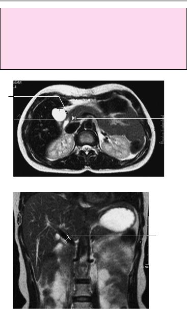
1b
1d
1c
Sagittal image at the level of the midclavicular line
Sagittal image through the left lobe of the liver
Moeller, Normal Findings in CT and MRI © 2000 Thieme
All rights reserved. Usage subject to terms and conditions of license.
Liver 139
Width of renal cortex: ! 4−5 mm
Diameter of abdominal aorta:
!Approximately 18−30 mm Inferior vena cava:
!Transverse diameter: up to 2.5 cm
Lymph nodes larger than 1 cm are suspicious for pathology.
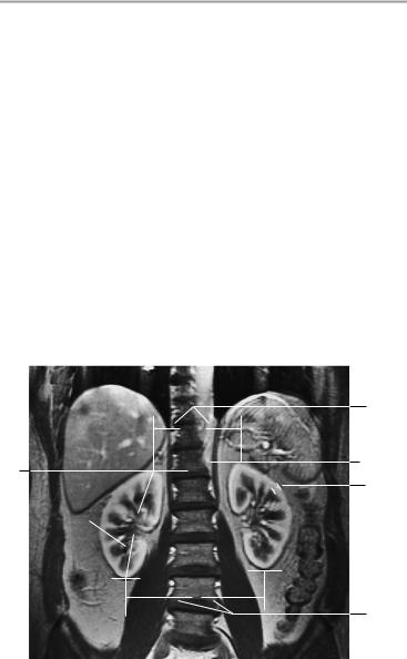
2b
2a |
|
|
|
|
|
|
|
|
|
3 |
Axial image
4
Coronal image
Moeller, Normal Findings in CT and MRI © 2000 Thieme
All rights reserved. Usage subject to terms and conditions of license.

140 MRI: Abdomen
Kidneys
Both kidneys appear normal in size and position, and the renal parenchyma displays normal width. There is no evidence of a mass. The calices are of normal shape, and the renal pelvis appears normal. The urinary drainage tract is unobstructed.
Postcontrast images show a normal time to corticomedullary equilibrium and undelayed, symmetrical contrast excretion into the renal pelves.
Other visualized upper abdominal organs, especially the adrenal glands, show no abnormalities.
Interpretation
Both kidneys appear normal.
Checklist
Kidneys |
! |
Paired |
|
|
! |
Position (see below) |
|
|
! |
Size (see below) |
|
|
! |
Contours: |
|
|
|
— Smooth |
|
|
! Parenchymal width and signal (see below) |
||
|
! Normal relation of cortex to medulla |
||
|
! |
Renal pelves: |
|
|
|
— |
Structure |
|
|
— |
Bilateral symmetry |
|
|
— Width |
|
|
! |
— |
Shape of calices |
|
Ureters: |
||
|
|
— One per side |
|
|
|
— Course |
|
|
|
— |
Width (see below) |
|
|
— No obstruction of urinary drainage |
|
|
! Perirenal and pararenal spaces: |
||
|
|
— |
Fat signal |
|
! Perirenal and pararenal fasciae: |
||
|
|
— |
Position |
|
|
— Width (no circumscribed thickening) |
|
Moeller, Normal Findings in CT and MRI © 2000 Thieme
All rights reserved. Usage subject to terms and conditions of license.
Kidneys 141
Adrenal glands |
! |
Shape |
|
! |
Size (see below) |
|
! |
Slender crura |
|
! |
No circumscribed expansion |
Retroperitoneal ! No abnormalities (mass, fluid) |
||
space |
|
|
Intestinal struc- |
! |
Normal |
tures (colon |
! |
Wall thickness |
haustrations, |
! No free extraintestinal or intra-abdominal air or |
|
small bowel) |
|
fluid |
Major vessels |
! |
Course |
|
! |
Caliber (see below) |
Soft tissues |
! No lymphadenopathy (see below) |
|
! |
Fat signal |
|
|
! |
Bilateral symmetry |
|
|
|
2
1b
1a
6
5a 
3
T1-weighted coronal image using breath-hold technique, early bolus phase
Moeller, Normal Findings in CT and MRI © 2000 Thieme
All rights reserved. Usage subject to terms and conditions of license.

142 MRI: Abdomen
Important Data
1Position of superior poles of kidneys: a Right: superior border of L1
b Left: inferior border of T12 (variable; right kidney is lower than left kidney by up to one vertebral body height)
2Distance between superior renal poles:
!Approximately 10 cm (4−16 cm)
3Distance between inferior renal poles:
!Approx. 13 cm (9−18.5 cm)
4Transverse renal axis:
!Posteriorly divergent angle of 120°
5Renal dimensions:
!Craniocaudal 8−13 cm (<1.5 cm craniocaudal difference in
renal sizes)
Transverse renal diameter at level of hilum: 5−6 cm (b = transverse) × 3−4 cm (c = anteroposterior)
6Renal cortical thickness:
!4−5 mm
7Time to corticomedullary equilibrium:
!1 minute
8Contrast excretion into the pyelocaliceal system:
!3 minutes
9Width of ureter:
!4−7 mm
10Gerota fascia (thickness):
!1−2 mm
11Abdominal aorta:
!Transverse diameter ca. 18−30 mm
12Inferior vena cava:
!Transverse diameter up to 2.5 cm
Lymph nodes larger than 1 cm are suspicious for pathology.
Moeller, Normal Findings in CT and MRI © 2000 Thieme
All rights reserved. Usage subject to terms and conditions of license.

Kidneys 143

 10
10
6
T1-weighted axial image without contrast medium
4
8
T1-weighted axial image after contrast administration
Moeller, Normal Findings in CT and MRI © 2000 Thieme
All rights reserved. Usage subject to terms and conditions of license.
144 MRI: Abdomen
12 |
11 |
||
|
|||
5b |
|
|
7 |
|
|||
|
|
|
|
5c |
|
|
|
|
T1-weighted axial image after contrast administration |
||
9
T1-weighted noncontrast axial image at the level of the ureters
Moeller, Normal Findings in CT and MRI © 2000 Thieme
All rights reserved. Usage subject to terms and conditions of license.