

Книги по МРТ КТ на английском языке / Normal Findings in CT and MRI
.pdfMale Pelvis 155
Male Pelvis
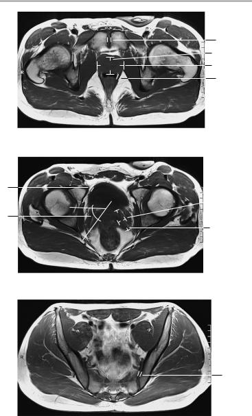
The pelvic inlet appears normal, with normal configuration of the iliac wings and iliopsoas muscles.
No abnormalities are found in imaged bowel structures, and there are no signs of wall thickening or mass lesions.
The distended urinary bladder appears normal and has normal wall thickness. The seminal vesicles are of normal size. The angle between the bladder and seminal vesicle is normal on each side. The prostate shows a normal size and configuration.
The vessels of the lesser pelvis are normal in their course and caliber. There is no evidence of lymphadenopathy.
The femoral heads are normally shaped and articulate normally with the acetabula. They have normal bone-marrow signal characteristics. The soft tissues show no abnormalities.
Interpretation
The lesser pelvis appears normal.
Checklist
Pelvic inlet |
! |
Configuration |
|
|
! |
Width |
|
|
! |
Symmetry |
|
|
! |
Iliac wings (shape) |
|
|
! |
Iliopsoas muscles: |
|
|
|
— |
Size |
|
|
— |
Signal characteristics |
|
|
— Symmetry |
|
Intestinal struc- ! Borders tures (especially ! Position
the cecum and ! Wall thickness (if with normal distension, see rectum) below)
!No circumscribed wall thickening
!Well-opacified lumen with no soft-tissue mass
Perirectal fat ! Signal characteristics (fat intensity)
!No infiltration
!No masses
Ischiorectal fossa ! Bilateral symmetry
!No masses
!No lymphadenopathy
Seminal vesicles ! Position (behind the bladder) ! Size (see below)
Moeller, Normal Findings in CT and MRI © 2000 Thieme
All rights reserved. Usage subject to terms and conditions of license.

156 MRI: Abdomen
|
! |
Symmetry |
|
|
! Angle between the bladder and seminal vesicle |
||
|
! |
(see below) is clear on each side |
|
Prostate |
Signal characteristics |
|
|
! |
Position (central at bladder outlet) |
||
|
! Configuration (rounded shape, |
intact capsule |
|
|
! |
and lobulation) |
|
|
Size (see below) |
|
|
|
! |
Homogeneous signal intensity |
|
|
! |
No calcifications |
|
|
! No unilateral nonhomogeneity after contrast ad- |
||
|
|
ministration |
|
Urinary bladder ! Adequate distension |
|
||
|
! |
Smooth outer contours |
|
Vessels |
! Wall thickness (see below) |
|
|
! |
Caliber (see below) |
|
|
Lymph node |
! |
Course |
|
! No lymphadenopathy |
|
||
stations |
|
|
|
Pelvic skeleton |
! |
Configuration |
|
|
! Margins (cortex smooth and sharp, no discon- |
||
|
|
tinuities) |
|
|
! Fat-equivalent signal intensity of bone marrow |
||
|
! No circumscribed areas of marrow replacement |
||
|
! Femoral heads rounded and |
centered in |
|
|
! |
acetabula |
|
|
Sacroiliac joints: |
|
|
|
|
— Smooth contours |
|
|
! |
— Width (see below) |
|
Subcutaneous |
Symphysis pubis |
|
|
! |
Density |
|
|
tissue and |
! |
Extent |
|
muscles |
! |
Borders |
|
|
! |
Symmetry |
|
|
|
|
|
Important Data
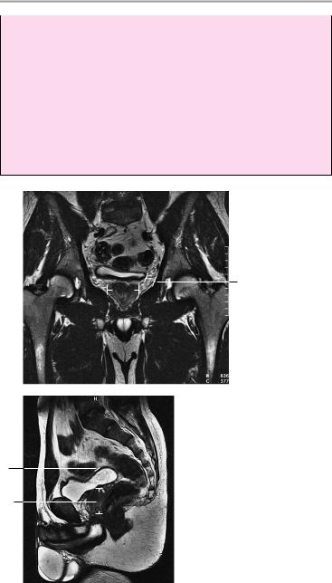
1Prostate (size varies with age, 20−70 years): a Anteroposterior diameter ca. 2.5−3 cm
b Lateral diameter: 3−5 cm
c Craniocaudal diameter: 3−5 cm
2Symphysis pubis:
!Width < 6 mm
Moeller, Normal Findings in CT and MRI © 2000 Thieme
All rights reserved. Usage subject to terms and conditions of license.
Male Pelvis 157
2
1a
1b
3
Axial image
4
 5b
5b
6
5a
Axial image
7
Axial image
Moeller, Normal Findings in CT and MRI © 2000 Thieme
All rights reserved. Usage subject to terms and conditions of license.
158 MRI: Abdomen
3Rectum:
!Wall thickness ! 5 mm
4Urinary bladder (well distended):
!Wall thickness ca. 3 mm
5Seminal vesicles (highly variable): a Length: up to 5 cm
b Width: up to 2 cm
6Angle between bladder and seminal vesicles:
!Open = normal
7Width of sacroiliac joint spaces:
!2−5 mm (anterior and inferior: 2−3 mm)
Coronal image
5b
 1b
1b
Midsagittal image
4
1c
Moeller, Normal Findings in CT and MRI © 2000 Thieme
All rights reserved. Usage subject to terms and conditions of license.

Testes 159
Testes
The scrotum and its contents display normal configuration.
The testes are symmetrical and of normal size with a homogeneous internal structure. Each testis is sharply demarcated by the tunica albuginea, which is of normal thickness.
The epididymis shows a normal size and position on each side and presents a normal internal structure.
The scrotal compartments appear normal on each side, with no sign of increased fluid.
The inguinal canal is normal in its shape, size, and course. The corpora cavernosa and corpus spongiosum are normal.
Interpretation
The testes appear normal.
Checklist
Scrotum |
! |
Size |
Testes |
! |
Configuration |
! |
Paired |
|
|
! |
Symmetrical |
|
! |
Size (see below) |
|
! |
Homogeneous internal structure (high T2- |
|
! |
weighted signal intensity) |
|
No circumscribed or diffuse change in signal in- |
|
Tunica albuginea ! |
tensity |
|
Smooth, sharp borders on all sides |
||
Epididymis |
! |
Normal thickness |
! |
Position |
|
(head and tail) |
! |
Size (bilateral symmetry) |
|
! |
Internal structure |
|
! |
Scrotal compartments have smooth, sharp |
|
! |
borders |
Inguinal canal |
No increased fluid |
|
! |
Shape |
|
|
! |
Size |
Corpora |
! |
Course |
! |
Size |
|
cavernosa |
! |
Bilateral symmetry |
Corpus |
! |
Honeycomb internal structure |
! |
Size |
|
spongiosum |
! |
Urethra |
Moeller, Normal Findings in CT and MRI © 2000 Thieme
All rights reserved. Usage subject to terms and conditions of license.
160 MRI: Abdomen
Important Data
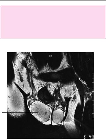
Testicular dimensions:
1Length:
!Up to ca. 4 cm
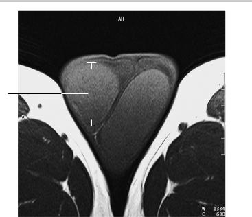
2Transverse diameter:
!Up to ca. 3 cm
1
T2-weighted coronal image
Moeller, Normal Findings in CT and MRI © 2000 Thieme
All rights reserved. Usage subject to terms and conditions of license.
Testes 161
2
T1-weighted axial image
Moeller, Normal Findings in CT and MRI © 2000 Thieme
All rights reserved. Usage subject to terms and conditions of license.

162
MRI: Spinal Column
Cervical Spine
The cervical spine shows a normal degree of lordosis with normal width of the bony spinal canal.
The vertebral bodies are normal in their number, shape, and interrelationships.
The atlantodental distance is normal. The articulating vertebral end plates present smooth margins. The intervertebral disk spaces are of normal height.
The intervertebral disks do not project past the posterior surface of the vertebral bodies in any imaged segment.
The spinal cord, including the craniocervical junction, displays normal position, configuration, width, and internal structure. The bone marrow of the vertebral bodies appears normal.
The prevertebral and paravertebral soft tissues show no abnormalities.
Interpretation
The cervical spine appears normal.
Checklist
Position |
! |
Normal cervical lordosis (no hypolordosis or hy- |
|
! |
perlordosis, no kyphotic deformity) |
|
No segmental malalignment |
|
|
! Normal position of the dens (see below) |
|
Bony spinal canal ! Width (see below)
! Smooth margins
Vertebral bodies ! Number (seven)
!Shape (square except for the dens)
!Position (straight alignment of posterior margins, no steps)
!End plates
—Continuous margins (no discontinuities)
—Smooth contours
—No circumscribed depression
—No marginal osteophytes
Moeller, Normal Findings in CT and MRI © 2000 Thieme
All rights reserved. Usage subject to terms and conditions of license.
|
|
Cervical Spine 163 |
Intervertebral |
! Width (see below) |
|
disk space |
! |
Normal signal characteristics: moderate to |
|
|
slightly hyperintense T2-weighted intensity (not |
|
! |
hypointense to other disks) |
|
No disk protrusion past posterior surface of adja- |
|
Spinal cord |
! |
cent vertebral bodies |
Configuration |
||
|
! |
Width |
|
! |
Signal characteristics |
|
! |
No circumscribed change in signal intensity |
Nerve roots |
! |
No circumscribed narrowing or expansion |
! |
Course |
|
|
! |
Passage through the neuroforamina |
|
! |
Dural tube: |
|
|
— Shape |
|
|
— Width |
|
|
— No circumscribed narrowing or expansion |
|
|
— Perimedullary contents of fluid signal inten- |
Neuroforamina |
! |
sity |
Configuration |
||
|
! |
Width |
Facet joints |
! |
No hypertrophy of uncovertebral joints |
! |
Shape |
|
|
! |
Position |
|
! |
Contours (smooth, continuous) |
|
! |
No hypertrophy |
|
! |
Vertebral arches intact |
Spinous |
! |
No shortening of pedicles |
! |
Shape |
|
processes |
! |
Position |
|
! |
Size |
|
! |
Bony structure |
|
! |
Fat-equivalent signal intensity of bone marrow |
|
! |
No circumscribed hypointense or hyperintense |
Soft tissues |
! |
areas |
Symmetrically arranged on both sides of the |
||
|
! |
vertebral bodies and spinous processes |
|
No masses |
|
|
! |
Prevertebral soft-tissue structures (especially |
|
|
the pharynx and thyroid gland; no masses) |
|
|
|
Moeller, Normal Findings in CT and MRI © 2000 Thieme
All rights reserved. Usage subject to terms and conditions of license.

164 MRI: Spinal Column
Important Data
1Atlantodental distance:
a Sagittal plane: approx. 1−3 mm (up to 5 mm in children) b Coronal and axial planes: dens is centered
2Craniovertebral angle (angle formed by the basilar line and the posterior tangent to C2):
!Normal range of 150° (flexion) to 180° (extension) (spinal compression occurs at less than 150°)
3Chamberlain’s line (line connecting the posterior border of
the hard palate with the posterior rim of the foramen magnum):
! Tip of the dens should project no more than 1 mm ± 6.6 mm above the line
4Retropharyngeal space:
!Up to 7 mm (at level of C2)
5Width of spinal cord:
!Sagittal > 6−7 mm
6Sagittal diameter:
!C1 ! 21 mm
!C2 ! 20 mm
!C3 ! 17 mm
!C4−C7 = 14 mm
7Height of intervertebral disk spaces:
!C2 < C3 < C4 < C5 < C6 ! C7
8Retrotracheal space:
!Up to 22 mm (at level of C6)
9Anteroposterior diameter of preodontoid space:
!< 2 mm
10Width of spinal canal:
!Transverse diameter at level of pedicles > 20−21 mm
Moeller, Normal Findings in CT and MRI © 2000 Thieme
All rights reserved. Usage subject to terms and conditions of license.