

Книги по МРТ КТ на английском языке / Normal Findings in CT and MRI
.pdfKnee Joint 205
Articularcartilage ! |
Thickness (see below) |
|
(patella, femoral |
! |
Signal characteristics |
condyles, tibial |
! |
Cartilage surface (smooth) |
plateau) |
|
|
Joint space |
! |
Width |
|
! |
No effusion |
|
! |
No intra-articular foreign bodies |
|
! |
No abnormal synovial folds (especially the |
Medial and |
! |
mediopatellar fold) |
Configuration (normal triangular cross section) |
||
lateral menisci |
! |
Internal structure (homogeneous, hypointense) |
(anterior horn, |
! |
Contours (smooth, intact surface) |
midportion, |
|
|
posterior horn) |
|
|
Anterior and |
! |
Continuity |
posterior cruciate ! |
Width |
|
ligaments |
! |
Course (see below) |
|
! |
Signal intensity (anterior cruciate ligament light |
|
|
and dark, posterior cruciate ligament uniformly |
Collateral |
! |
dark) |
Position |
||
ligaments |
! |
Width |
|
! |
Continuity |
Soft tissues and |
! |
Low signal intensity |
! |
No masses (e.g., Baker cyst, popliteal cyst, gan- |
|
imaged vessels |
! |
glion) |
|
No varices |
|
|
|
|
Important Data
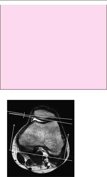
Patella:
Shape: Wiberg I−III (see drawing)
Centering
1Patellar tilt angle (formed by a line parallel to the lateral patellar articular surface and a line parallel to the posterior aspect of the femoral condyles [may be drawn in sections at various levels, if necessary]):
!> 8°
Moeller, Normal Findings in CT and MRI © 2000 Thieme
All rights reserved. Usage subject to terms and conditions of license.
206 MRI: Joints
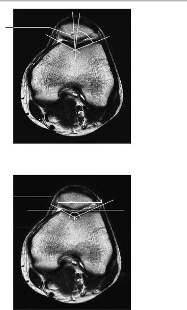
2Congruence angle (formed by the bisector of the notch angle and a line connecting the patellar apex with the deepest point of the notch):
!6° to -6°
3Notch angle:
!135−145° (average ca. 138°)
4Lateral displacement:
!< 5% (i.e., less than 5% of the patella is lateral to a line that is perpendicular to the line joining the femoral condyles at the level of the lateral condyle)
5Patellar ligament:
a Length: 3.5−5.5 cm b Width: 2.5−3 cm c Thickness: 7 mm
6Ratio of length of patellar ligament to height of patella = 0.8−1.2 (> 1.2 = high-riding patella)
7Cartilage:
a Patella: 3−4 ± 1 mm
b Femoral condyles and tibial plateau: ca. 2.2 ± 0.6 mm
1
Axial image
Moeller, Normal Findings in CT and MRI © 2000 Thieme
All rights reserved. Usage subject to terms and conditions of license.
Knee Joint 207
2
1/2
Axial image
4 |
|
|
95% |
5% |
|
|
|||||
|
|
|
|
|
. |
3 |
|
|
|
|
|
|
|
|
|
|
|
Axial image
Moeller, Normal Findings in CT and MRI © 2000 Thieme
All rights reserved. Usage subject to terms and conditions of license.
208 MRI: Joints
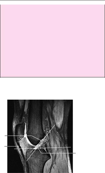
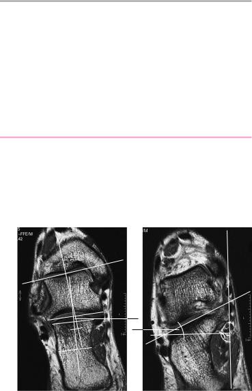
8Anterior cruciate ligament:
!Length: ca. 38 mm
!Width: ca. 11 mm
aAngle formed by tangents to the tibial plateau and the anterior surface of the anterior cruciate ligament: 55°
bAngle formed by Blumensaat’s line (dashed) and the anterior surface of the anterior cruciate ligament: 1.6°
cAngle of posterior cruciate ligament: ca. 123° (abnormal at ca. 106°)
dLine of posterior cruciate ligament should intersect the dis-
tal femur.
Abnormal c and d are indirect signs of anterior cruciate ligament rupture.
Posterior cruciate ligament:
!Length: ca. 38 mm
!Width: ca. 18 mm
8b
5c
8a
Sagittal image at the level of the anterior cruciate ligament
Moeller, Normal Findings in CT and MRI © 2000 Thieme
All rights reserved. Usage subject to terms and conditions of license.
Knee Joint 209
A |
|
|
|
|
|
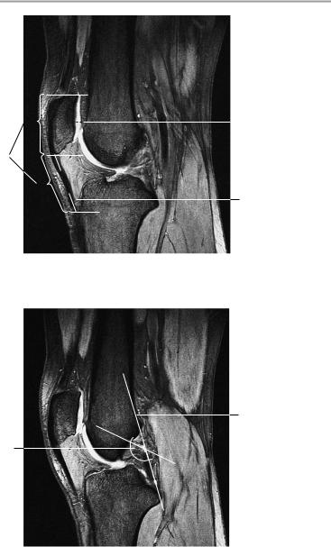
7a |
6
B
5a
Sagittal image at the level of the patellar ligament
8d
8c
Sagittal image at the level of the posterior cruciate ligament
Moeller, Normal Findings in CT and MRI © 2000 Thieme
All rights reserved. Usage subject to terms and conditions of license.
210 MRI: Joints
7b
Coronal image
I |
II |
III |
IV |
lateral medial
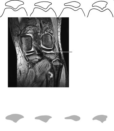
Wiberg classification of patellar shapes (right knee). (From Möller, T.B.: Röntgennormalbefunde. Thieme, Stuttgart 1996.)
Moeller, Normal Findings in CT and MRI © 2000 Thieme
All rights reserved. Usage subject to terms and conditions of license.

Ankle and Subtalar Joints 211
Ankle and Subtalar Joints
The bones comprising the ankle joint show normal position and configuration, with normal development of the ankle mortise.
The bone marrow signal, trabecular pattern, and epiphyseal lines are all normal.
The joint space is of normal width. The cortex shows normal thickness and smooth contours, especially along the tibial and talar articular surfaces. There are no subchondral signal changes and no osteophytes.
The lateral and medial ligaments are normal in their course, width, and signal characteristics.
The talocalcaneal and talonavicular joints appear normal. The interosseous ligament between the talus and calcaneus is intact. The Achilles tendon is normal in its course, width, and signal characteristics, and the preachilles fat is clear. The tendons and plantar aponeurosis are unremarkable.
The soft tissues show no abnormalities.
Interpretation
The ankle joint and subtalar joint appear normal.
Checklist
Skeleton ! Medial and lateral malleoli (ankle mortise), talus, calcaneus, tarsal bones:
—Configuration
—Position (shape, centering—see below)
—Normal bone marrow signal
—Epiphyseal plate closure after age 18
—Normal trabecular pattern
Articular surfaces ! Congruence
!Cortex:
—Cortical thickness (uniform, no circumscribed expansion)
!Contours: smooth and sharp, no subchondral signal changes (especially on the medial side = 60% site of predilection for osteochondritis dissecans), no discontinuities
!Articular cartilage (if visible):
—Thickness
—Smooth surface
Moeller, Normal Findings in CT and MRI © 2000 Thieme
All rights reserved. Usage subject to terms and conditions of license.

212 MRI: Joints
Ligaments |
! |
Lateral ligaments (in order of trauma frequency: |
|
|
|
anterior fibulotalar ligament, fibulocalcaneal |
|
|
|
ligament, posterior fibulotalar ligament): |
|
|
|
— Course (not wavy) |
|
|
|
— |
Signal intensity |
|
|
— Width |
|
|
|
— Smooth contours |
|
|
|
— |
Continuity |
|
! |
— |
No periligamentous fluid |
|
Medial (deltoid) ligament: |
||
|
|
— Course |
|
|
|
— |
Signal intensity |
|
|
— |
Width (see below) |
|
|
— Smooth contours |
|
|
! |
— |
Continuity |
|
Interosseous ligament: |
||
|
|
— Course |
|
|
! |
— |
Continuity |
|
Achilles tendon: |
||
|
|
— Course |
|
|
|
— |
Width (see below) |
|
|
— Shape (transverse oval cross section) |
|
|
|
— |
Signal characteristics |
|
|
— Continuity (especially 2−6 cm above the cal- |
|
|
|
|
caneal attachment = site of predilection for |
|
|
|
tears) |
Subtalar joint |
! |
— Normal-appearing preachilles fat |
|
Configuration |
|||
(talocalcaneal |
! |
Position |
|
joint, talonavi- |
! Width of joint space |
||
cular joint) |
|
|
|
Soft tissues |
! |
Tendons: |
|
|
|
— Flexor hallucis longus tendon is particularly |
|
|
|
|
important (especially in the tarsal tunnel be- |
|
|
|
hind the medial malleolus, which is a site of |
|
|
|
predilection for tendinitis and rupture) |
|
|
— Tibialis posterior (its navicular attachment is |
|
|
|
|
a site of predilection for rupture) |
|
|
— Course |
|
|
|
— Signal intensity (uniformly hypointense, no |
|
|
|
|
central signal change) |
|
|
— Width |
|
|
|
— |
No discontinuities |
Moeller, Normal Findings in CT and MRI © 2000 Thieme
All rights reserved. Usage subject to terms and conditions of license.
Ankle and Subtalar Joints 213
—Wall of tendon sheath (no fluid increase or wall thickening)
!Plantar aponeurosis and calcaneonavicular ligament:
—Shape
—Width (see below)
—Hypointense in all MRI sequences
—No circumscribed expansion or nodularity
—No subcutaneous edema
!Normal tarsal canal
!Soft tissues
!Blood vessels
|
1 |
2 |
. |
|
Fig. 1: Semicoronal image showing all of the posterior articular surface of the subtalar joint and portions of the medial and lateral malleoli
Fig. 2: Semicoronal image showing the posterior articular surface, all of the tarsal canal, and the sustentaculum
Moeller, Normal Findings in CT and MRI © 2000 Thieme
All rights reserved. Usage subject to terms and conditions of license.

214 MRI: Joints
Important Data
Position
Semicoronal image plane:
1Calcaneal valgus angle = relation of the talar axis (line connecting the bisectors of the corners of the ankle and subtalar joint surfaces) to the axis of the calcaneus (line connecting the bisectors of the corners of the subtalar joint and a line parallel to it through the narrowest part of the calcaneus):
!Approximately 0° ± 10°
2Sustentacular angle (formed by a line connecting the corners of the lateral posterior joint surface and sustentaculum and a line perpendicular to a tangent to the sustentaculum and medial calcaneal tuberosity):
!18−28°
Axial image plane (image is acquired 4 cm above the level where the lateral part of the talus is first visualized):
3Plantar talocalcaneal angle (formed by a line connecting the lateral corner of the posterior articular surface of the talus and the medial corner of its medial articular surface with a line bisecting the calcaneal articular surface and the midpoint of a parallel line through the caudal third of the calcaneus):
!60−70°
4Calcaneocuboid angle (angle between the longitudinal axes of the cuboid and calcaneus):
!Approximately 20−35°
5Arch angle (angle between tangents to the inferior calcaneal border and the soft-tissue sole):
!20−30°
6Achilles tendon:
!Anteroposterior diameter < 6 mm
7Lateral ligaments:
!Width of the anterior talofibular ligament and calcaneofibular ligament: 2−3 mm
!Angle between the longitudinal axes of the first and second metatarsals = 7.4 ° ± 2.6° (> 9° is suspicious for hallux valgus)
!Relation of calcaneus to talus: 1.8−2.1
8Boehler’s angle (formed by a line connecting the posterosuperior and anterosuperior prominences of the calcaneus and a line through the sustentaculum tali):
!20−40° (signifies calcaneal integrity)
Moeller, Normal Findings in CT and MRI © 2000 Thieme
All rights reserved. Usage subject to terms and conditions of license.