

Книги по МРТ КТ на английском языке / Normal Findings in CT and MRI
.pdfCervical Arteries 235
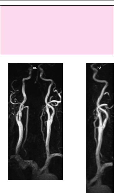
Common carotid ! Origin (usually the left artery arises directly from
artery |
|
the aortic arch while the right artery arises with |
|
|
the subclavian artery from the brachiocephalic |
|
! |
trunk) |
|
Symmetry |
|
|
! |
Course |
|
! |
Caliber (symmetry) |
|
! |
Signal characteristics |
|
! |
No excessive tortuosity |
Carotid |
! |
No circumscribed dilatation |
! |
Usually occurs at C4/5 or C3/4 level |
|
bifurcation |
! |
Shape |
|
! No circumscribed narrowing, especially at the |
|
|
|
origin of the internal carotid artery |
Internal carotid ! Position (no displacement) |
||
artery |
! |
Course |
|
! Caliber (slight proximal dilatation due to the |
|
|
|
carotid sinus, right−left symmetry) |
|
! Signal characteristics (homogeneous intralumi- |
|
|
|
nal signal, no filling defect) |
|
! Contours (smooth, no circumscribed constric- |
|
|
|
tion or ulceration) |
|
! No circumscribed narrowing (especially in the |
|
|
|
proximal segment) with poststenotic dilatation |
External carotid |
! Symmetrical appearance of the carotid siphons |
|
! |
Position |
|
artery |
! |
Course |
|
! |
Caliber |
Vertebral artery |
! |
Signal characteristics |
! Origin (from the subclavian artery or, rarely, |
||
|
! |
from the aortic arch) |
|
Position |
|
|
! Course (no excessive tortuosity) |
|
|
! Caliber (no general or circumscribed luminal di- |
|
|
|
latation) |
|
! Caliber discrepancy (usually left > right) is com- |
|
|
|
mon |
|
! Signal characteristics (homogeneous intralumi- |
|
|
! |
nal signal, no filling defect) |
|
Contours (smooth) |
|
|
|
|
Moeller, Normal Findings in CT and MRI © 2000 Thieme
All rights reserved. Usage subject to terms and conditions of license.
236 MRI: Special Investigations
Important Data
Sites of predilection for stenosis:
!Internal carotid artery:
—Carotid bifurcation (ca. 2/3 of all carotid stenoses)
—At entrance to the carotid siphon
—Within the carotid siphon
!Vertebral artery
—Origin from the subclavian artery
—Passage through dura at craniocervical junction
Moeller, Normal Findings in CT and MRI © 2000 Thieme
All rights reserved. Usage subject to terms and conditions of license.

237
References
Allen, K. S., H. Y. Kressel, P. P. Arger, H. M. Pollack: Age-related changes of the prostate: evaluation by MR Imaging. Amer. J. Roentgenol. 152 (1989) 77–81 Berli, A., R. Putz, M. Schumacher: Maße und Varianten im Bereich des Canalis opticus. Radiologe 32 (1992) 436–440 Biggemann, W., W. Frobin, P. Brinckmann: Physiologisches Muster lumbaler Bandscheibenhöhen. Fortschr. Röntgenstr.
167 (1997) 1
Brown, H. K. et al.: Uterine junctional zone: correlation between histologic findings and MR Imaging. Radiology 179 (1991) 409–413
Buthiau, D., D. L. Kaech: CT und MR in der klinischen Praxis. Huber, Bern 1996 Chan, T. W., M. K. Dalinka, J. B. Kneeland, A. Chervrot: Biceps tendon dislocation: evaluation with MR Imaging. Radiology
179 (1991) 649–652
Claussen, C., B. Lochner: Dynamische Computertomographie. Springer, Berlin 1983
Dähnert, W.: Radiology Review Manual. Williams & Wilkins, Baltimore 1996 Dihlmann, W.: Computertomographie des lumbalen Diskusprolapses und der Vertebralkanalstenose. Z. Rheumatol.
43 (1984) 153–159
Elster, A. D.: Modern imaging of the pituitary. Radiology 187 (1993) 1–14
Frahm, R., E. Drescher: Topographische Anatomie, Radiologie und Pathologie der Handwurzel und des Handgelenkes. Schnetztor, Konstanz 1988
Frahm, R., H. Fritz, E. Drescher: Winkelmessung des Rückfußes im CT. Fortschr. Röntgenstr. 151 (1989) 77–81
Friedmann, G., E. Bücheler, P. Thurn: Ganzkörper-Computertomographie. Thieme, Stuttgart 1981
Gentili, A., L. L. Seeger, L. Yao, H. M. Do: Anterior cruciate ligament tear: indirect signs at MR Imaging. Radiology 193 (1994) 835–840
Graßhoff, H., C. Buhtz, I. Gellerich, Ch. v. Knorre: CT-Diagnostik bei der Instabili-
tät des Schultergelenkes. Fortschr. Röntgenstr. 155 (1991) 523–526
Gürtler, K.-F., R. W. Janzen, J. Hageman, H. F. Otto: CT des Mediastinums bei Myastenia gravis pseudoparalytica. Fortschr. Röntgenstr. 136 (1982) 35−40 Hamm, B., T. Römer, M. Albig, R. Felix, K.-J. Wolf: Magnetische Resonanztomographie der Ovarialtumoren. Fortschr.
Röntgenstr. 146 (1987) 429–438 Harnsberger, H. R.: Handbook of Head and
Neck Imaging. Mosby, St. Louis 1995 Hosten, N., Ch. Schubert, M. Cordes, R. Schneider, R. Felix: Kernspintomographie der Orbita bei endokriner Orbitopathie. Röntgenpraxis 41 (1988) 400–
405
Hübener, K.-H.: Computertomographie des Körperstammes. Thieme, Stuttgart 1985
Jend, H.-J., H.-Ch. Tödt: Arbeitsbuch Computertomographie. Schnetztor, Konstanz 1989
Kahn, Th.: Leber-Galle-Pankreas. Thieme, Stuttgart 1996
Klaue, K., C. W. Durnin, R. Ganz: The acetabular rim syndrome. J. Bone Jt Surg. B 73-B (1991) 423–429
Kock, C.: Sagittale Weiten des cervikalen Wirbelkanales im Computertomogramm. Radiologe 26 (1986) 239–241 Lange, S.: Niere und ableitende Harnwege.
Thieme, Stuttgart 1993
Lee, M. J., W. Mayo-Smith, P. Hahn, M. Goldberg, G. Boland, S. Saini, N. Papanicolaou: MR Imaging of the adrenal gland. Radiographics 14 (1994)
Lörcher, U., H. Schmidt, K. H. Hering: HR–CT der Lunge. Thieme, Stuttgart 1996
Maier, W.: Hochauflösende CT des Pankreas. In Bargon, G.: Symposium über bildgebende Verfahren in der Pankreasdiagnostik. Schnetztor, Konstanz 1986 Maier, W.: Zur Wertigkeit der Nativ-CT bei der akuten Pankreatitis. Fortschr. Rönt-
genstr. 150 (1989) 458–461
Möller, T. B.: Röntgennormalbefunde, 2, Aufl. Thieme, Stuttgart 1996
Moeller, Normal Findings in CT and MRI © 2000 Thieme
All rights reserved. Usage subject to terms and conditions of license.
238 |
References |
|
|
|
|
|
|
|
|
|
|
|
|
||
Möller, |
T. B., |
E. |
Reif: |
MR-Atlas |
des |
Schumacher, |
K. A., |
J. M. |
Friedrich: |
Die |
|||||
muskuloskelettalen Systems. Blackwell, |
Computertomographie in der Diagnos- |
||||||||||||||
Berlin 1993 |
|
|
|
|
|
tik der Nierenerkrankungen. In Bargon, |
|||||||||
Möller, T. B., E. Reif: Taschenatlas der Ein- |
G.: Symposium über bildgebende Ver- |
||||||||||||||
stelltechnik, 2. Aufl., Thieme, Stuttgart |
fahren in der Diagnostik der Nieren und |
||||||||||||||
1995 |
|
|
|
|
|
|
|
oberen Harnwege. Schnetztor, Konstanz |
|||||||
Möller, |
T. B., |
E. Reif: |
Taschenatlas |
der |
1987 |
|
|
|
|
|
|
|
|||
Schnittbildanatomie, |
|
Bd. I, 2. |
Aufl., |
Scotti, G. et al.: MR Imaging of cavernous |
|||||||||||
Thieme, Stuttgart 1997 |
|
|
sinus involvement by pituitary ade- |
||||||||||||
Moore, S. G., G. S. Bisset III, M. J. Siegel, J. S. |
nomas, Amer. J. Roentgenol. 151 (1988) |
||||||||||||||
Donaldson: Pediatric |
musculoskeletal |
799–806 |
|
|
|
|
|
|
|||||||
MR Imaging. Radiology 179 (1991) 345– |
Scoutt, L. M. et al.: Junctional zone of the |
||||||||||||||
360 |
|
|
|
|
|
|
|
uterus: correlation of MR Imaging and |
|||||||
Mühlberger, V.: Kardio-CT. Röntgenpraxis |
histologic examination of hysterectomy |
||||||||||||||
39 (1985) 329–352 |
|
|
|
specimens. Radiology 179 (1991) 403– |
|||||||||||
Munk, P. L., C. A. Helms: MRI of the Knee. |
407 |
|
|
|
|
|
|
|
|||||||
Lippincott-Raven, Philadelphia 1996 |
Smith, D. K.: Anatomic features of the car- |
||||||||||||||
Murphey, |
M. D., |
L. H. |
Wetzel, |
J. M. |
pal scaphoid: validation of biometric |
||||||||||
Bramble, E. Levine, K. M. Simpson, H. B. |
measurements |
and |
symmetry |
with |
|||||||||||
Lindsley: Sacro iliitis: MR Imaging find- |
tree-dimensional MR Imaging. Radi- |
||||||||||||||
ings. Radiology 180 (1991) 239–244 |
ology 187 (1993) 187–191 |
|
|||||||||||||
Nugent, R. A. et al.: Graves orbitopathy: |
Stern, E. J., C. M. Graham, W. R. Webb, G. |
||||||||||||||
correlation of CT and clinical findings. |
Gamsu: Normal trachea during forced |
||||||||||||||
Radiology 177 (1990) 675–682 |
|
expiration: dynamic CT measurements. |
|||||||||||||
Outwater, |
E. K., D. G. Mitchell: Normal |
Radiology 187 (1993) 27–31 |
|
||||||||||||
ovaries and functional cysts: MR ap- |
Stiskal, M., A. Neuhold, R. Weinstabl, F. M. |
||||||||||||||
pearance. Radiology 198 (1996) 397– |
Kainberger, B. Gisinger: MR-tomo- |
||||||||||||||
402 |
|
|
|
|
|
|
|
graphische |
Befunde |
bei Achillodynie. |
|||||
Pickuth, D.: Sonographie – systematisch. |
Fortschr. Röntgenstr. 153 (1990) 9–13 |
||||||||||||||
Bon-Med, Lorch 1993 |
|
|
|
Sugimoto, H., T. Shinozaki, T. Ohsawa: Tri- |
|||||||||||
Pommeranz, S.: Gamuts & Perls in MRI. |
angular |
fibrocartilage |
in asymtomatic |
||||||||||||
MRI-EFI Publications, Cincinnati 1993 |
subjects: investigation of abnormal MR |
||||||||||||||
Putz, R.: Anatomie des Retroperitoneal- |
signal intensity. Radiology 191 (1994) |
||||||||||||||
raumes: In Frommhold, P., P. Gerhard: |
194–197 |
|
|
|
|
|
|
||||||||
Tumoren im Retroperitonealraum. Klin- |
Sugimura, |
K., |
B. M. |
Carrington, |
J. M. |
||||||||||
isch-radiologisches |
Seminar, Bd. 16. |
Quivey, |
H. |
|
Hricak: |
Postirradiation |
|||||||||
Thieme, Stuttgart 1987 |
|
|
changes in the pelvis: assessment with |
||||||||||||
Reiser, M., M. Nägele: Aktuelle Gelenkdi- |
MR Imaging. Radiology 175 (1990) 805– |
||||||||||||||
agnostik. Thieme, Stuttgart 1992 |
|
813 |
|
|
|
|
|
|
|
||||||
Richards, R. D., D. J. Sartoris, M. N. Pathria, |
Takashi Ohnishi, et al.: Levator palpebrae |
||||||||||||||
D. Resnick: Hill-Sachs lesion and nor- |
superioris muscle: MR evaluation of en- |
||||||||||||||
mal humeral groove: MR Imaging fea- |
largement as a cause of upper eyelid re- |
||||||||||||||
tures |
allowing |
their |
differentation. |
traction in graves diseases. Radiology |
|||||||||||
Radiology 190 (1994) 665–668 |
|
188 (1993) 115–118 |
|
|
|
||||||||||
Robertson, |
P. L., M. E. |
Schweitzer, |
A. R. |
Tomczak, R. et al.: Messung des femoralen |
|||||||||||
Bartolozzi, A. Ugoni: Anterior cruciate |
Torsionswinkels von Kindern durch MR |
||||||||||||||
ligament tear: evaluation of multiple |
im Vergleich zu CT und Ultraschall. |
||||||||||||||
signs with MR Imaging. Radiology 193 |
Fortschr. Röntgenstr. 163 (1995) 224– |
||||||||||||||
(1994) 829–834 |
|
|
|
|
228 |
|
|
|
|
|
|
|
|||
Schild, H. H., F. Schweden: Computerto- |
Uhlenbrock, D.: MRT und MRA des Kopfes. |
||||||||||||||
mographie in der Urologie. Thieme, |
Thieme, Stuttgart 1996 |
|
|
||||||||||||
Stuttgart 1989 |
|
|
|
|
Vahlensieck, M., M. Reiser: MRT des Be- |
||||||||||
Schneider, B., J. Laubenberger, M. Wildner, |
wegungsapparats. |
Thieme, Stuttgart |
|||||||||||||
V. Exne, M. Langer: Kernspintomo- |
1997 |
|
|
|
|
|
|
|
|||||||
graphisches |
Messungsverfahren |
von |
Vannier, M. W. et al.: Brain surface cortical |
||||||||||||
Femurantetorsion |
und Tibiatorsion. |
sulcal |
lengths: |
quantification |
with |
||||||||||
Fortschr. Röntgenstr. 163 (1995) 229– |
three-dimensional MR Imaging. Radi- |
||||||||||||||
231 |
|
|
|
|
|
|
|
ology 180 (1991) 479–484 |
|
||||||
Moeller, Normal Findings in CT and MRI © 2000 Thieme
All rights reserved. Usage subject to terms and conditions of license.
|
References 239 |
|
Wegener, O. H.: Ganzkörpercomputerto- |
Woerner, H., G. Brill, T. Frenzel, H. Stoll, M. |
|
mographie. Blackwell, Berlin 1992 |
Tesseraux: Pelvimetrie mittels Kern- |
|
Wiesen, E. J., J. R. Crass, E. M. Bellon, G. G. |
spintomographie. Fortschr. Röntgenstr. |
|
Ashmead, A. M. Cohen: Improvement in |
149 (1988) 378–382 |
|
CT Pelvimetry. Radiology 178 (1991) |
Zaunbauer, W., S. Däpp, M. Haertel: Anat- |
|
259–262 |
omische Normalmaße |
im zervikalen |
|
Computertomogramm. |
Radiologe 25 |
|
(1985) 521–524 |
|
Moeller, Normal Findings in CT and MRI © 2000 Thieme
All rights reserved. Usage subject to terms and conditions of license.

240
Moeller, Normal Findings in CT and MRI © 2000 Thieme
All rights reserved. Usage subject to terms and conditions of license.

241
Index
A
abdomen CT, 36−71
MRI, 128−61 acetabulum, MRI, 200, 203
Achilles tendon, MRI, 212, 214 acromioclavicular joint, MRI, 187
width, 188 acromion, MRI, 187 adrenal glands
CT, 38, 46, 52, 57, 61−3 dimensions, 39, 47, 58, 62
MRI, 130, 141, 145−9 dimensions, 132, 138, 146−7
ankle joint, MRI, 211−16 dimensions, 214
anterior cruciate ligament, MRI, 205 dimensions, 208
aorta
abdominal aorta
CT, diameter, 39, 52, 58 MRI, 223
dimensions, 132, 139, 142, 224, 229
CT, diameter, 33, 62 MRI, 169
bifurcation, 224, 229 diameter, 120
aortic arch, MRI, 234 arteries
basilar, MRI, 218 carotid
bifurcation, 235 common, MRI, 116, 235 external, MRI, 235 internal
CT, 12
MRI, 96, 217, 235 stenosis sites, 236
cerebral, MRI
anterior, 217−18 middle, 217 posterior, 218
cervical, MRI, 234−6 communicating arteries, MRI
anterior, 218 posterior, 218
mesenteric CT, 51 MRI, 224
popliteal, MRI, 229 pulmonary, diameter
CT, 33 MRI, 120
renal arteries, MR angiography, 223−6 dimensions, 224−6
spinal, MRI, 224 subclavian, MRI, 234
superficial femoral, MRI, 229 vertebral, MRI, 235
stenosis sites, 236 see also aorta; vessels
atlantodental distance, 164 auditory canal
external, MRI, 101, 184 internal
CT, 13
dimensions, 6, 15 MRI, 100−3
dimensions, 93, 102 axilla, MRI, 125
B
basal ganglia CT, 4
MRI, 88
basilar artery, MRI, 218 biceps tendon, MRI, 187
diameter, 188
Moeller, Normal Findings in CT and MRI © 2000 Thieme
All rights reserved. Usage subject to terms and conditions of license.

242 Index
bicipital groove, MRI, 188 bile ducts
CT, 37, 45, 51 width, 47, 53
MRI, 129, 134 cholangiopancreatography, 231, 232
intrahepatic, 232 width, 131, 136, 233
bladder, see urinary bladder Boehler’s angle, MRI, 214 bone marrow signal, MRI
hip joint, 201 knee joint, 204
brachiocephalic trunk, MRI, 234 brain stem
CT, 4 MRI, 88
breast, MRI, 125−7
C
calcaneal valgus angle, MRI, 214 calcaneocuboid angle, MRI, 214 calvarium, CT, 5
carotid artery, see arteries carpal tunnel, MRI, 196 cavernous sinus
CT, 12
MRI, 96 cecum
female CT, 64
MRI, 150 male
CT, 68 MRI, 155
cerebellopontine angle area CT, 14
MRI, 101 cerebellum CT, 4
MRI, 87, 89, 101
cerebral arteries, see arteries cerebral cortex
CT, 4, 6
MRI, 88
cerebrum, MRI, 87, 101 cervical spaces
CT, 27 MRI, 113
cervical spine CT, 28, 72−5
dimensions, 74−5 MRI, 162−7
dimensions, 164 cervix
CT, 65
MRI, 151, 162−7 dimensions, 152
Chamberlain’s line, 114, 164 chest
CT, 31−5 MRI, 118−27
cholangiopancreatography, 231−3 clavicle
CT, 32
MRI, 119, 187 cochlea
CT, 13−14 MRI, 89, 101
colon haustrations CT, 38, 52
MRI, 130, 141
conus medullaris, MRI, 173 corpora cavernosa, MRI, 159 corpus callosum
CT, 4 MRI, 88
corpus spongiosum, MRI, 159 cortical sulcation
CT, 3 MRI, 87−8
costophrenic sinus CT, 37, 45
MRI, 130, 135 cranial nerves, MRI VII (facial), 100
VIII (vestibulocochlear), 100 craniovertebral angle, 164 CSF spaces
CT, 12, 15
MRI, 96, 101, 102 cubital tunnel, MRI, 191
Moeller, Normal Findings in CT and MRI © 2000 Thieme
All rights reserved. Usage subject to terms and conditions of license.

Index 243
cystic duct, MRI, 232 length, 233
D
deltoid muscle, MRI, 187 diaphragm
CT, 32, 62
MRI, 119 duodenum, CT, 51 dural sack, MRI, 169 dural tube, CT, 80−1
E
elbow joint, MRI, 191−4 dimensions, 192
epididymis, MRI, 159 esophagus
CT, 27 MRI, 113
wall thickness, 116 ethmoid cells
CT, 23 MRI, 108
external auditory canal, MRI, 101, 184 external capsule
CT, 4 MRI, 88
eye muscles, see muscles
F
facet joints CT
cervical spine, 73 lumbar spine, 81 thoracic spine, 77
MRI
cervical spine, 163 lumbar, 173 thoracic spine, 169
facial nerve, MRI, 100 fat
peripancreatic, CT, 51 perirectal
female
CT, 64 MRI, 150
male CT, 68
MRI, 155 retrobulbar, CT, 17
femoral head, MRI, 200, 203 femoral shaft, MRI, 201 frontal sinuses, see sinuses
G
gallbladder CT, 37, 45, 52
dimensions, 39, 47 MRI, 129, 135
cholangiopancreatography, 231−2 dimensions, 131, 136, 233
gerota fascia, thickness CT, 58, 62
MRI, 142, 147 glenoid, MRI, 183−4, 187 glenoid angle, MRI, 188 globe
CT, 17 diameter, 19 position, 6, 19
MRI, 104 diameter, 106 position, 93, 107
glottis, MRI, 116
H
head
CT, 3−30 MRI, 87−117
heart CT, 32
dimensions, 33−4 MRI, 119, 126
dimensions, 120 hip joint, MRI, 200−3
dimensions, 203 humerus, MRI, 191
humeral head, 186
Moeller, Normal Findings in CT and MRI © 2000 Thieme
All rights reserved. Usage subject to terms and conditions of license.

244 Index
I
iliac wings, MRI, 179 iliopsoal muscles, MRI, 150
infraspinatus muscle, MRI, 187 infundibulum
CT, 12
MRI, 96
inguinal canal, MRI, 159 interhemispheric fissure
CT, 3 MRI, 87
internal auditory canal CT, 13
dimensions, 6, 15 MRI, 100−3
dimensions, 93, 102 internal capsule
CT, 4 MRI, 88
intervertebral disk space CT
cervical spine, 73 lumbar spine, 80, 82 thoracic spine, 76, 78
MRI
cervical spine, 163, 164 lumbar spine, 172−3, 174 thoracic spine, 168, 170
ischiorectal fossa female
CT, 64 MRI, 150
male CT, 68
MRI, 155
J
joints, MRI, 178−216 Jones-Thomson ratio
CT, 78, 82
MRI, 174
K
kidneys
CT, 37−8, 52, 56−60, 62 dimensions, 38, 47, 58
MRI, 130, 135, 140−4, 146 dimensions, 132, 138−9, 142, 146− 7,
224
renal arteries, angiography, 223−6 knee joint, MRI, 204−10
kyphotic angle, MRI, 170
L
lacrimal gland CT, 18, 19 MRI, 105, 107
larynx CT, 27
MRI, 113
left atrium, dimensions CT, 33
MRI, 120 ligamenta flava
CT, 82 MRI, 174
ligaments, MRI
ankle joint, 212, 214 elbow joint, 192 knee joint, 205, 206
dimensions, 208 wrist joint, 196
liver
CT, 36−7, 44−9, 51−2, 61 dimensions, 39, 46
MRI, 129, 134−9, 146 dimensions, 131, 136 intrahepatic bile ducts, 232
lordosis, MRI, 174 lumbar spine
CT, 80−3 dimensions, 82
MRI, 172−7, 179−80 dimensions, 174
lumbosacral angle CT, 82
MRI, 174, 180
Moeller, Normal Findings in CT and MRI © 2000 Thieme
All rights reserved. Usage subject to terms and conditions of license.