

Книги по МРТ КТ на английском языке / Normal Findings in CT and MRI
.pdfCervical Soft Tissues 115
|
|
|
|
|
|
|
|
|
Ch |
||
4 |
|
|
|
|
|
|
|
|
|
1 |
|
|
|
|
|
|
|
||||||
5 |
|
|
|
|
|
2 |
|||||
|
|
|
|||||||||
|
|
|
|
|
|
|
|
|
|
|
|
6 |
3 |
|
Midsagittal image
7a
7c
Sagittal image
Moeller, Normal Findings in CT and MRI © 2000 Thieme
All rights reserved. Usage subject to terms and conditions of license.
116 MRI: Head and Neck
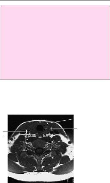
Lumina of upper respiratory tract (normal respiration):
4Laryngeal inlet (hyoid level):
!Approximatley 19 ± 4 mm
5Glottis:
!Approximately 21 ± 4 mm
6Trachea:
!Approximately 17 ± 3 mm
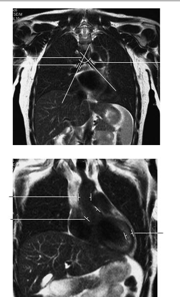
7Dimensions of thyroid gland: a Length: 3.5−6 cm
b Width: 1.5−2 cm
c Depth: 1−2 cm
Vascular calibers (at level of thyroid gland):
8Common carotid artery:
!6−10 mm
9Esophagus:
!Wall thickness 3 mm
 7c
7c
7b
8
9
Axial image
Moeller, Normal Findings in CT and MRI © 2000 Thieme
All rights reserved. Usage subject to terms and conditions of license.

Cervical Soft Tissues 117
7a
8
Coronal image
Moeller, Normal Findings in CT and MRI © 2000 Thieme
All rights reserved. Usage subject to terms and conditions of license.

118
MRI: Chest
Thoracic Organs
Both lungs are normally aerated and are applied to the chest wall on all sides. The pleurae show normal homogeneous signal intensity, and there are no fluid collections.
The pulmonary structure is normal and presents normal vascular markings. There are no intrapulmonary nodules or patchy densities.
The mediastinum is centered and of normal width. There is no evidence of masses in the anterior, central, or posterior compartment.
The hilar region on each side is unremarkable, and the main bronchi appear normal. There is no lymphadenopathy and there are no perihilar masses.
The heart is orthotopic and has a normal configuration. The cardiac chambers are of normal size.
Major intrathoracic vessels are unremarkable, and imaged portions of the supra-aortic vessels appear normal.
The thoracic skeleton and thoracic soft tissues show no abnormalities.
Interpretation
The thoracic organs appear normal.
Checklist
Lungs |
! |
Anatomy (paired and symmetrical) |
|
! Fully apposed to the chest wall |
|
|
! |
No pleural thickening |
|
! No fluid collection (patchy or circumscribed) |
|
|
! |
Normal aeration |
|
! Normal low signal of the lung parenchyma |
|
|
! |
Normal pulmonary structure |
|
! Vascular markings diminish from center to pe- |
|
|
! |
riphery |
|
No pulmonary nodules |
|
Mediastinum |
! No larger densities (e.g., plaques or infiltrates) |
|
! |
Configuration |
|
|
! |
Position: |
Moeller, Normal Findings in CT and MRI © 2000 Thieme
All rights reserved. Usage subject to terms and conditions of license.
|
|
Thoracic Organs |
119 |
|
|
— Centered |
|
|
|
— Width (see below) |
|
|
|
— No masses in the anterior, central, or poste- |
|
|
! |
rior compartment |
|
|
Hilar region: |
|
|
|
|
— No masses |
|
|
! |
— No lymphadenopathy |
|
|
Main bronchi: |
|
|
|
|
— Anatomy |
|
|
|
— Course |
|
|
! |
— Width (see below) |
|
|
Heart: |
|
|
|
|
— Position (centered slightly left of midline) |
|
|
|
— Configuration |
|
|
|
— Size of cardiac chambers (see below) |
|
Vessels |
|
— Normal myocardial thickness (see below) |
|
! Intrathoracic vessels (ascending aorta, |
aortic |
||
|
|
arch, descending aorta, vena cava): |
|
|
|
— Anatomy |
|
|
! |
— Size |
|
|
Supra-aortic vessels (subclavian artery, brachio- |
||
|
|
cephalic trunk, left common carotid artery): |
|
|
|
— Anatomy |
|
Diaphragm |
! |
— Size |
|
Shape (bell-shaped, no contour abnormalities, |
|||
|
! |
costophrenic angle is sharp and clear) |
|
|
Position (at approximately the level of the 10th− |
||
|
! |
11th posterior rib) |
|
|
Width (no circumscribed widening, no defect) |
||
Thoracic skeleton ! |
Position |
|
|
(ribs, clavicle, |
! |
Structure and signal characteristics |
|
sternum, scapula) ! |
Contours and symmetry |
|
|
|
! |
No bony expansion or destruction |
|
|
! |
Thoracic spine: |
|
|
|
— Position and shape of thoracic vertebrae |
|
|
|
— Spinal cord |
|
Thoracic soft |
! |
— Signal characteristics of thoracic vertebrae |
|
Normal |
|
||
tissues |
! |
Symmetrical |
|
|
|
|
|
Moeller, Normal Findings in CT and MRI © 2000 Thieme
All rights reserved. Usage subject to terms and conditions of license.

120 MRI: Chest
Important Data
1Angle of tracheal bifurcation:
!Approximately 55−65°
2Diameter of main bronchi:
aRight approx. 15 mm
bLeft approx. 13 mm
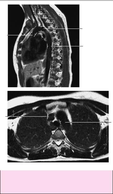
3 Diameter of aorta:
!< 4 cm
aAscending aorta:
a1 At level of pulmonary trunk bifurcation: 3.2 cm ± 0.5 cm a2 At level of aortic root: 3.7 cm ± 0.3 cm
bAortic arch: 1.5 cm ± 1.2 cm
cDescending aorta: 2.5 cm ± 0.4 cm
Ratio of ascending to descending aortic diameters = 1.5:1
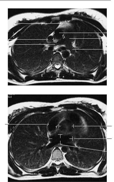
4Diameter of superior vena cava:
a At level of aortic arch: 1.4 cm ± 0.4 cm
b At level of pulmonary trunk bifurcation: 2 cm ± 0.4 cm
5Diameter of pulmonary arteries:
a Pulmonary trunk: 2.4 cm ± 0.2 cm
b Proximal right pulmonary artery: 1.9 cm ± 0.3 cm c Left pulmonary artery: 2.1 cm ± 0.4 cm
6Mediastinum:
!Thymus 1−2 cm in transverse diameter
Heart:
Dimensions of cardiac chambers:
7Right atrium:
!Maximum transverse diameter: 4.4 cm a At level of aortic root: 1.9 cm ± 0.8 cm b At level of mitral valve: 3.2 cm ± 1.2 cm c At center of ventricles: 2.8 cm ± 0.4 cm
8Left atrium:
a Maximum anteroposterior diameter: 4−5 cm
a1 At level of aortic root: 2.4 cm ± 4.5 cm a2 At level of mitral valve: 2.9 cm ± 4.9 cm
b Maximum transverse diameter: 9 cm
b1 At level of aortic root: 5.5 cm ± 8.4 cm b2 At level of mitral valve: 4.9 cm ± 9.1 cm
9Angle between midsagittal plane and septum = 38° (increases in response to pressure loading or volume loading of the ventricles)
10Thickness of ventricular septum:
!Approximately 5−10 mm
Moeller, Normal Findings in CT and MRI © 2000 Thieme
All rights reserved. Usage subject to terms and conditions of license.
Thoracic Organs 121
1 |
|
|
|
|
|
|
2b |
|
|
|
|
|
|
||
2a |
|
|
|
|
|
||
|
|
|
|
|
|||
|
|
|
|
|
|
||
Coronal image
3a1
3a2
12
Coronal image
Moeller, Normal Findings in CT and MRI © 2000 Thieme
All rights reserved. Usage subject to terms and conditions of license.
122 MRI: Chest
Sagittal image
3b
3a1
3c
4a
3b
Axial image at level of aortic arch
11Thickness of pericardium:
!1−2 mm
12Thickness of myocardium:
!10−12 mm
Moeller, Normal Findings in CT and MRI © 2000 Thieme
All rights reserved. Usage subject to terms and conditions of license.
Thoracic Organs 123
6 |
|
|
|
|
|
|
|
|
|
|
|
|
3a1 |
|
|
|
|
|
|
|
|
|
|
|
|
||||
|
|
|
|
|
|
|
|
|
|
|
|
|||
|
|
|
|
|
|
|
|
|
|
|
||||
|
|
|
|
|
|
|
|
|
|
|
|
|
||
|
|
|
|
|
|
|
|
|
|
|
|
|
||
4b |
|
|
|
|
|
|
|
|
|
|
|
|
5a |
|
|
|
|
|
|
|
|
|
|
|
|
|
|||
|
|
|
|
|
|
|
||||||||
|
|
|
|
|
|
|
|
|
|
|
||||
5b |
|
|
|
|
|
|
|
|
|
5c |
||||
|
|
|
|
|
|
|
|
|
|
|
||||
|
|
|
|
|
|
|
|
|
|
|
|
|
|
|
|
|
|
|
|
|
|
|
|
|
|
|
|
|
|
|
|
|
|
|
|
|
|
|
|
|
|
|
|
|
Axial image at level of pulmonary trunk bifurcation
7a
3a2
8b1
 8a1
8a1
Axial image at level of aortic root
Moeller, Normal Findings in CT and MRI © 2000 Thieme
All rights reserved. Usage subject to terms and conditions of license.
124 MRI: Chest
7b |
8a2 |
|
|
|
|
8b2 |
|
3c |
|
|
|
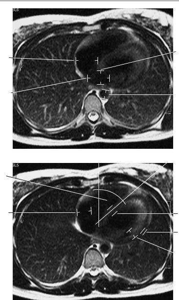
Axial image at level of mitral valve
9
7c 10
11
 12
12
Axial image through center of ventricles
Moeller, Normal Findings in CT and MRI © 2000 Thieme
All rights reserved. Usage subject to terms and conditions of license.