

Книги по МРТ КТ на английском языке / Normal Findings in CT and MRI
.pdfTemporomandibular Joint 185
Important Data
1Posterior ligament:
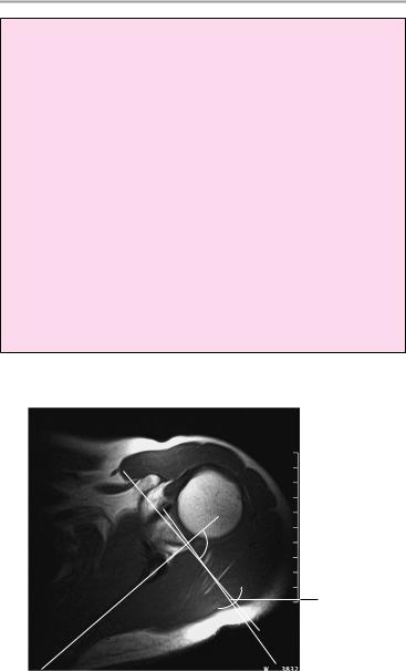
!With jaw in resting position, between 11 and 12 o’clock
11 12
1
Parasagittal image with the mouth closed
Parasagittal image with the mouth open
Moeller, Normal Findings in CT and MRI © 2000 Thieme
All rights reserved. Usage subject to terms and conditions of license.

186 MRI: Joints
Shoulder Joint
The humeral head has normal configuration and articulates properly with the normally developed glenoid. The articular surfaces are smooth and show normal cortical thickness. The width of the joint space is normal. The bone marrow displays homogeneous, fat-equivalent signal intensity.
The glenoid labrum is intact on all sides.
The acromioclavicular joint has normal configuration, with no hypertrophy. The subacromial fat is intact.
The muscles comprising the rotator cuff show normal course and configuration. In particular, the supraspinatus muscle is normal in its position, width, and signal characteristics and shows a normal musculotendinous junction.
The intact biceps tendon appears normal and occupies a normal position in the bicipital groove.
The other muscles that cover the shoulder joint appear normal, as do imaged portions of the lungs and soft tissues.
Interpretation
The shoulder joint appears normal.
Checklist
Humeral head ! Position (centered in the shoulder joint; does not ride high in the glenoid)
!Configuration (rounded cross section). (Caution: the bicipital groove appears anteriorly and the tuberosity posteriorly, but the highest axial section is always circular—useful for excluding a proximal depressed fracture = Hill-Sachs lesion)
!Contours (smooth and sharp)
!No osteophytes, especially in fovea area
!Bone marrow signal:
—Homogeneous, fat-equivalent intensity (in humeral head and shaft)
—“Adolescent” bone marrow signal before age 25 years
—No subchondral signal changes
—Normal articular cartilage
Joint space ! Width (see below)
! No increase of intra-articular fluid
Moeller, Normal Findings in CT and MRI © 2000 Thieme
All rights reserved. Usage subject to terms and conditions of license.

|
|
Shoulder Joint 187 |
Glenoid |
! |
Size congruent with humeral head |
|
! |
Smooth articular surface |
|
! |
Cortex (thickness, no discontinuities) |
|
! |
No osteophytes |
|
! |
No subchondral erosion |
|
! |
Bone marrow signal |
|
! |
Articular cartilage |
|
! |
Glenoid labrum is triangular about its whole cir- |
|
|
cumference and is firmly attached to the glenoid. |
|
|
(Caution: variant often seen in the anterosupe- |
Acromion, |
! |
rior quadrant should not be mistaken for a tear!) |
Normal development of the acromion (straight, |
||
clavicle |
|
curved, hook-shaped, upslope angle—see below) |
|
! |
and clavicle |
|
Smooth, sharp margins |
|
Acromioclavi- |
! |
Normal bone-marrow signal |
! |
Configuration |
|
cular joint |
! |
Width (see below) |
|
! |
No hypertrophy |
|
! |
Normal subacromial fat layer |
|
! |
Subacromial bursa is not fluid-filled, fat stripe of |
Rotator cuff |
! |
bursa is visible and undisplaced |
Configuration |
||
(supraspinatus, |
! |
Position |
infraspinatus, |
! |
Course (over humeral head) |
subscapularis, |
! |
Homogeneous signal intensity of tendon |
teres minor |
! |
No hyperintense signal (on T2-weighted images) |
muscles) |
! |
No peritendinous fluid |
Biceps tendon |
! |
Long tendon segment runs in bicipital groove |
|
! |
Hypointense |
|
! |
No discontinuities |
|
! |
Normal width |
|
! |
No increase of fluid in long biceps tendon sheath |
|
! |
No fluid in other bursae (especially the subcora- |
Muscles covering ! |
coid and subdeltoid) |
|
Shape |
||
the shoulder |
! |
Position |
joint (especially |
! |
Signal intensity |
the deltoid) |
|
|
Lungs, soft tissues
Moeller, Normal Findings in CT and MRI © 2000 Thieme
All rights reserved. Usage subject to terms and conditions of license.
188 MRI: Joints
Important Data
1Glenoid angle:
!Approx. 5° of retroversion (i.e., angle between the glenoid and a perpendicular to the scapular long axis is slightly open posteriorly, but the range of variation is large)
2Joint space:
!Shoulder joint: < 6 mm
3Acromioclavicular joint:
!Width < 1 cm
4Angle of acromion upslope (oblique sagittal image plane):
!10−40°
5Diameter of biceps tendon:
!Approximately 4−6 mm
6Bicipital groove:
!Width: 7−9 mm
!Depth: 4−7 mm
The bicipital groove starts at least 20 mm below the tip of the humeral head. (This differentiates the groove from a Hill-Sachs lesion, which often occurs at a higher level.)
.
1
Axial image
Moeller, Normal Findings in CT and MRI © 2000 Thieme
All rights reserved. Usage subject to terms and conditions of license.
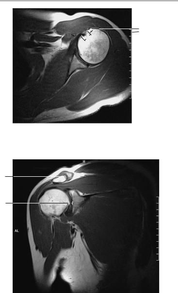
Shoulder Joint 189
6
5
Axial image
3
2
Paracoronal image parallel to the supraspinatus muscle
Moeller, Normal Findings in CT and MRI © 2000 Thieme
All rights reserved. Usage subject to terms and conditions of license.
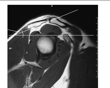
190 MRI: Joints
4 
Parasagittal image at right angles to the supraspinatus muscle
Moeller, Normal Findings in CT and MRI © 2000 Thieme
All rights reserved. Usage subject to terms and conditions of license.

Ellbow Joint 191
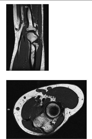
Elbow Joint
The elbow joint shows normal configuration with normal articulation of the bone ends. The articular surfaces are smooth and congruent, especially in the radiohumeral and ulnohumeral joints, with no discontinuities. There are no osteophytes or subchondral joint changes.
The joint spaces are of normal width. The olecranon fossa is clear, and there are no intra-articular loose bodies.
The cortex of the tubular bones is of normal thickness. The bone-mar- row signal is normal.
Imaged ligamentous structures appear normal, particularly the annular ligament.
The ulnar, radial, and median nerves display a normal course and diameter.
The imaged muscles show no abnormalities.
Interpretation
The elbow joint appears normal.
Checklist
Radius, ulna, |
! Normal configuration |
|
humerus |
! |
Articulation |
Joint |
! |
Position (see below) |
! |
Articular surfaces are smooth and congruent, |
|
|
|
especially in the radiohumeral and ulnohumeral |
|
! |
joints |
|
Normal cortical thickness |
|
|
! No discontinuities in the articular surfaces |
|
|
! |
No marginal osteophytes |
|
! No subchondral joint changes |
|
|
! |
Normal olecranon fossa |
|
! |
Joint spaces: |
|
|
— Width |
|
|
— No loose bodies |
|
|
— No effusion |
Cubital tunnel |
! |
— No synovial folds in radiohumeral joint |
Shape |
||
|
! |
Depth |
|
! |
Retinaculum |
Moeller, Normal Findings in CT and MRI © 2000 Thieme
All rights reserved. Usage subject to terms and conditions of license.

192 MRI: Joints
Other bony |
! Cortex of tubular bones (width, contours) |
|
structures |
! Bone marrow signal (normal for age, fat-equiv- |
|
Muscles and |
! |
alent; adolescent signal before age 25) |
Position |
||
ligaments |
! |
Width |
|
! |
Signal characteristics |
|
! No thinning or discontinuities |
|
|
! Muscle and tendon attachments: signal inten- |
|
|
|
sity, width, no discontinuities, no signs of tennis |
|
|
elbow: |
|
|
— No widening or abnormal enhancement of |
|
|
extensor tendon (next to radial epicondyle of |
|
|
humerus) |
|
|
— No increased water content of anconeus |
|
|
muscle |
|
! Bursae (e.g., radial bicipital bursa proximal to ten- |
|
|
|
don insertion, olecranon bursa) are not fluid- |
|
|
filled |
|
! Ligaments are intact (e.g., medial collateral liga- |
|
|
|
ment complex = posterior, transverse ligament |
|
|
and functionally important anterior ligament; |
|
|
lateral collateral ligament complex = medial col- |
|
|
lateral ligament, annular ligament, lateral ulnar |
|
|
collateral ligament, joint capsule) |
|
! No circumscribed thinning or expansion |
|
|
! |
Smooth borders |
|
! |
Homogeneous signal intensity |
Soft tissues |
! No increase in peritendinous fluid |
|
! Ulnar, radial and median nerves: |
||
|
|
— Course |
|
! |
— Diameter |
|
Normal ulnar groove |
|
|
|
|
Important Data
1Carrying angle of elbow:
!162°
2Angle of trochlear axis to ulnar axis:
!79°
3Angle of trochlear axis to humeral axis:
!83°
Moeller, Normal Findings in CT and MRI © 2000 Thieme
All rights reserved. Usage subject to terms and conditions of license.
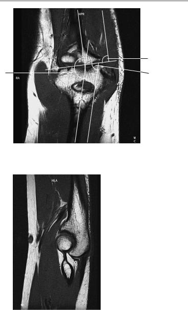
Ellbow Joint 193
. 2
1 |
3 |
Coronal image
Sagittal image at level of olecranon
Moeller, Normal Findings in CT and MRI © 2000 Thieme
All rights reserved. Usage subject to terms and conditions of license.
194 MRI: Joints
Sagittal image at level of radius
Axial image at level of radial head
Moeller, Normal Findings in CT and MRI © 2000 Thieme
All rights reserved. Usage subject to terms and conditions of license.