

Книги по МРТ КТ на английском языке / Normal Findings in CT and MRI
.pdfThoracic Organs 35
6
7a
8a
8b
Contrast bolus scan at level of aortic root
10
9
11
12
Contrast bolus scan through center of ventricles
Moeller, Normal Findings in CT and MRI © 2000 Thieme
All rights reserved. Usage subject to terms and conditions of license.

36
CT: Abdomen
Upper Abdominal Organs
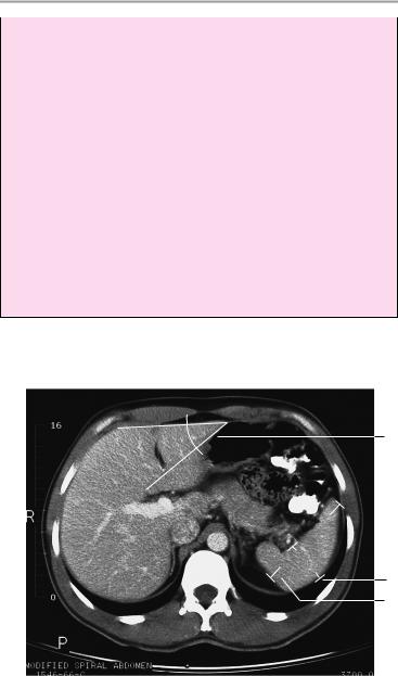
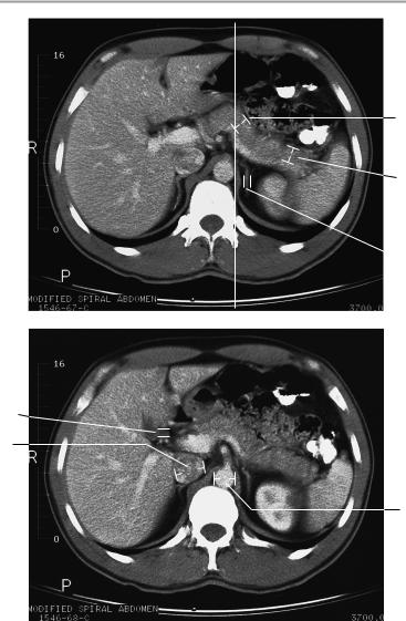
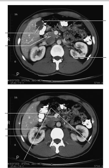
The liver is normally positioned and has normal size and smooth borders.
Its internal structure and attenuation values are normal. The intrahepatic and extrahepatic bile ducts and gallbladder are unremarkable. The spleen is orthotopic and of normal size. It has smooth outer contours and a homogeneous internal structure.
The pancreas is normal in size, position, and internal structure with smooth, lobulated outer contours. The pancreatic duct is unobstructed. Both kidneys show normal size and position. The renal parenchyma shows normal width and structure.
The renal pelvis and calices show a normal configuration. The urinary drainage tract is unobstructed.
The adrenal glands are unremarkable.
Major blood vessels appear normal, and there is no evidence of lymphadenopathy.
Imaged portions of the lung and soft tissues are normal.
Interpretation
The upper abdominal organs appear normal at CT.
Checklist
Liver |
! |
Position |
|
! |
— Directly below the right hemidiaphragm |
|
Size (see below) |
|
|
! |
Borders: |
|
|
— Smooth |
|
|
— Sharp |
|
! Normal attenuation values (see below) |
|
|
! Homogeneous internal parenchymal structure |
|
|
! |
No focal abnormalities |
|
! |
Intrahepatic bile ducts: |
|
|
— Course (centrifugal) |
|
|
— Width (general rule: ducts should no longer |
Moeller, Normal Findings in CT and MRI © 2000 Thieme
All rights reserved. Usage subject to terms and conditions of license.
|
|
Upper Abdominal Organs 37 |
|
|
be visible after contrast administration—see |
|
|
below) |
|
|
— No calculi |
|
! |
— No air |
|
Extrahepatic bile ducts: |
|
|
|
— Course (from porta hepatis to head of pan- |
|
|
creas) |
|
|
— Width (see below) |
|
|
— Contents of homogeneous fluid density |
|
|
— No calculi |
|
! |
— No air |
|
Gallbladder: |
|
|
|
— Size (see below) |
|
|
— Smooth outer contours |
|
|
— Normal wall thickness (see below) |
|
! |
— No pericholecystic fluid |
|
Gallbladder contents: |
|
|
|
— Homogeneous |
|
|
— Fluid density |
|
|
— No calculi (hypodense or hyperdense) |
|
|
— No air |
|
! Porta hepatis occupied by the hepatic artery, |
|
|
! |
common bile duct, and portal vein |
|
No masses |
|
|
! |
No lymphadenopathy |
|
! Costophrenic sinus clear and aerated on both |
|
Spleen |
! |
sides |
Size (see below) |
||
|
! |
Smooth outer contours |
|
! |
Homogeneous internal structure |
Pancreas |
! Attenuation values (see below) |
|
! |
Size normal for age (see below) |
|
|
! |
Normal lobulation |
|
! |
Smooth outer contours |
|
! |
Pancreatic duct unobstructed |
|
! |
No peripancreatic fluid |
Kidneys |
! Normal para-aortic region |
|
! |
Paired |
|
|
! |
Position (see below) |
|
! |
Size (see below) |
|
! |
Smooth contours |
|
! Width of parenchyma (see below) |
|
|
! |
Density (see below) |
Moeller, Normal Findings in CT and MRI © 2000 Thieme
All rights reserved. Usage subject to terms and conditions of license.

38 CT: Abdomen
|
! |
Width of cortex and medulla |
|
! |
Renal pelvis (anatomy, symmetry, size, no widen- |
|
! |
ing, contents of homogeneous fluid density) |
|
Calices (shape, width, homogeneous contents) |
|
Ureters |
! |
Enhancement characteristics (see below) |
! |
Not duplicated |
|
|
! |
Course |
|
! |
No obstruction of urinary drainage |
|
! |
Normal-appearing periureteral fat |
|
! |
Near-simultaneous opacification of both ureters |
Adrenal glands |
! |
after contrast administration |
Shape |
||
|
! |
Size (see below) |
|
! |
Slender crura |
|
! |
No circumscribed hypodense (cyst, adenoma), |
Intestinal |
! |
isodense or hyperdense expansion |
Anatomy |
||
structures (colon |
! |
Shape |
haustrations, |
! |
Wall thickness |
small bowel) |
! |
Homogeneous opacification after oral contrast |
|
! |
administration |
|
No free extraintestinal or intra-abdominal air or |
|
Major vessels |
! |
fluid |
Position |
||
|
! |
Size (see below) |
|
! |
Luminal opacification after contrast administra- |
|
! |
tion |
|
No large (intimal) calcifications |
|
|
! |
No mural thrombosis |
Lymph node |
! |
No dissection |
! |
No lymphadenopathy |
|
stations (espe- |
|
|
cially retrocrural, |
|
|
mesenteric, |
|
|
paraaortic) |
|
|
Lung segments |
! |
Configuration |
|
! |
Complete aeration |
|
! |
No adhesions |
Soft tissues |
! |
No pulmonary nodules |
! |
Anatomy |
|
|
! |
Symmetry |
|
! |
Density |
|
|
|
Moeller, Normal Findings in CT and MRI © 2000 Thieme
All rights reserved. Usage subject to terms and conditions of license.

Upper Abdominal Organs 39
Important Data
Dimensions:
1 Liver:
aAngle of left hepatic border: ca. 45°
bLeft lobe (anteroposterior diameter measured on the paravertebral line): up to 5 cm
!Caudate lobe/right lobe (CL/RL) = 0.37 ± 0.16 (e.g., 0.88 ± 0.2 in cirrhosis)
2Spleen:
a Depth (D): 4−6 cm b Width (W): 7−10 cm
c Length (L): 11−15 cm (reconstruction)
Splenic index: D×W×L = between 160 and 440
3Pancreas:
a Head up to 3.5 cm b Body up to 2.5 cm c Tail up to 2.5 cm
4Adrenal glands (variable):
!Crural thickness < 10 mm
5Gallbladder:
a Horizontal diameter up to 5 cm (> 5 cm is suspicious for hydrops)
bWidth of gallbladder wall:
!1−3 mm
cWidth of common bile duct:
!! 8 mm (after cholecystectomy: ! 10 mm)
6 Inferior vena cava:
! Transverse diameter up to 2.5 cm
7Abdominal aorta:
!Transverse diameter ca. 18−30 mm
8Kidneys:
a Anteroposterior diameter ca. 4 cm
b Transverse diameter 5−6 cm; craniocaudal diameter (= highest to lowest section) 8−13 cm
c Transverse renal axis: posteriorly divergent angle of 120° d Width of renal cortex: 4−5 mm
e Width of ureter: 4−7 mm Position of superior poles of kidneys:
!Right: superior border of L1
!Left: inferior border of T12
Moeller, Normal Findings in CT and MRI © 2000 Thieme
All rights reserved. Usage subject to terms and conditions of license.
40 CT: Abdomen
Time to corticomedullary equilibrium: ! 1 minute
Contrast excretion into the pyelocaliceal system: ! 3 minutes
Gerota fascia (thickness): ! 1−2 mm
Lymph nodes larger than 1 cm are suspicious for pathology.
Attenuation values:
!Liver: 65 ± 10 HU
!Spleen: 45 ± 5 HU
!Pancreas: 40 ± 10 HU
!Fat: -65 to -100 HU
!Kidneys: 30−45 HU without contrast medium; renal cortex after contrast medium: approx. 140 HU
!Adrenal glands: 25−40 HU without contrast medium
!Muscle: 45 ± 5 HU
!Blood vessels: approx. 40−55 HU without contrast medium
!Gallbladder contents: 0−25 HU
1a
2a
2b
Moeller, Normal Findings in CT and MRI © 2000 Thieme
All rights reserved. Usage subject to terms and conditions of license.
Upper Abdominal Organs 41

 1b
1b
3b
3c
 4
4
5c
6
7
Moeller, Normal Findings in CT and MRI © 2000 Thieme
All rights reserved. Usage subject to terms and conditions of license.
42 CT: Abdomen
5b
5a
3a
8d
8c
8a
8b
Moeller, Normal Findings in CT and MRI © 2000 Thieme
All rights reserved. Usage subject to terms and conditions of license.

Upper Abdominal Organs 43
8e 
Moeller, Normal Findings in CT and MRI © 2000 Thieme
All rights reserved. Usage subject to terms and conditions of license.

44 CT: Abdomen
Liver
The liver is orthotopic and of normal size, with smooth borders and normal attenuation values. It presents a normal internal structure with no focal abnormalities.
The intrahepatic and extrahepatic bile ducts are normal in their course, width, and contents.
The gallbladder appears normal, presenting smooth borders and homogeneous contents.
The porta hepatis shows no abnormalities.
Other visualized upper abdominal organs are unremarkable.
Interpretation
The liver appears normal at CT.
Moeller, Normal Findings in CT and MRI © 2000 Thieme
All rights reserved. Usage subject to terms and conditions of license.