

Книги по МРТ КТ на английском языке / Normal Findings in CT and MRI
.pdfPancreas 55
1 
5 
Moeller, Normal Findings in CT and MRI © 2000 Thieme
All rights reserved. Usage subject to terms and conditions of license.

56 CT: Abdomen
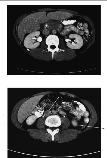
Kidneys
Both kidneys appear normal in size and position, with normal width and density of the renal parenchyma. There is no evidence of a mass. The calices are of normal shape. The renal pelvis is normal and free of stones, and there is no obstruction of urinary drainage.
Contrast-enhanced scans show a normal time to corticomedullary equilibrium and timely, symmetrical contrast excretion into the renal pelves with no filling defects.
The perirenal and pararenal spaces are unremarkable.
Other visualized upper abdominal organs, especially the adrenal glands, show no abnormalities.
Interpretation
Both kidneys appear normal at CT.
Checklist
Kidneys |
! |
Anatomy: |
|
|
|
— |
Paired |
|
|
— |
Position (see below) |
|
! |
— |
Size (see below) |
|
Organ contours: |
||
|
! |
— Smooth and sharp |
|
|
Width of parenchyma |
||
|
! |
Density (see below) |
|
|
! Normal relation of cortex to medulla |
||
|
! |
Renal pelves: |
|
|
|
— Structure and shape of caliceal groups |
|
|
|
— |
Bilateral symmetry |
|
! |
— No expansion |
|
|
Ureters: |
||
|
|
— One per side |
|
|
|
— Course |
|
|
|
— |
Width (see below) |
|
|
— |
No obstruction |
|
! Periand pararenal spaces: |
||
|
|
— |
Fat attenuation |
|
|
— No masses |
|
|
|
— No increase in soft-tissue structures |
|
|
|
— |
No fluid |
Moeller, Normal Findings in CT and MRI © 2000 Thieme
All rights reserved. Usage subject to terms and conditions of license.

Kidneys 57
|
! Periand pararenal fasciae: |
|
|
|
— Course |
|
|
— Width (no diffuse or circumscribed thicken- |
Adrenal glands |
! |
ing) |
Shape |
||
|
! |
Size (see below) |
|
! |
Slender crura |
|
! |
No circumscribed expansion |
Retroperitoneal ! No mass, fluid, or increased density |
||
space |
|
|
Intestinal |
! |
Colon haustrations, small bowel |
structures |
! |
Wall thickness |
|
! |
Homogeneous contrast enhancement |
|
! No free extraintestinal or intra-abdominal air or |
|
Vessels |
! |
fluid |
Course |
||
|
! |
Size (see below) |
Soft tissues |
! No lymphadenopathy (see below) |
|
! |
Density |
|
|
! |
Symmetry |
|
! Muscles (size, internal structure, borders) |
|
|
! Fat (density, no soft-tissue or fluid infiltration) |
|
|
|
|
Moeller, Normal Findings in CT and MRI © 2000 Thieme
All rights reserved. Usage subject to terms and conditions of license.

58 CT: Abdomen
Important Data
1Distance between renal poles:
!Superior poles: ca. 10 cm (4−16 cm) apart
!Inferior poles: ca. 13 cm (9−18.5 cm) apart
2Transverse renal axis:
!Posteriorly divergent angle of 120°
3Transverse renal diameter at level of hilum:
!5−6 cm (a, transverse) x 3−4 cm (b, anteroposterior)
4Width of cortex:
!4−5 mm
5Width of ureter:
!4−7 mm
6Gerota fascia (thickness):
!1−2 mm
Position of superior poles of kidneys:
!Right: superior border of L1
!Left: inferior border of T12 (variable; note that the difference does not exceed 1.5 vertebral body heights)
Renal dimensions:
!Craniocaudal (= highest to lowest section!) 8−13 cm Right−left disparity in renal sizes:
!Craniocaudal < 1.5 cm
Renal attenuation values:
!35−45 HU without contrast medium
!Renal cortex ca. 140 HU after contrast administration Time to corticomedullary equilibrium:
!1 minute
Contrast excretion into the pyelocaliceal system: ! 3 minutes
7Size of adrenal glands (variable):
!Crural thickness < 10 mm
Density of normal adrenal glands: 25−40 HU without contrast medium
8Abdominal aorta:
!Transverse diameter ca. 18−30 mm
9Inferior vena cava:
!Transverse diameter up to 2.5 cm
Vascular density: ca. 40−55 HU without contrast medium Lymph nodes larger than 1 cm are suspicious for pathology.
Moeller, Normal Findings in CT and MRI © 2000 Thieme
All rights reserved. Usage subject to terms and conditions of license.

Kidneys 59
7
1a
Early bolus phase
3b
2
4
3a
Early bolus phase
Moeller, Normal Findings in CT and MRI © 2000 Thieme
All rights reserved. Usage subject to terms and conditions of license.
60 CT: Abdomen
Late phase with corticomedullary equilibrium and opacification of the renal pelvis
9
8
5
6
1b
Late phase with ureteral opacification
Moeller, Normal Findings in CT and MRI © 2000 Thieme
All rights reserved. Usage subject to terms and conditions of license.

Adrenal Glands 61
Adrenal Glands
Both adrenal glands present normal size and position with normally developed crura. There is no evidence of a mass and no circumscribed expansion.
The adrenal compartment is unremarkable.
Postcontrast scans show normal adrenal enhancement characteristics and dynamics.
Other visualized upper abdominal organs, especially the kidneys, show no abnormalities.
Interpretation
Both adrenal glands appear normal at CT.
Checklist
Adrenal glands |
! |
Paired |
|
! |
Position (superior and anterior to the kidneys) |
|
! |
Shape |
|
! |
Size (see below) |
|
! |
Borders (smooth, sharp) |
|
! |
Slender adrenal crura showing no circumscribed |
|
! |
hypodense, isodense, or hyperdense expansion |
|
No calcifications |
|
|
! |
Adrenal compartment: |
|
|
— Fat attenuation |
|
! |
— No mass |
|
Enhancement characteristics: |
|
|
|
— Uniform increase in density |
|
|
— No hypodense or hyperdense lesions within |
Liver |
! |
the adrenal crura |
Size (see below) |
||
|
! |
Borders: |
|
|
— Smooth |
|
! |
— Sharp |
|
Homogeneous internal parenchymal structure |
|
|
! |
Intrahepatic and extrahepatic bile ducts |
Spleen |
! |
Costophrenic sinus clear and aerated on each side |
! |
Size (see below) |
|
|
! |
Smooth outer contours |
Pancreas |
! |
Homogeneous internal structure |
! |
Size |
|
|
! |
Pancreatic duct |
Moeller, Normal Findings in CT and MRI © 2000 Thieme
All rights reserved. Usage subject to terms and conditions of license.

62 CT: Abdomen
Kidneys |
! |
Paired |
|
! |
Position (see below) |
|
! |
Size (see below) |
Stomach and |
! |
Smooth contours |
! |
Position |
|
bowel |
! |
Size |
|
! |
No masses |
Major vessels |
! |
No infiltration |
! Transverse diameter |
||
Lymph nodes |
! |
Flow |
! No lymphadenopathy |
||
Diaphragm |
! No circumscribed widening |
|
|
! Lungs in the costophrenic sinus (no effusion or |
|
opacities)
Vertebral bodies ! Margins, bony structure
Soft tissues
Important Data
Dimensions
1Adrenal glands (variable):
!Crural thickness < 10 mm
!Density (without contrast medium): 25−40 HU Position of superior poles of kidneys:
!Right: superior border of L1
!Left: inferior border of T12
Transverse renal axis:
!Posteriorly divergent angle of 120° Renal dimensions:
!Craniocaudal: 8−13 cm
!Anteroposterior: ca. 4 cm
!Transverse: 5−6 cm
Gerota fascia (thickness):
!1−2 mm Spleen:
!Width: 7−10 cm
!Depth: 4−6 cm
!Length: 11−15 cm Diameter of abdominal aorta:
!Approximately 18−30 mm
Moeller, Normal Findings in CT and MRI © 2000 Thieme
All rights reserved. Usage subject to terms and conditions of license.
Adrenal Glands 63
1
1
Moeller, Normal Findings in CT and MRI © 2000 Thieme
All rights reserved. Usage subject to terms and conditions of license.

64 CT: Abdomen
Female Pelvis
The pelvic inlet appears normal, with normal configuration of the iliac wings and iliopsoas muscles.
Imaged bowel structures, especially the cecum and rectum, show no abnormalities with no evidence of wall thickening or mass lesions. The perirectal fat and ischiorectal fossa are unremarkable. The uterus is orthotopic with smooth borders. It displays a normal configuration and appropriate development for age. The myometrium shows homogeneous density. The uterine cavity is normally developed, and the adnexa are unremarkable. The vaginal fornix is normal.
The adequately distended urinary bladder has smooth outer contours and normal wall thickness. The vessels of the lesser pelvis are normal in course and caliber. There are no signs of lymphadenopathy.
The appearance of the pelvic skeleton, especially the femoral heads, sacroiliac joints, and symphysis pubis, is normal. There are no significant soft-tissue abnormalities.
Interpretation
The lesser pelvis appears normal at CT.
Checklist
Pelvic inlet |
! |
Configuration |
|
! |
Width |
|
! |
Symmetry |
|
! |
Iliac wings (shape) |
|
! |
Iliopsoas muscles: |
|
|
— Size |
|
|
— Density |
Intestinal |
! |
— Symmetry |
Position |
||
structures |
! |
Wall thickness (when normally distended, see |
(especially the |
|
below) |
cecum and |
! |
No circumscribed wall thickening |
rectum) |
! |
Well-opacified lumen with no soft-tissue mass |
Perirectal fat |
! |
Density (fat attenuation) |
|
! |
No infiltration |
|
! |
No masses |
Ischiorectal fossa ! |
Bilateral symmetry |
|
|
! |
No masses |
|
! |
No lymphadenopathy |
Moeller, Normal Findings in CT and MRI © 2000 Thieme
All rights reserved. Usage subject to terms and conditions of license.