

Книги по МРТ КТ на английском языке / Normal Findings in CT and MRI
.pdf
|
Petrous Pyramids |
15 |
Rest of |
! Cerebrum (especially the temporal lobe) |
and |
neurocranium |
cerebellum: |
|
—Configuration
—Sulcation
—Cortical markings (arbor vitae) well defined
—Width of sulci
—No circumscribed widening or narrowing
—Homogeneous density of cortex and white
matter (no hypodense or hyperdense changes)
CSF spaces ! Prepontine cistern ! Fourth ventricle
Important Data
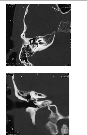
1Internal auditory canal:
!Approx. 5−10 mm, with ca. 1 mm difference between the right and left sides
1
Axial scan through the internal auditory canal
Moeller, Normal Findings in CT and MRI © 2000 Thieme
All rights reserved. Usage subject to terms and conditions of license.
16 CT: Head and Neck
Axial scan for evaluating the auditory ossicles
Coronal scan through the internal auditory canal
Moeller, Normal Findings in CT and MRI © 2000 Thieme
All rights reserved. Usage subject to terms and conditions of license.

Orbit 17
Orbit
The orbits are symmetrical and of normal size, with normal development of the orbital cone. The configuration of the smooth, sharply defined orbital walls is normal. There are no foci of bone destruction and there is no circumscribed widening of bony or soft-tissue components of the orbital walls.
The globes are symmetrical and show normal size and position. The ocular contents are of normal density. The ocular wall is smooth, sharply defined, and of normal thickness. The optic nerve shows a normal course and caliber on each side.
The eye muscles are normally positioned and display normal width and course. The retrobulbar fat and ophthalmic vein are unremarkable. Imaged portions of the neurocranium and paranasal sinuses show no abnormalities.
Interpretation
The orbit and its contents appear normal.
Checklist
Orbits |
! |
Symmetrical |
|
! |
Normal size |
Orbital walls |
! |
Normal orbital cone |
! Smooth, sharp borders |
||
|
! |
No bone destruction |
|
! No circumscribed widening of bone or soft- |
|
Globe |
! |
tissue components |
Position (see below) |
||
|
! |
Symmetry |
|
! |
Size (see below) |
Ocular contents |
! |
Spherical |
! |
Density |
|
Ocular wall |
! Borders (smooth and sharp) |
|
Optic nerve |
! |
Uniform thickness |
! Normal caliber (see below) |
||
Eye muscles |
! |
Course |
! |
Position |
|
|
! |
Width (see below) |
Retrobulbar fat |
! |
Course |
! |
Clear |
|
|
! |
No masses |
Moeller, Normal Findings in CT and MRI © 2000 Thieme
All rights reserved. Usage subject to terms and conditions of license.
18 CT: Head and Neck
Ophthalmic vein |
! |
Course |
Lacrimal gland |
! |
Caliber (see below) |
! |
Size |
|
|
! |
Symmetry |
|
! No unilateral or bilateral enlargement |
|
|
! |
Position (see below) |
|
! No excavation or destruction of adjacent bone |
|
|
! |
Homogeneous internal structure |
|
! |
No hypodense areas |
Neurocranium |
! |
Smooth borders |
! |
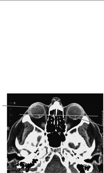
Temporal lobes |
|
Paranasal |
! |
Frontal lobes |
! |
Maxillary sinuses |
|
sinuses |
! |
Ethmoid cells |
|
|
|
1
 2
2
Axial scan
Moeller, Normal Findings in CT and MRI © 2000 Thieme
All rights reserved. Usage subject to terms and conditions of license.

Orbit 19
Important Data
1Diameter of globe:
!Axial plane:
—Right: 28.6 ± 1.2 mm
—Left: 29.4 ± 1.4 mm
!Sagittal plane (reconstruction):
—Right: 27.8 ± 1.2 mm
—Left: 28.2 ± 1.2 mm
2Position of globe:
!Posterior margin is 9.9 mm ± 1.7 mm behind the interzygomatic line
3Optic nerve (axial plane):
a Retrobulbar segment: 5.5 mm ± 0.8 mm
bNarrowest point (at approxinately mid-orbit): 4.2 mm ± 0.6 mm
4Ophthalmic vein:
!1.8 mm ± 0.5 mm (axial plane, 4 mm slice thickness)
!2.7 mm ± 1 mm (coronal plane)
5Eye muscles
a Superior rectus: 3.8 mm ± 0.7 mm b Oblique: 2.4 mm ± 0.4 mm
c Lateral rectus: 2.9 mm ± 0.6 mm d Medial rectus: 4.1 mm ± 0.5 mm e Inferior rectus: 4.9 mm ± 0.8 mm
Lacrimal gland: less than half of the gland is anterior to the frontozygomatic process.
Moeller, Normal Findings in CT and MRI © 2000 Thieme
All rights reserved. Usage subject to terms and conditions of license.
20 CT: Head and Neck
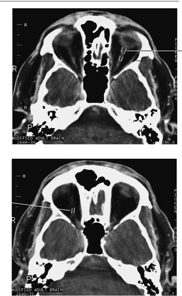
3a
 3b
3b
Axial scan
4 
Axial scan
Moeller, Normal Findings in CT and MRI © 2000 Thieme
All rights reserved. Usage subject to terms and conditions of license.
Orbit 21
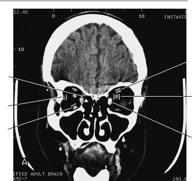
 5a
5a
5b
3a
5c
5e
5d
Coronal scan
Moeller, Normal Findings in CT and MRI © 2000 Thieme
All rights reserved. Usage subject to terms and conditions of license.

22 CT: Head and Neck
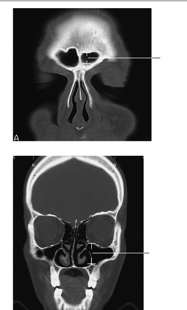
Paranasal Sinuses
The frontal sinuses are normally developed, clear and pneumatized with smooth wall contours.
The ethmoid cells have a normal appearance and intact bony walls, especially on the orbital side. There are no areas of wall erosion or mucosal thickening.
The sphenoid sinus is normally developed and has a coarse honeycomb structure. There are no fluid collections or mucosal swelling.
The maxillary sinuses are bilaterally symmetrical and have smooth walls of normal thickness. The sinuses are clear and aerated with no bone destruction. The nasal septum is on the midline, and the turbinates are normally developed.
The nasal cavity, pharynx, and imaged parapharyngeal structures show no abnormalities.
Interpretation
The paranasal sinuses appear normal.
Moeller, Normal Findings in CT and MRI © 2000 Thieme
All rights reserved. Usage subject to terms and conditions of license.

Paranasal Sinuses 23
Checklist
Frontal sinuses |
! |
Anatomy |
|
! |
Wall contours (smooth) |
Ethmoid cells |
! |
Pneumatization |
! |
Anatomy |
|
|
! |
Pneumatization |
|
! Bony structures (especially bordering the orbit: |
|
|
! |
boundaries are smooth, sharp, and intact) |
|
No wall erosions |
|
|
! |
No mucosal thickening |
Sphenoid sinus ! Anatomy (coarse honeycomb structure) |
||
|
! |
Clear and pneumatized |
|
! |
No fluid collection |
|
! |
No mucosal swelling |
|
! Bony structures (smooth, intact walls, no ero- |
|
|
|
sion) |
Maxillary |
! No extrinsic wall indentations |
|
! |
Anatomy |
|
sinuses |
! |
Size (bilaterally symmetrical) |
|
! Bony structures (smooth, intact contours, walls |
|
|
! |
of normal width, no bone erosion or destruction) |
|
Pneumatization |
|
|
! No tooth roots projecting through maxillary |
|
Nasal cavity |
|
sinus floor |
! Anatomy (symmetry) |
||
|
! |
Size |
|
! |
Aeration (clear) |
|
! Nasal septum on the midline |
|
|
! Nasal turbinates (three on each side: superior, |
|
Pharynx and |
|
middle, inferior) are normally developed |
! Anatomy (symmetry) |
||
parapharyngeal |
! |
Size |
structures |
! |
Wall thickness |
|
! No foreign bodies, calcifications, or masses |
|
|
|
|
Moeller, Normal Findings in CT and MRI © 2000 Thieme
All rights reserved. Usage subject to terms and conditions of license.
24 CT: Head and Neck
1
Coronal scan
3b
Coronal scan
Moeller, Normal Findings in CT and MRI © 2000 Thieme
All rights reserved. Usage subject to terms and conditions of license.