

Книги по МРТ КТ на английском языке / Normal Findings in CT and MRI
.pdfCervical Spine 165
1a |
|
|
|
|
|
|
|
|
|
|
|
|
3 |
||
|
|
|
|
|
|
|
|
|
|
|
|||||
4 |
|
|
|
|
|
|
|
|
|
|
|||||
|
|
|
|
|
|
|
|
|
|
|
|
|
|
5 |
|
|
|
|
|
|
|
|
|
|
|
|
|
|
|
||
|
|
|
|
|
|
|
|
||||||||
|
|
|
|
|
|
|
|
|
6 |
||||||
|
|
|
|
|
|
|
|
||||||||
7
8
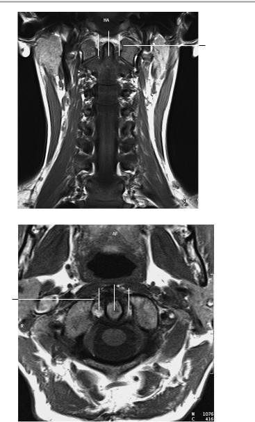
T2-weighted midsagittal image
2
T2-weighted sagittal image
Moeller, Normal Findings in CT and MRI © 2000 Thieme
All rights reserved. Usage subject to terms and conditions of license.
166 MRI: Spinal Column
1/2 1/2
1b
Coronal image
1/2 1/2
1b
Axial image at the level of the dens
Moeller, Normal Findings in CT and MRI © 2000 Thieme
All rights reserved. Usage subject to terms and conditions of license.
Cervical Spine 167
9
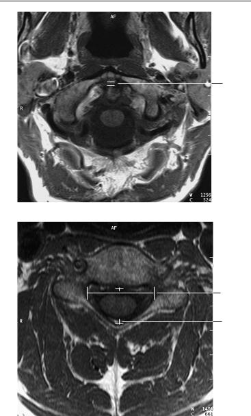
Axial image at the level of the dens
10
6
Axial image at the level of the laminae
Moeller, Normal Findings in CT and MRI © 2000 Thieme
All rights reserved. Usage subject to terms and conditions of license.

168 MRI: Spinal Column
Thoracic Spine
The thoracic spine shows a normal degree of kyphosis with a normal width of the bony spinal canal.
The vertebral bodies are normal in their number, shape, and interrelationships. The articulating vertebral end plates present smooth margins. The intervertebral disk spaces are of normal height, and the disks do not project past the posterior surface of the vertebral bodies in any segment.
The spinal cord displays normal course, configuration, width, and internal structure.
The bone marrow of the vertebral bodies appears normal.
The prevertebral and paravertebral soft tissues show no abnormalities.
Interpretation
The thoracic spine appears normal.
Checklist
Position |
! |
Thoracic kyphosis (see below) |
|
! |
No segmental malalignment |
Bony spinal canal ! |
Width (see below) |
|
Vertebral bodies |
! |
Smooth margins |
! |
Number (12) |
|
|
! |
Shape (square) |
|
! |
Position (straight alignment of posterior mar- |
|
! |
gins, no step) |
|
End plates |
|
|
|
— Continuous margins |
|
|
— No circumscribed depression |
Intervertebral |
! |
— Smooth contours, no marginal osteophytes |
Width (see below) |
||
disk space |
! |
Normal signal characteristics: moderate to |
|
|
slightly hyperintense T2-weighted signal inten- |
|
|
sity (not hypointense to other disks); “nuclear |
|
! |
cleft” signifies an adult disk |
|
No disk protrusion past posterior surface of adja- |
|
Spinal cord |
! |
cent vertebral bodies |
Configuration |
||
|
! |
Width |
|
! |
Signal characteristics |
Moeller, Normal Findings in CT and MRI © 2000 Thieme
All rights reserved. Usage subject to terms and conditions of license.
|
|
Thoracic Spine |
169 |
|
! No circumscribed signal changes |
|
|
Nerve roots |
! No circumscribed narrowing or expansion |
|
|
! |
Course |
|
|
Dural sack |
! Passage through the neuroforamina |
|
|
! |
Shape |
|
|
|
! |
Width |
|
|
! No circumscribed narrowing or expansion |
|
|
Neuroforamina |
! Contents of fluid intensity |
|
|
! |
Configuration |
|
|
Facet joints |
! |
Width |
|
! |
Shape |
|
|
|
! |
Position |
|
|
! |
Contours (smooth, continuous) |
|
|
! |
No hypertrophy |
|
|
! |
Vertebral arches intact |
|
|
! |
Pars interarticularis intact |
|
|
! |
No cleft anomalies |
|
Spinous |
! No shortening of pedicles |
|
|
! |
Shape |
|
|
processes |
! |
Position |
|
|
! |
Size |
|
|
! |
Bony structure |
|
|
! Fat-equivalent signal intensity of bone marrow |
||
|
! No circumscribed hypointense or hyperintense |
||
Soft tissues |
|
areas |
|
! Symmetrically arranged on both sides of |
the |
||
|
! |
vertebral bodies and spinous processes |
|
Aorta |
No masses |
|
|
! |
Prevertebral soft-tissue structures |
|
|
|
|
|
|
Moeller, Normal Findings in CT and MRI © 2000 Thieme
All rights reserved. Usage subject to terms and conditions of license.
170 MRI: Spinal Column
Important Data
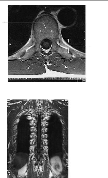
1Kyphotic angle (of Stagnara):
!Angle formed by a line parallel to the vertebral end plates of T3 and T11 = 25°
Width of spinal canal:
2Transverse diameter at level of pedicles:
!> 20−21 mm
3Sagittal diameter:
!T1−T11 = 13−14 mm
!T12 = 15 mm
4Width of intervertebral disk spaces:
!Smallest at T1
!T6−T11: ca. 4−5 mm
!Largest at T11−T12
4
3
3
1 |
|
|
|
4 |
|
|
|
||
|
11
 4
4
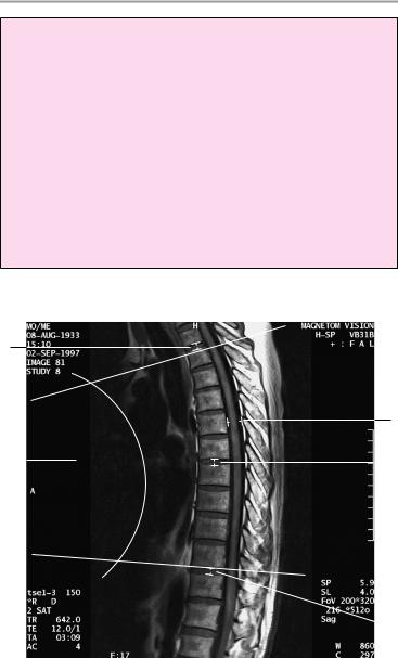
T2-weighted midsagittal image
Moeller, Normal Findings in CT and MRI © 2000 Thieme
All rights reserved. Usage subject to terms and conditions of license.
Thoracic Spine 171
2
3
Axial image at the level of the laminae
Coronal image
Moeller, Normal Findings in CT and MRI © 2000 Thieme
All rights reserved. Usage subject to terms and conditions of license.

172 MRI: Spinal Column
Lumbar Spine
The lumbar spine shows a smooth lordotic curve with a normal promontory angle. The bony spinal canal displays normal width.
The vertebral bodies are normal in their number, shape, and interrelationships. The articulating vertebral end plates present smooth margins. The intervertebral disk spaces are of normal height, and the disks do not project past the posterior surface of the vertebral bodies in any segment.
The conus medullaris terminates normally at the L1 level and divides into its filaments.
The dural tube appears normal in its lumbar portion and evaluable sacral portion.
The bone marrow of the vertebral bodies appears normal. The imaged soft tissues show no abnormalities.
Interpretation
The lumbar spine appears normal.
Checklist
Position |
! |
Lumbar lordosis (see below) |
|
! Lumbosacral angle (see below) |
|
Bony spinal |
! |
No segmental malalignment |
! Width (see below) |
||
canal |
! |
Smooth margins |
Vertebral bodies |
! |
Number (five) |
|
! |
Shape (square) |
|
! Position (straight alignment of posterior mar- |
|
|
! |
gins, no step) |
|
End plates |
|
|
|
— Continuous margins |
|
|
— No circumscribed depression |
|
|
— Smooth contours |
Intervertebral |
|
— No marginal osteophytes |
! Width (see below) |
||
disk space |
! |
Normal signal characteristics: moderate to |
|
|
slightly hyperintense T2-weighted signal inten- |
|
|
sity (not hypointense to other disks); “nuclear |
|
|
cleft” signifies an adult disk |
Moeller, Normal Findings in CT and MRI © 2000 Thieme
All rights reserved. Usage subject to terms and conditions of license.
|
|
|
Lumbar Spine 173 |
|
! |
No disk protrusion past posterior surface of adja- |
|
|
|
cent vertebral bodies (posterior disk contours on |
|
|
|
axial images: concave at L1−L4, straight at L4/5, |
|
Conus medullaris ! |
slightly convex at L5/S1) |
||
Configuration |
|||
|
! |
Width |
|
|
! |
No circumscribed narrowing or expansion |
|
|
! |
Position (terminates at approximately the L1 |
|
|
! |
level) |
|
|
Normal division into filaments |
||
|
! |
Signal characteristics |
|
|
! |
Filaments: |
|
|
|
— Course (sweeping, not straight; no posterior |
|
|
|
|
adhesions) |
|
|
— Width |
|
Nerve roots |
! |
— No circumscribed mass |
|
Course |
|||
|
! |
Passage through neuroforamina |
|
|
! |
Dural sac: |
|
|
|
— Shape |
|
|
|
— Width |
|
|
|
— No circumscribed narrowing or expansion |
|
Bony portions of |
! |
— Contents of fluid intensity |
|
Neuroforamina: |
|||
vertebral bodies |
|
— |
Configuration |
|
! |
— Width |
|
|
Facet joints: |
||
|
|
— Shape |
|
|
|
— |
Position |
|
|
— |
Contours (smooth, continuous) |
|
! |
— No hypertrophy of facet joints |
|
|
Vertebral arches intact |
||
|
! |
Pars interarticularis intact |
|
|
! |
No cleft anomalies |
|
|
|
— No shortening of pedicles |
|
|
|
— |
Spinous processes: |
|
|
— Shape |
|
|
|
— |
Position |
|
|
— |
Size |
|
! |
— |
Bony structure |
|
Fat-equivalent signal intensity of bone marrow |
||
|
|
— No circumscribed hypointense or hyper- |
|
|
|
|
intense areas |
Moeller, Normal Findings in CT and MRI © 2000 Thieme
All rights reserved. Usage subject to terms and conditions of license.

174 MRI: Spinal Column
Soft tissues ! Symmetrically arranged on both sides of the vertebral bodies and spinous processes
!Prevertebral soft-tissue structures
!No masses
Aorta, iliac vessels
Important Data
1Width of intervertebral disk space and height of lumbar intervertebral disks:
!8−12 mm
!Increases from L1 to L4/5
!Usually decreases at L5/S1, but may be the same as or greater than L4/5
2Lordosis (static axis):
!Plumb line from center of L3 should intersect S1
3Lumbosacral angle (S1/horizontal plane) = 26−57°
4Width of spinal canal: sagittal diameter:
! 16−18 mm (simple formula: not less than 15 mm; 11− 15 mm = relative stenosis, less than 10 mm = absolute stenosis)
5Width of spinal canal: transverse diameter (at level of pedicles):
!L1−L4: > 20−21 mm
!L5: > 24 mm
6Jones-Thomson ratio (= A × B/C × D):
!Between 1/2 and 1/4.5 = normal (denominator > 4.5 = spinal stenosis)
7Lateral recess (sagittal diameter):
!> 4−5 mm
8Ligamenta flava:
!Width < 6 mm
Moeller, Normal Findings in CT and MRI © 2000 Thieme
All rights reserved. Usage subject to terms and conditions of license.