

Книги по МРТ КТ на английском языке / Normal Findings in CT and MRI
.pdfPituitary 95
Pituitary
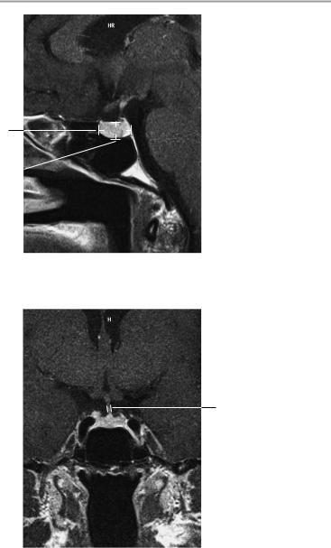
The size, position, and configuration of the sella are normal. The floor and walls of the sella are smooth and well-defined.
The pituitary is normal in size, shape, and position. The pituitary tissue shows normal signal characteristics both before and after contrast injection, with no circumscribed abnormalities of signal intensity.
The infundibulum is centered and of normal size.
The optic chiasm and suprasellar CSF spaces appear normal.
The cavernous sinus and imaged portions of the internal carotid artery and carotid siphon are unremarkable.
Evaluable portions of the neurocranium show no abnormalities. The sphenoid sinus is clear and pneumatized.
Interpretation
The pituitary appears normal.
Checklist
Sella |
! |
Position |
|
! |
Size |
|
! Configuration (U-shaped) |
|
|
! Walls steep, not splayed |
|
Pituitary |
! Borders smooth, sharp, and of normal width |
|
! |
Position (centered in the sella) |
|
|
! Configuration (bean-shaped) |
|
|
! Superior border straight or slightly concave |
|
|
! |
(convex only during puberty or pregnancy) |
|
Size (see below) |
|
|
! Delineation of adenohypophysis and neurohy- |
|
|
|
pophysis (sagittal image) |
|
! Pituitary tissue homogeneous on noncontrast |
|
|
! |
images |
|
Homogeneous contrast enhancement |
|
|
! No circumscribed hypointense or hyperintense |
|
|
|
areas (especially on coronal images, no signal |
|
|
difference between left and right halves of pitui- |
tary)
! Dynamic sequence (if performed) shows no time differential in the enhancement of different pituitary areas
Moeller, Normal Findings in CT and MRI © 2000 Thieme
All rights reserved. Usage subject to terms and conditions of license.

96 MRI: Head and Neck
Infundibulum |
! |
Position (centered) |
Optic chiasm |
! |
Size (see below) |
! |
Position |
|
|
! |
Size (see below) |
Suprasellar CSF |
! |
Symmetry |
! |
Symmetrical |
|
spaces (chias- |
! |
Not constricted |
matic cistern) |
|
|
Cavernous |
! |
Symmetry |
sinuses |
! |
Size |
Internal carotid |
! |
No infiltration |
! |
Symmetry |
|
arteries |
! |
Size (especially in siphon area) |
|
! No circumscribed or generalized narrowing or |
|
Neurocranium |
! |
expansion |
Temporal lobe |
||
|
! |
Hypothalamus |
Sphenoid sinus |
! Floor of third ventricle |
|
! Smooth margins, normal width (especially of the |
||
|
|
roof), clear and pneumatized |
|
|
|
Important Data
Pituitary
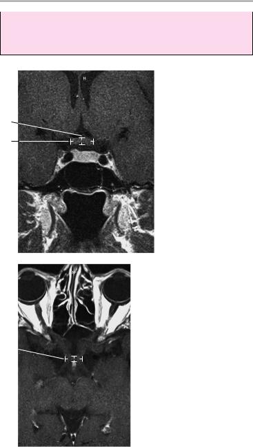
1Sagittal diameter:
!Men and postmenopausal women: < 8 mm
!Women of childbearing age: < 10 mm
2Height in sagittal plane:
!2−6 mm (Caution: normal size variations during
—Pregnancy: up to 12 mm
—Puberty: up to 10 mm in girls, up to 8 mm in boys
3Pituitary stalk:
!< 4 mm
Moeller, Normal Findings in CT and MRI © 2000 Thieme
All rights reserved. Usage subject to terms and conditions of license.
Pituitary 97
1
2
Sagittal image
3
Coronal image
Moeller, Normal Findings in CT and MRI © 2000 Thieme
All rights reserved. Usage subject to terms and conditions of license.
98 MRI: Head and Neck
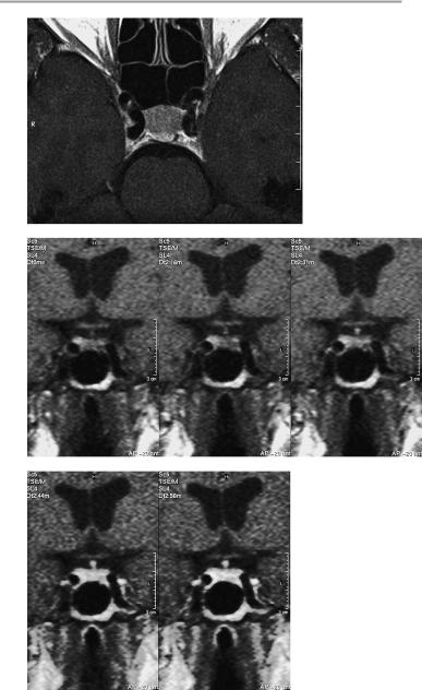
4Optic chiasm:
!Coronal: a, width 9−18 mm; b, height 3−6 mm
!Axial: c, width 12−27 mm; d, depth 4−9 mm
Coronal image
4b
4a
Axial image
4c
 4d
4d
Moeller, Normal Findings in CT and MRI © 2000 Thieme
All rights reserved. Usage subject to terms and conditions of license.
Pituitary 99
Axial image
Coronal image: dynamic examination following intravenous contrast administration (Gd-DTPA: gadolinium diethylenetriamine pentaacetic acid)
Moeller, Normal Findings in CT and MRI © 2000 Thieme
All rights reserved. Usage subject to terms and conditions of license.

100 MRI: Head and Neck
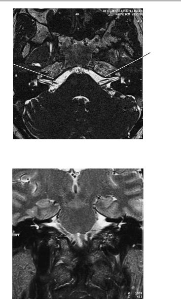
Internal Auditory Canals, Petrous Pyramids
The petrous pyramids appear normal and symmetrical. The internal acoustic meatus is of normal width, and its walls are smooth and sharply defined. The vestibulocochlear nerve on each side shows normal course and diameter. Contrast administration is not followed by abnormal rise of signal intensity within the nerve, especially its intrameatal portion.
The cochlea and semicircular canals appear normal. The mastoid air cells are clear and pneumatized. The tympanic cavity and external auditory canal are normal.
The cerebellopontine angle area shows normal configuration on each side.
The brain stem shows normal configuration and normal signal characteristics, with normal emergence of the nerves of the auditory canal. The cerebellopontine angle cistern is clear and symmetrical on each side.
The other imaged portions of the neurocranium are unremarkable.
Interpretation
The internal auditory canals appear normal.
Checklist
Petrous pyramids ! |
Configuration |
|
||
|
! |
Bilateral symmetry |
|
|
|
! |
Internal auditory canals: |
|
|
|
|
— Shape |
|
|
|
|
— Course |
|
|
|
|
— |
Width (see below) |
|
Vestibulocochlear ! |
— |
Borders (smooth, sharp) |
|
|
Course (straight, continuous) |
|
|||
nerve (cranial |
! Width (uniform, no right-left discrepancy, no |
|||
nerve VIII) |
! |
circumscribed expansion) |
|
|
|
Enhancement characteristics |
(nonenhancing, |
||
Facial nerve |
! |
especially within the meatus) |
|
|
Course (starts parallel to vestibulocochlear |
||||
(cranial nerve VII) |
|
nerve) |
|
|
|
! Width (uniform, no right-left |
discrepancy, no |
||
|
! |
circumscribed expansion) |
|
|
|
Enhancement characteristics (nonenhancing) |
|||
Moeller, Normal Findings in CT and MRI © 2000 Thieme
All rights reserved. Usage subject to terms and conditions of license.
|
|
|
Internal Auditory Canals, Petrous Pyramids 101 |
|
Cochlea and |
! |
Anatomy |
|
|
semicircular |
! |
Configuration |
||
canals |
! |
Smooth borders |
||
Mastoid cells, |
! |
Anatomy |
||
mastoid antrum, |
! |
Pneumatization |
||
tympanic cavity |
! |
Borders (wall thickness, smooth and continuous |
||
|
! |
contours) |
||
|
No masses |
|||
|
! |
Not opacified by material of soft-tissue or fluid |
||
External auditory ! |
signal intensity |
|||
Anatomy |
||||
canal |
! |
Course |
||
|
! |
Width |
||
|
! |
Borders |
||
Cerebellopontine ! |
Brain stem: |
|||
angle area |
|
— Shape |
||
|
|
— |
Signal intensity (homogeneous) |
|
|
! |
— |
No focal abnormalities |
|
|
Vestibulocochlear and facial nuclei (motor root |
|||
|
|
in medial eminence on floor of fourth ven- |
||
|
|
tricle): |
||
|
|
— No demyelination |
||
|
! |
— No masses |
||
|
Sites of entry of vestibulocochlear nerve (enters |
|||
|
|
pons and medulla at lateral extension of medul- |
||
|
|
lopontine sulcus) and facial nerve: |
||
|
! |
— |
Bilaterally symmetrical |
|
|
CSF spaces: |
|||
|
|
— Cerebellopontine angle cistern (symmetrical, |
||
|
|
|
fluid intensity) |
|
|
|
— No masses |
||
|
|
— |
Well delineated |
|
Rest of neuro- |
! |
— |
No vascular loops |
|
Cerebrum (especially the temporal lobe) and |
||||
cranium |
|
cerebellum: |
||
|
|
— |
Configuration |
|
|
|
— |
Sulcation |
|
|
|
— Cortical markings (arbor vitae) not effaced |
||
|
|
— |
Width of sulci |
|
|
|
— No circumscribed narrowing or expansion |
||
|
|
— Homogeneous signal intensity of cortex and |
||
white matter (no hypointense or hyperintense changes)
Moeller, Normal Findings in CT and MRI © 2000 Thieme
All rights reserved. Usage subject to terms and conditions of license.
102
CSF spaces ! Prepontine cistern ! Fourth ventricle
Important Data
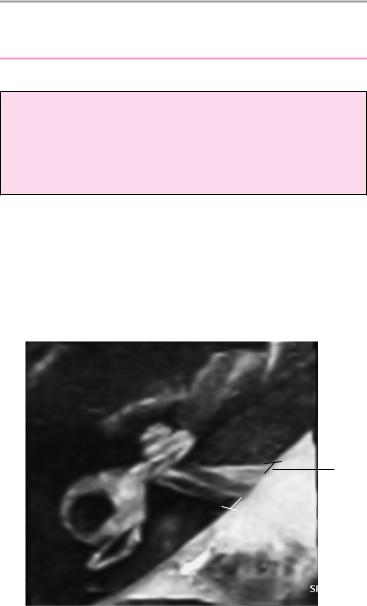
1Internal auditory canal:
!Approximately 5−10 mm
2Difference between right and left internal auditory canals:
!Approximately 1 mm
1
Axial image
Moeller, Normal Findings in CT and MRI © 2000 Thieme
All rights reserved. Usage subject to terms and conditions of license.
Internal Auditory Canals, Petrous Pyramids 103
2
2
Axial image
Coronal image
Moeller, Normal Findings in CT and MRI © 2000 Thieme
All rights reserved. Usage subject to terms and conditions of license.

104 MRI: Head and Neck
Orbit
The orbits are symmetrical and of normal size, with normal development of the orbital cones. The orbital walls show a normal configuration with smooth, sharp margins. No foci of bone destruction, no circumscribed expansion of the bony or soft-tissue components of the orbital wall are evident.
The globes are symmetrical and of normal size and position, and the ocular contents show normal signal characteristics. The ocular walls are smooth, sharply defined, and of normal thickness. The optic nerve has normal course and caliber on each side.
The eye muscles are normally positioned and display normal course and width. The retrobulbar fat, ophthalmic vein, and lacrimal gland are unremarkable.
Evaluable portions of the neurocranium and paranasal sinuses show no abnormalities.
Interpretation
The orbits and orbital contents appear normal.
Checklist
Orbits |
! |
Shape (orbital cone) |
|
! |
Size |
|
! |
Symmetry |
|
! |
Orbital walls: |
|
|
— Borders (smooth and sharp) |
|
|
— No bone destruction |
|
|
— No circumscribed expansion of bony or soft- |
Globe |
! |
tissue components of the orbital wall |
Shape (spherical) |
||
|
! |
Size (see below) |
|
! |
Position (see below) |
|
! |
Symmetry |
|
! |
Ocular contents: |
|
! |
— Signal intensity (fluid-equivalent) |
|
Ocular wall: |
|
|
|
— Borders (smooth and sharp) |
|
! |
— Thickness |
|
Retrobulbar fat (clear) |
|
|
! |
No masses |
Moeller, Normal Findings in CT and MRI © 2000 Thieme
All rights reserved. Usage subject to terms and conditions of license.