

Книги по МРТ КТ на английском языке / Normal Findings in CT and MRI
.pdfAdrenal Glands 145
Adrenal Glands
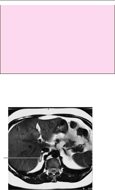
Both adrenal glands present normal size and position with normally developed crura. There is no evidence of a mass or circumscribed expansion.
The adrenal compartment appears normal.
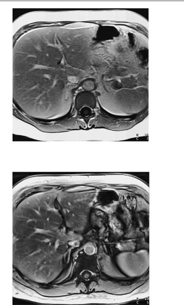
Postcontrast images show normal adrenal enhancement characteristics and dynamics.
No abnormalities are found in other visualized upper abdominal organs, especially the kidneys.
Interpretation
Both adrenal glands appear normal.
Checklist
Adrenal glands ! Paired
!Position (superior and anterior to kidneys)
!Shape, size (see below)
!Borders (smooth, sharp)
!Signal characteristics of normal adrenals (T1: slightly hypointense to liver; T1 fat-saturated: isointense; T2: hypointense; T2 fat-saturated: hyperintense)
!No circumscribed hypointense, isointense or hyperintense expansion of adrenal crura (e.g., T2weighted signal is increased in many pheochromocytomas) or circumscribed hypointense or hyperintense lesions (e.g., calcifications, fat deposits)
!Enhancement characteristics:
—Adenomas show moderate signal increase that usually returns to initial level by 10 minutes postinjection
—Most malignant tumors still show intense enhancement after 15 minuntes
!Chemical shift imaging:
—In-phase and out-of-phase T1-weighted images show fat intensity (decreased signal) in benign disease
Moeller, Normal Findings in CT and MRI © 2000 Thieme
All rights reserved. Usage subject to terms and conditions of license.

146 MRI: Abdomen
|
! |
Adrenal compartment: |
|
|
— Fat intensity |
Liver |
! |
— No masses |
Size (see below) |
||
|
! |
Borders: |
|
|
— Smooth |
|
|
— Sharp |
|
! Homogeneous internal parenchymal structure |
|
|
! Intrahepatic and extrahepatic bile ducts |
|
|
! Costophrenic sinus is clear and aerated on each |
|
Spleen |
! |
side |
Size (see below) |
||
|
! |
Smooth outer contours |
Pancreas |
! |
Homogeneous internal structure |
! |
Size |
|
Kidneys |
! |
Pancreatic duct |
! |
Paired |
|
|
! |
Position (see below) |
|
! |
Size (see below) |
Stomach and |
! |
Smooth contours |
! |
Position |
|
bowel |
! |
Size |
|
! |
No masses |
Major blood |
! |
No infiltration |
! Transverse diameter (see below) |
||
vessels |
! |
Flow |
Lymph nodes |
! No lymphadenopathy |
|
Soft tissues |
|
|
|
|
|
Important Data
Dimensions:
1Adrenal glands (variable):
!Crural thickness < 10 mm Kidneys:
Position of superior poles of kidneys:
!Right: superior border of L1
!Left: inferior border of T12 Transverse renal axis:
!Posteriorly divergent angle of 120°
Moeller, Normal Findings in CT and MRI © 2000 Thieme
All rights reserved. Usage subject to terms and conditions of license.
Adrenal Glands 147
Thickness of renal cortex: ! 4−5 mm
Renal dimensions:
!Craniocaudal diameter: 8−13 cm
!Anteroposterior diameter: ca. 4 cm
!Transverse diameter: 5−6 cm Gerota fascia (thickness):
!1−2 mm
Spleen:
!Width: 7−10 cm
!Depth: 4−6 cm
!Length: 11−15 cm Diameter of abdominal aorta:
!Approximately 18−30 mm
1
T2-weighted axial image through the adrenal glands
Moeller, Normal Findings in CT and MRI © 2000 Thieme
All rights reserved. Usage subject to terms and conditions of license.
148 MRI: Abdomen
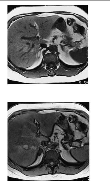
T1-weighted axial image, noncontrast and in-phase
T1-weighted axial image, noncontrast and out-of-phase
Moeller, Normal Findings in CT and MRI © 2000 Thieme
All rights reserved. Usage subject to terms and conditions of license.
Adrenal Glands 149
T1-weighted axial image, postcontrast and in-phase
T1-weighted axial image, postcontrast and out-of-phase
Moeller, Normal Findings in CT and MRI © 2000 Thieme
All rights reserved. Usage subject to terms and conditions of license.

150 MRI: Abdomen
Female Pelvis
The pelvic inlet appears normal, with normal configuration of the iliac wings and iliopsoas muscles.
No abnormalities are found in imaged bowel structures, and there are no signs of wall thickening or mass lesions.
The uterus is anteverted and has normal internal structure. The adnexa appear normal on both sides.
The adequately distended urinary bladder appears normal and has a normal wall thickness.
The vessels of the lesser pelvis are normal in course and caliber. There is no apparent lymphadenopathy.
The femoral heads are normally shaped and articulate normally with the acetabula. They have normal bone-marrow signal characteristics. The soft tissues show no abnormalities.
Interpretation
The lesser pelvis appears normal.
Checklist
Pelvic inlet |
! |
Configuration |
|
! |
Width |
|
! |
Symmetry |
|
! |
Iliac wings (shape) |
Iliopsoas muscles ! |
Size |
|
|
! |
Signal characteristics |
Intestinal struc- |
! |
Symmetry |
! |
Position |
|
tures (especially |
! |
Wall thickness (if with normal distension, see |
the cecum and |
|
below) |
rectum) |
! |
No circumscribed wall thickening |
Perirectal fat |
! |
Well-opacified lumen with no soft-tissue mass |
! |
Signal characteristics (fat intensity) |
|
|
! |
No infiltration |
|
! |
No masses |
Ischiorectal fossa ! |
Bilateral symmetry |
|
|
! |
No masses |
Uterus |
! |
No lymphadenopathy |
! |
Position |
|
|
! |
Size (see below) |
|
! |
Borders (smooth outer contours) |
Moeller, Normal Findings in CT and MRI © 2000 Thieme
All rights reserved. Usage subject to terms and conditions of license.
|
|
|
Female Pelvis |
151 |
|
|
! |
Signal characteristics |
|
|
|
|
! |
Uterine cavity: |
|
|
|
|
|
— |
Configuration |
|
|
|
|
— |
Size |
|
|
Cervix, vagina |
! |
— |
Signal characteristics |
|
|
Position |
|
|
|||
|
! |
Size |
|
|
|
Ovaries |
! |
Borders |
|
|
|
! |
Position |
|
|
||
|
! |
Size (see below) |
|
|
|
|
! |
Signal characteristics |
|
|
|
|
! |
Symmetry |
|
|
|
|
! No masses of soft-tissue or fluid signal intensity |
||||
Urinary bladder ! Adequate distention |
|
|
|||
|
! |
Outer contours: |
|
|
|
|
|
— Smooth |
|
|
|
Vessels |
! |
— Wall thickness (see below) |
|
|
|
Caliber (see below) |
|
|
|||
|
! |
Course |
|
|
|
Lymph node |
! No significant intimal calcifications |
|
|
||
! No lymphadenopathy |
|
|
|||
stations |
|
|
|
|
|
Pelvic skeleton |
! |
Configuration |
|
|
|
|
! Margins (cortex smooth and sharp, no discon- |
||||
|
|
tinuities) |
|
|
|
|
! Fat-equivalent signal intensity of bone marrow |
||||
|
! No circumscribed areas of marrow replacement |
||||
|
! Femoral heads rounded and centered |
in |
|||
|
! |
acetabula |
|
|
|
|
Sacroiliac joints: |
|
|
||
|
|
— Smooth contours |
|
|
|
|
|
— Normal width (see below) |
|
|
|
Subcutaneous |
! Symphysis pubis (see below) |
|
|
||
! |
Signal characteristics |
|
|
||
tissue and |
! |
Extent |
|
|
|
muscles |
! |
Borders |
|
|
|
|
! |
Symmetry |
|
|
|
|
|
|
|
|
|
Moeller, Normal Findings in CT and MRI © 2000 Thieme
All rights reserved. Usage subject to terms and conditions of license.
152 MRI: Abdomen
Important Data
Pelvic dimensions:
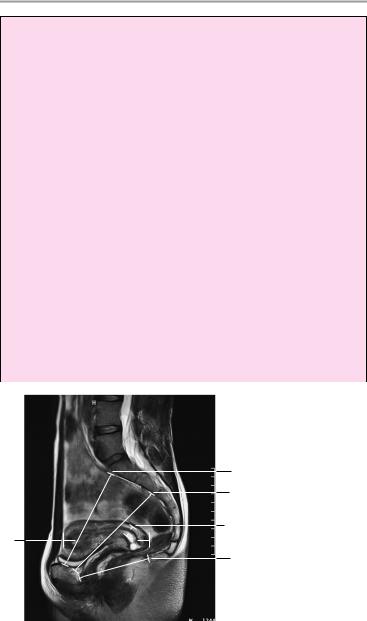
1True conjugate:
!Approximately 11 cm
2Pelvic cavity:
!> 12 cm
3Pelvic outlet:
!Approximately 9 cm
4Transverse diameter (transverse interspinous distance):
!Approximately 13 cm
5Uterus (variable):
!Prepubescent: a, length up to 3 cm; b, transverse diameter ca. 1 cm
!Nullipara: a, length up to 8 cm; b, transverse diameter ca. 4 cm
!Multipara: a, length up to 9.5 cm; b, transverse diameter ca. 5.5 cm
!Postmenopausal: a, length up to 6 cm; b, transverse diameter ca. 2 cm
(Transverse diameter of upright uterus = well distended bladder ! 5 cm)
6Uterine cervix:
a Craniocaudal ! 2 cm
b Transverse diameter ! 3 cm
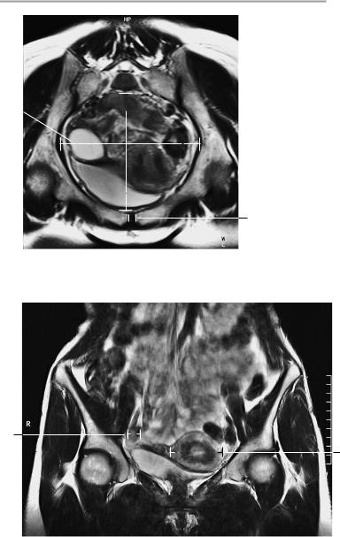
Midsagittal image through the lower abdomen.
1
2
6a
5a
3
Moeller, Normal Findings in CT and MRI © 2000 Thieme
All rights reserved. Usage subject to terms and conditions of license.
Female Pelvis 153


 11 4
11 4 1
1
10
Paracoronal image along the true conjugate (line 1 in Fig. on left [= midsagittal section through the lower abdomen]).
7b
5b
Coronal image
Moeller, Normal Findings in CT and MRI © 2000 Thieme
All rights reserved. Usage subject to terms and conditions of license.
154 MRI: Abdomen
7Ovaries:
!Prepubescent: a, length up to 2.5 cm; b, transverse diameter ca. 2.5 cm
!Sexual maturity: a, length up to 4 cm; b, transverse diameter ca. 2.5 cm
!Postmenopausal: a, length up to 3 cm; b, transverse diameter ca. 1.5 cm
8Urinary bladder (well distended):
!Wall thickness ca. 3 cm
9Rectum:
!Wall thickness ! 5 mm
10Symphysis pubis:
!Width < 6 mm
11Cartilage thickness of sacroiliac joint spaces:
!2−5 mm (anterior and inferior: 2−3 mm)
Axial image
5b
7b
6b
 7a
7a
Axial image
8
9
Moeller, Normal Findings in CT and MRI © 2000 Thieme
All rights reserved. Usage subject to terms and conditions of license.