

Книги по МРТ КТ на английском языке / Neuro Imaging Redcases
.pdf68 |
RadCases.thieme.com |
|
RadCases Neuro Imaging |
|
|
■ Imaging Findings |
|
|
|
A |
B |
C |
|
D |
(A) Axial computed tomography (CT) scan of the head shows a slightly hyperdense lesion in the right frontal region (arrowhead) with adjacent cortical thickening (arrows). (B) Axial T1-weighted image (WI) shows an isointense lesion in the right frontal region (arrowhead) that e aces the adjacent sulci.
(C) Di usion-WI shows increased signal in the right frontal lesion (arrow). (D) Coronal T1WI with contrast shows di use enhancement of the mass. There is thickening of the adjacent skull (arrow). The arrowhead shows the dural tail.
■ Di erential Diagnosis
•Meningioma: Meningioma is the most common extraaxial intracranial neoplasm. It is a well-circumscribed globular peripheral mass with marked enhancement. It causes “cortical buckling” of the underlying brain and has broad attachment to the dura mater. Calcifications occur in 25% of cases. A dural tail and hyperostosis of the adjacent calvarium are typically seen. Peripheral vasogenic edema occurs in 60% of cases.
•Epidural metastasis: This presents as thickening of the dura mater with di use enhancement. Extension to the inner table of the skull is common. The lesions are infiltrating rather than well-defined globular lesions with cortical buckling, as in meningioma.
■Essential Facts
•Symptomatic meningiomas are more common in female patients between 40 and 60 years of age.
•Definitions:
•Typical meningioma: World Health Organization (WHO) grade I
•Atypical meningioma: WHO grade II
•Malignant meningioma: WHO grade III
■ Other Imaging Findings
•CT: 75% are hyperdense and 25% calcify.
•Magnetic resonance imaging (MRI):
•T1: isointense to brain
•T2: variable intensity
•Fluid-attenuated inversion recovery: best for evaluation of peripheral edema
•T1 with contrast: di use enhancement with dural tail
•Angiogram: Shows the “mother-in-law” sign: “arrives early, leaves late.” Evaluate for involvement of venous sinus.
¸Pearls & ˚ Pitfalls
¸Meningiomas can be multiple if associated with muta-
tions.
¸ Less frequently, meningiomas may have cystic degeneration, necrosis, or hemorrhage.
˚Always review bone windows on CT to evaluate for cortical thickening, inner table invasion, or fractures.
˚MRI with contrast is the imaging modality of choice for characterizing and evaluating the extent of a meningioma.
69
Case 35
A B
C 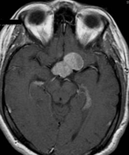
 D
D
■ Clinical Presentation
A 43-year-old with headache and bitemporal hemianopsia.
70 |
RadCases.thieme.com |
RadCases Neuro Imaging |
|
■ Imaging Findings |
|
A B C D
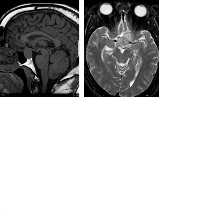
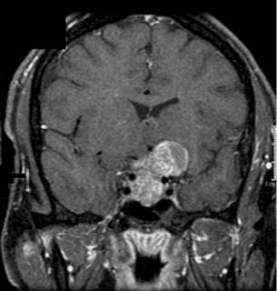
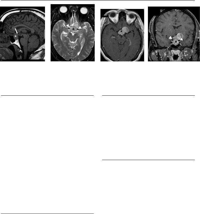
(A) Sagittal T1-weighted image (WI) shows a sellar mass with suprasellar extension (asterisk). The mass is isointense to brain and is displacing the optic chiasm superiorly (arrow). (B) Axial T2WI shows a heterogeneous mass in the suprasellar region (asterisk) that encases the internal carotid arteries without obstructing their flow (arrowheads). (C) Axial T1WI with contrast shows avid enhancement of the mass (asterisk). (D) Coronal T1WI with contrast show the encasement of the internal carotid arteries (arrowheads) by the mass (asterisk).
■ Di erential Diagnosis
•Pituitary macroadenoma: This is a well-defined, enhancing sellar mass that can extend to the suprasellar region. It is a “plastic” tumor that narrows as it crosses the diaphragma sellae, giving the typical “snowman” configuration.
Pituitary macroadenomas encase without narrowing the internal carotid arteries. Cavernous extension is common. Central necrosis is seen as cystic formation. There are no calcifications. The mean age at presentation is 20 to 40 years.
•Craniopharyngioma: This tumor has a bimodal age distribution, with a first peak in childhood/early adolescence and a second peak in older adults. It is mostly suprasellar; only 21% are intrasellar. Calcifications are typical. Craniopharyngiomas have a cystic component and a solid component that enhances. They have increased signal on precontrast T1WIs.
•Meningioma: Sellar region meningiomas start in the suprasellar region and rarely a ect the sella. The normal gland can be separated from the lesion. Meningioma enhances avidly and demonstrates a dural tail. Meningioma narrows the internal carotid arteries as it grows. It may calcify.
■Essential Facts
•Pituitary macroadenoma is a benign lesion.
•Ten percent of intracranial neoplasms are pituitary macroadenomas.
•Seventy-five percent have abnormal hormonal activity, usually involving prolactin.
•It is rare in children.
•The capsule of the adenoma represents the normal pituitary gland.
■ Other Imaging Findings
•Computed tomography (CT): The sella is widened without calcifications. CT can detect hemorrhage in apoplexy.
•Magnetic resonance imaging:
•T1: isointense to brain
•T2: heterogeneous signal, with necrosis appearing as eccentric cyst
•T1 with contrast: di use enhancement
•Fluid-attenuated inversion recovery: high signal in comparison with brain
•T2* can detect hemorrhage.
¸Pearls & ˚ Pitfalls
¸Macroadenomas are said to be “giant” if they exceed 4
cm in diameter.
¸It is crucial for the clinician to evaluate for cavernous sinus, internal carotid artery, and optic chiasm involvement.
¸High intensity of the optic nerves has been associated
with visual impairment.
˚It is extremely rare to find a pituitary adenoma that does not involve the sella.

71
Case 36
A B C
■ Clinical Presentation
A 38-year-old woman with progressive bilateral lower extremity paresthesia.
Further Work-up
D

72 |
RadCases.thieme.com |
|
RadCases Neuro Imaging |
|
|
■ Imaging Findings |
|
|
|
A |
B |
C |
|
D |
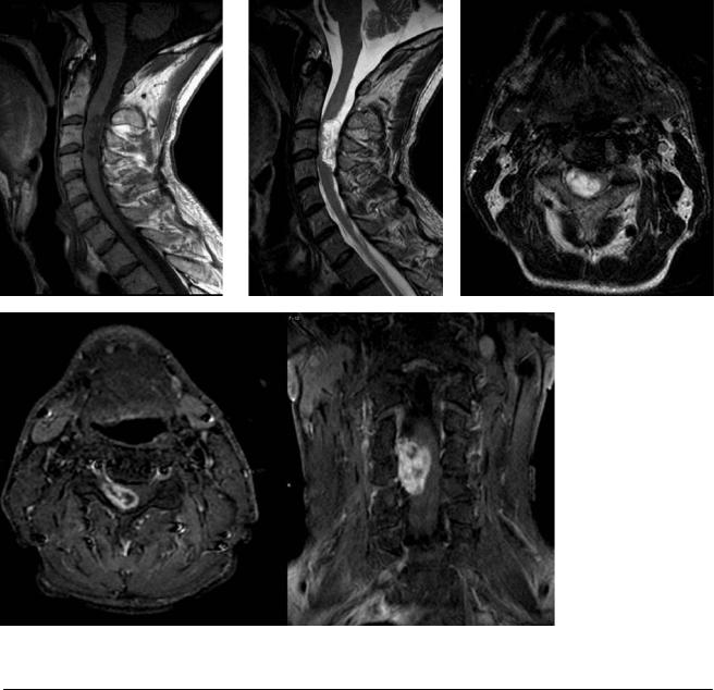
(A) Axial T1-weighted image (WI) of the cervical spine shows a syrinx (arrow) proximal to an ill-defined mass that thickens the spinal cord (arrowhead). (B) Sagittal T2WI of the cervical spine shows an ill-defined intramedullary mass (arrowhead) that is hyperintense to the spinal cord and proximal syringomyelia (arrow). (C) Sagittal T1WI of the cervical spine with contrast shows an enhancing mass at the C2-C3 level (arrow). (D) Axial T1WIs of the cervical spine with contrast, without and with fat suppression, show a thickened spinal cord with a di use intramedullary enhancing mass (arrow).
■ Di erential Diagnosis
•Intramedullary astrocytoma: This accounts for 30% of intraspinal gliomas. It is the most common intramedullary tumor in children and the second most common in adults. The mean age at presentation is 29 years. It is an infiltrating, enhancing tumor, more frequently located in the thoracic cord (70%) and rare in the filum terminale. It tends to be eccentric rather than central and expands the cord. The “cap sign” is not associated with cord astrocytomas.
•Ependymoma: This is the most common (60%) of intramedullary tumors in adults. The mean age at presentation is 39 years (10 years later than astrocytomas). Ependymomas are well-defined intraspinal masses, not infiltrative, and more frequently located in the cervical region. They have a central location and are associated with cysts or necrosis. Polar cysts and syrinxes are common. Hemorrhage is common; the “cap sign” is hemosiderin in the cephalic
or caudal margin. Spinal canal widening (11%) can occur. They enhance intensely.
•Hemangioblastoma: This is an intramedullary enhancing mass. It is associated with Von Hippel-Lindau disease, which is usually located in the posterior aspect of the spi-
nal cord. Hemangioblastomas are adjacent to subpial cysts. Syrinx is less likely because of the eccentric location. They occur most frequently in the thoracic cord. They are typically small lesions. They may have flow voids of prominent vessels.
■ Essential Facts
•Patients with intraspinal astrocytomas are less likely to survive than are patients with ependymomas.
•The lesion has poorly defined margins and extends beyond the imaging boundaries.
•Polar and intratumoral cysts and syrinxes are common.
•They span four or fewer spinal segments.
■ Other Imaging Findings
•Computed tomography: A thickened spinal cord is associated with syrinx. The spinal canal can be remodeled (less frequent than in ependymomas).
•Magnetic resonance imaging:
•T1: solid portion isointense to cord, associated cyst/ syrinx with low intensity
•T2: hyperintense
•T1 with contrast: moderate enhancement with illdefined borders
¸Pearls & ˚ Pitfalls
¸Can be exophytic
¸Rarely associated with bleeding
˚Fewer than 4% of spinal astrocytomas located in the conus medullaris
73
Case 37
A B
C D
■ Clinical Presentation
A 60-year-old woman with the history of a lump in her left breast.
74 |
RadCases.thieme.com |
RadCases Neuro Imaging |
|
■ Imaging Findings |
|
A B C D
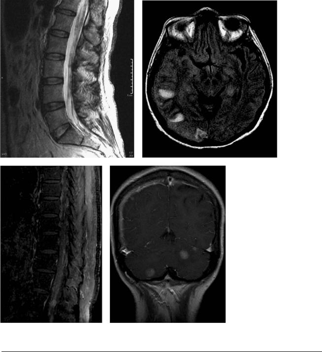
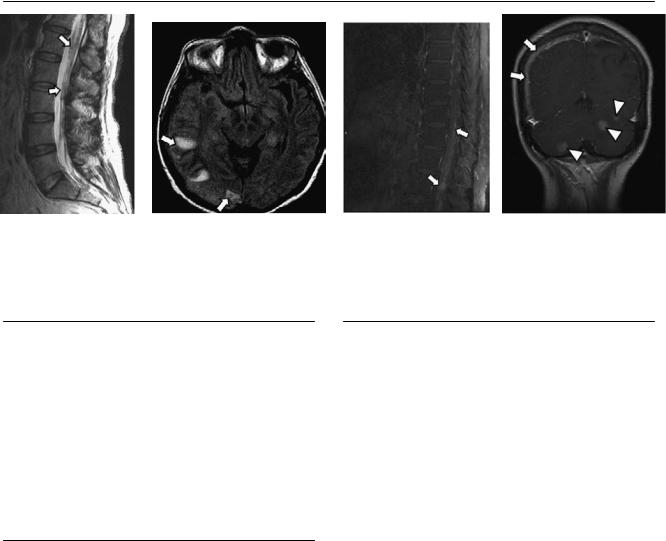
(A) Sagittal T2-weighted image (WI) of the lumbar spine shows nodular thickening of the nerve roots (arrows). (B) Axial fluid-attenuated inversion recovery (FLAIR) image of the brain shows three foci of increased signal in the subcortical white matter on the right (arrows). (C) Sagittal T1WI of the spine after contrast with fat saturation shows di use nodular enhancement of the nerve roots (arrows). (D) Coronal T1WI of the brain with contrast shows thick meningeal enhancement on the right (arrows) along with foci of nodular enhancement in the posterior fossa (arrowheads).
■ Di erential Diagnosis
•Meningeal carcinomatosis: “Sugarcoating” and nodular enhancement of the nerve roots are typical findings of meningeal carcinomatosis. It can result from hematogenous spread of extracranial disease or from “drop” metastasis of intracranial disease. It often causes thickening of the nerve roots. The conus medullaris is also involved. The entire neuraxis should be assessed.
•Meningitis and cerebritis: The combination of leptomeningeal enhancement and brain parenchymal lesions can be the result of infection, leading to this diagnosis.
■ Essential Facts
•Meningeal carcinomatosis can be secondary to metastasis from primary tumors of the central nervous system or from distant tumors.
•Cytology of the cerebrospinal fluid is useful to confirm the diagnosis.
•Hematogenous brain metastases are typically located in the subcortical white matter.
•Tumors that typically have leptomeningeal spread include lung carcinoma, breast carcinoma, and lymphoma.
•Meningeal metastases tend to be isointense to gray matter, so that they are hard to identify without contrast.
•Nodular enhancement of the nerve roots is typical.
•FLAIR can show increased signal in the sulci of patients with meningeal carcinomatosis; however, this does not help to di erentiate it from meningitis.
¸Pearls & ˚ Pitfalls
¸Remember to scan the entire neuraxis in patients with
suspected meningeal carcinomatosis.
¸Skull involvement is possible and appears as an enhancing lesion invading the diploic space.
¸Di usion-WIs will be bright in hematogenous parenchymal abscesses and can help di erentiate them from
metastases.
˚Both magnetic resonance imaging with contrast and cerebrospinal fluid analysis yield false-negative results for the detection of leptomeningeal carcinomatosis and should be used in combination to increase their sensitivity.
75
Case 38
A B
C 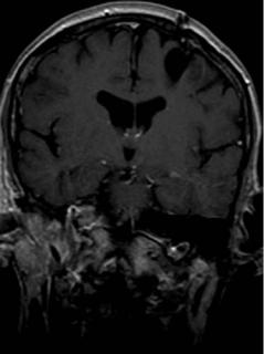
 D
D
■ Clinical Presentation
An adult woman with severe intermittent headaches.
76 |
RadCases.thieme.com |
RadCases Neuro Imaging |
|
■ Imaging Findings |
|
A B C D
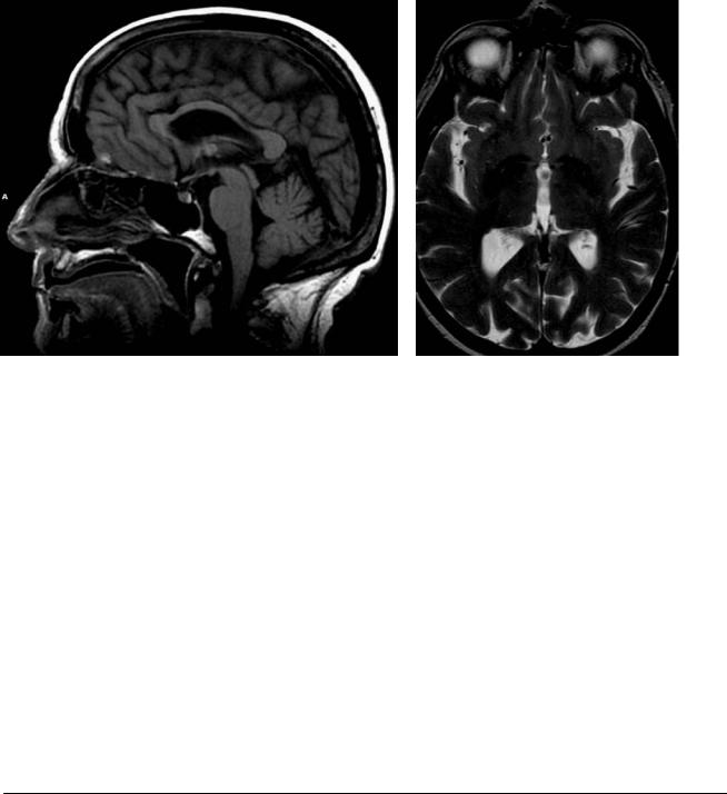
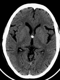
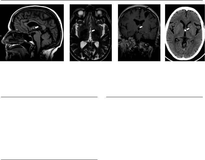
(A)Sagittal T1-weighted image (WI) shows a round mass in the roof of the 3rd ventricle (arrow). The mass is isointense to the gray matter.
(B)Axial T2WI shows the lesion as isointense to cerebrospinal fluid (arrow). (C) Coronal T1WI with contrast shows the lesion in the roof of the 3rd ventricle (arrow); there is no enhancement. (D) Computed tomography (CT) of the head without contrast shows a hyperdense lesion in the 3rd ventricle (arrow) adjacent to the anterior pillars of the fornix.
■ Di erential Diagnosis
•Colloidal cyst: Colloidal cysts are histologically benign lesions attached to the roof of the 3rd ventricle. They obstruct the foramen of Monro and cause hydrocephalus. They do not have enhancement or calcifications.
•Flow artifact: Flow artifact in the foramen of Monro on fluid-attenuated inversion recovery and T2WIs is usually seen in the lateral ventricles, not the 3rd ventricle. This is not seen on CT or magnetic resonance (MR) sequences with a short echo time. There is no hydrocephalus.
¸Pearls & ˚ Pitfalls
¸Many small colloidal cysts can be hard to identify on MR.
CT without contrast can be the best diagnostic modality in these cases.
˚ Rarely, colloid cysts can present with rim enhancement.
■ Essential Facts
•Benign cysts are characterized histologically by an epithelial lining with mucous goblet cells.
•They apparently originate from the endoderm, sharing characteristics with Rathke cleft cysts.
•Most colloidal cysts are hyperdense on CT.
•The MR appearance is variable, and the cysts can be difficult to identify.
•Half of them show increased T1 signal.
•There is no enhancement.
77
Case 39
A B C
D
■ Clinical Presentation
A 27-year-old woman with progressive paresthesia in the right shoulder and arm.