

Книги по МРТ КТ на английском языке / Neuro Imaging Redcases
.pdf48 |
RadCases.thieme.com |
|
RadCases Neuro Imaging |
|
|
■ Imaging Findings |
|
|
|
A |
B |
C |
|
D |
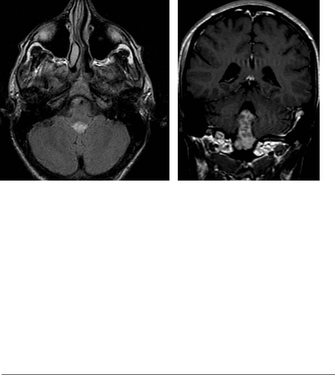
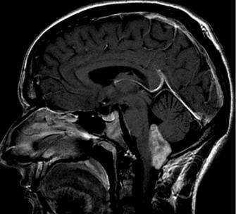
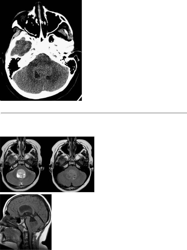
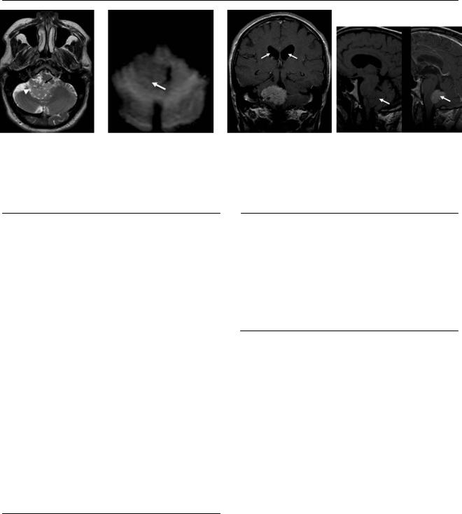
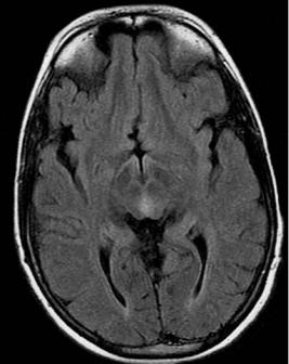
(A) Sagittal T1-weighted image (WI) shows the irregular anterior border of the pons (arrows). (B) Coronal T2WI demonstrates displacement of the pons to the right (arrow). A lesion in the left cerebellopontine angle (CPA) has a slightly higher signal than that of cerebrospinal fluid (CSF; asterisk). (C) Axial T1WI shows a lesion in the left CPA that has a signal like that of CSF and displaces the pons to the right (arrow). (D) Di usion-WI demonstrates high signal in the mass located in the left CPA (arrow).
■ Di erential Diagnosis
•Epidermoid cyst: Ectodermal inclusion cysts are composed of epithelial cells that form a keratohyalin matrix as a consequence of desquamation. The location is usually away from the midline in the basal subarachnoid cisterns and ventricles. The cysts engulf vessel and nerves. Epidermoid cysts have a signal like that of CSF on T1 and T2WIs but have a heterogeneous signal on fluid-attenuated inversion recovery (FLAIR) images. They have a high signal on dif- fusion-WIs secondary to T2 “shine through.” They do not enhance.
•Dermoid cyst: Dermoid cysts are also ectodermal inclusion cysts that, in addition to squamous epithelium, contain skin appendages that form cholesterol. They have a heterogeneous signal, with areas of high T1 signal secondary to fatty content. They tend to have a midline location. Dermoid cysts can rupture and cause chemical meningitis. Typically, there is no enhancement, although the meninges may enhance if the cysts rupture. A supratentorial location is more common. Dermoid cysts do not have a signal on di usion-WIs.
•Arachnoid cyst: These are CSF-filled cysts. They have a signal like that of CSF in all sequences. There is no signal on di usion-WIs. There is no enhancement. The middle cranial fossa is the most common location (50%), followed by the CPA (10%). Arachnoid cysts displace cortical vessels away from the calvarium.
■ Essential Facts
•The CPA is most common location for epidermoid cysts (45%), followed by the 4th ventricle (15%).
•The lesion encases arteries without compromising the lumina.
•On computed tomography, they resemble CSF and rarely calcify (20%).
•On magnetic resonance imaging, the lesions are isointense to CSF on T1WIs and slightly hyperintense to CSF on T2WIs. On FLAIR images, they show a signal slightly higher than that of CSF. On di usion-WIs, they show T2 “shine through.”
¸Pearls & ˚ Pitfalls
¸Very rare degeneration to squamous cell carcinoma has
been seen.
¸Dermoid cysts are more common in the supratentorial territory, whereas epidermoid cysts are more frequent in
the infratentorial region.
˚The high signal of epidermoid cysts on di usion-WIs is caused by T2 “shine through,” not restricted di usion.
˚Rare cases of epidermoid cysts with a high protein content have been reported as hyperdense with a high signal on T1 and T2WIs.
49
Case 25
A B
C 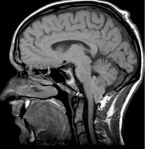
 D
D
■ Clinical Presentation
A 28-year-old man presenting with ataxia, headache, nausea, and vomiting.
50 |
RadCases.thieme.com |
|
RadCases Neuro Imaging |
|
|
■ Imaging Findings |
|
|
|
A |
B |
C |
|
D |
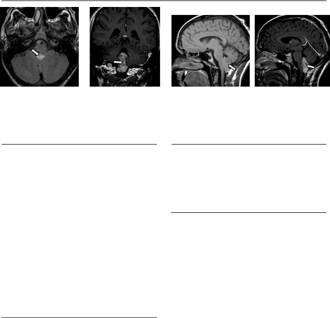
(A) Axial fluid-attenuated inversion recovery (FLAIR) image shows a well-defined hyperdense lesion occupying the 4th ventricle (arrow). (B) Coronal T1weighted image (WI) with contrast shows heterogeneous enhancement of the 4th ventricular mass (arrow). (C) Sagittal T1WI demonstrates an isointense mass (arrow) that occupies the lower aspect of the 4th ventricle. (D) Sagittal T1WI with contrast shows the heterogeneous enhancement of the mass (arrow).
■ Di erential Diagnosis
•Ependymoma: Sixty percent of intraventricular ependymomas are in the 4th ventricle. They can extend through the foramen of Luschka into the subarachnoid space. They are isodense to brain on noncontrasted computed tomography (CT). Punctate or coarse calcifications are present in 40 to 80%. There is moderate enhancement except for cystlike regions. Hydrocephalus and hemorrhage are common. There is no gender predilection.
•Subependymoma: Subependymomas have no infiltrative pattern. These occur mostly in the 4th ventricle and are usually smaller than 2 cm. Males are more frequently affected. Subependymomas have slight to no enhancement. Thirty percent calcify; coarse calcifications are unusual.
•Medulloblastoma: These are hyperdense on CT. They originate in the roof of the 4th ventricle. They have intense enhancement and do not extend through the foramen of Luschka. Calcifications are less common. There is a male predominance. They have early subarachnoid spread.
■ Other Imaging Findings
•CT: isodense mass with calcifications
•MRI:
•T1: isointense
•T2: slightly hyperintense on pseudocystic areas
•Hyperintense to cerebrospinal fluid on FLAIR images
•Mild enhancement
¸Pearls & ˚ Pitfalls
¸If a lesion arises from the floor of the 4th ventricle, favor
the diagnosis of ependymoma over medulloblastoma.
¸Ependymomas present in an older age range than do medulloblastomas.
¸Ependymomas are isodense to gray matter on CT, whereas medulloblastomas tend to have a higher attenuation. Most supratentorial ependymomas are extraventricular.
■ Essential Facts
•Sixty percent of ependymomas occur in the posterior fossa.
•They are the third most common tumors of the posterior fossa in children (after pilocytic astrocytomas and medulloblastomas).
•They arise from the floor of the 4th ventricle.
•Supratentorial ependymomas are typically extraventricular, most commonly seen in children. They have a better survival rate.
51
Case 26
A
■ Clinical Presentation
A 7-year-old girl with headaches.
Further Work-up
B
C 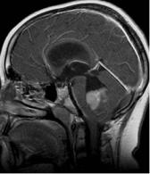 D
D
52 |
RadCases.thieme.com |
|
RadCases Neuro Imaging |
|
|
■ Imaging Findings |
|
|
|
A |
B |
C |
|
D |
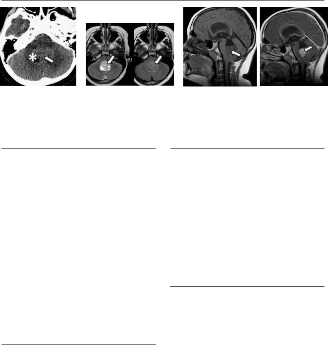
(A) Computed tomography (CT) scan of the head without contrast shows a slightly hyperdense mass in the area of the 4th ventricle (arrow); there is a cystic part of the lesion (asterisk). (B) Axial T2-weighted and fluid-attenuated inversion recovery (FLAIR) images show a mass centered in the 4th ventricle that has intermediate signal with cystic components (arrows). (C) Sagittal T1-weighted image (WI) without contrast shows the mass (arrow), which is slightly hypointense to the white matter, occupying the lower aspect of the 4th ventricle. Note the dilatation of the entire ventricular system.
(D) Sagittal T1WI with contrast shows enhancement of the mass (arrow).
■ Di erential Diagnosis
•Medulloblastoma: These are the most common posterior fossa tumors and the second most frequent brain tumors in children (after astrocytomas). They are rare in young adults. Medulloblastomas are highly malignant neuroepithelial tumors. The cerebellum is the most frequent location (95%); most of them occur in the vermis (75%). Hyperattenuation on CT is typical. Medulloblastomas arise from the roof of the 4th ventricle and almost always enhance.
•Ependymoma: These are midline cerebellar enhancing masses. Calcifications are frequent (medulloblastomas rarely calcify). They are hypoattenuated on CT (this helps to di erentiate them from medulloblastomas). They typically extend through the foramen of Luschka to the subarachnoid space. They arise from the floor of the 4th ventricle (medulloblastomas usually arise from the roof).
•Pilocytic astrocytoma: These are the most common pediatric brain tumors. They are cystic with a mural nodule. They have low attenuation on CT (medulloblastomas are typically hyperattenuated). The most common location is the cerebellar hemisphere.
■ Essential Facts
•Arise from undi erentiated cells of the posterior medullary velum
•Lateral location more frequent in older children
•Brainstem infiltration in 33%
•Recurrence rates are high.
•World Health Organization grade IV
■ Other Imaging Findings
•CT:
•Well-defined vermian mass
•Surrounding vasogenic edema (97%)
•High density because of compact cells (90%)
•Hydrocephalus
•Homogeneous enhancement
•Cyst formation (50%)
•Rarely calcify (20%)
•Magnetic resonance imaging:
•Hypointensse relative to white matter on T1-weighted images
•Heterogeneous on T2WIs
•High choline peak with reduced N-acetylaspartate on spectroscopy
¸Pearls & ˚ Pitfalls
¸Basal nevus syndrome is the association of dural calcifi-
cations, keratocysts of the jaw, and medulloblastoma.
¸Some authors suggest that medulloblastomas are infratentorial primitive neuroectodermal tumors, but there is controversy because genetic di erences between these
tumors have been noted.
˚When a medulloblastoma is diagnosed, scan the entire neuraxis for spinal lesions.
˚Five percent of patients with a medulloblastoma have a concomitant nevoid basal cell carcinoma syndrome.

53
Case 27
A 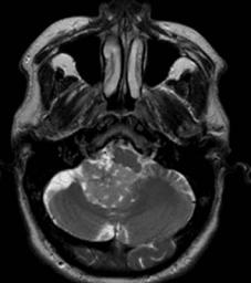 B
B  C
C
D
■ Clinical Presentation
A 52-year-old with increasing headaches.
54 |
RadCases.thieme.com |
|
RadCases Neuro Imaging |
|
|
■ Imaging Findings |
|
|
|
A |
B |
C |
|
D |
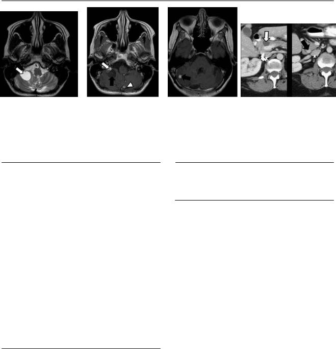
(A) Axial T2-weighted image (WI) of the brain shows a heterogeneous mass in the 4th ventricle (arrow) that extends to the right cerebellopontine angle (CPA) through the foramen of Luschka (arrowhead). (B) On axial di usion-WI, the mass (arrow) shows no restriction to di usion. (C) Coronal T1WI with contrast demonstrates avid enhancement of the 4th ventricle mass and dilatation of the lateral ventricles (arrows). (D) Sagittal T1WIs without and with contrast show a mass with intensity similar to that of the white matter that avidly enhances with gadolinium (arrows).
■ Di erential Diagnosis
•Choroid plexus papilloma: These are rare neuroectodermal tumors that account for fewer than 1% of intracranial neoplasms. They are more common in the 4th ventricle in adults, and more common in the atria in children. They are small, cauliflower-like masses with peripheral lobula-
tions. From the 4th ventricle, they may extend through the foramen of Luschka to the subarachnoid space. They have marked enhancement and are associated with hydrocephalus.
•Medulloblastoma: These are the most common posterior fossa tumors and the second most frequent brain tumors in children (after astrocytomas). They are rare in young adults. They are highly malignant neuroepithelial tumors. The 4th ventricle is the most frequent location (95%). Hyperattenuation on CT is typical. Medulloblastomas arise from the roof of the 4th ventricle and almost always enhance. They rarely extend to the basal cisterns.
•Ependymoma: These are midline cerebellar enhancing masses. Calcifications are frequent. Ependymomas are hypoattenuated on CT. They arise from the floor of the 4th ventricle and typically extend through the foramen of Luschka into the subarachnoid space. They do not have a lobulated appearance.
■ Other Imaging Findings
•CT:
•High attenuation with calcifications in 25% of cases
•Heterogeneous enhancement
•Magnetic resonance imaging:
•Hypointense on T1 and hyperintense on T2
•Bright on fluid-attenuated inversion recovery imaging
•Avid enhancement
¸Pearls & ˚ Pitfalls
¸Seeding can occur with both choroid plexus papillomas
and choroid plexus carcinomas.
¸ It is necessary to screen the whole neuraxis.
˚The lobulated peripheral appearance is the best way to di erentiate choroid plexus papillomas from ependymomas.
■ Essential Facts
•Choroid plexus papillomas are located where choroid plexus is present.
•The lateral ventricle is the most common site (50%), followed by the 4th ventricle (40%).
•Imaging cannot di erentiate choroid plexus papilloma from its histologically aggressive counterpart: choroid plexus carcinoma.
55
Case 28
A B C
■ Clinical Presentation
A 54-year-old woman with a recent history of vertigo.
Further Work-up
D
56 |
RadCases.thieme.com |
|
RadCases Neuro Imaging |
|
|
■ Imaging Findings |
|
|
|
A |
B |
C |
|
D |
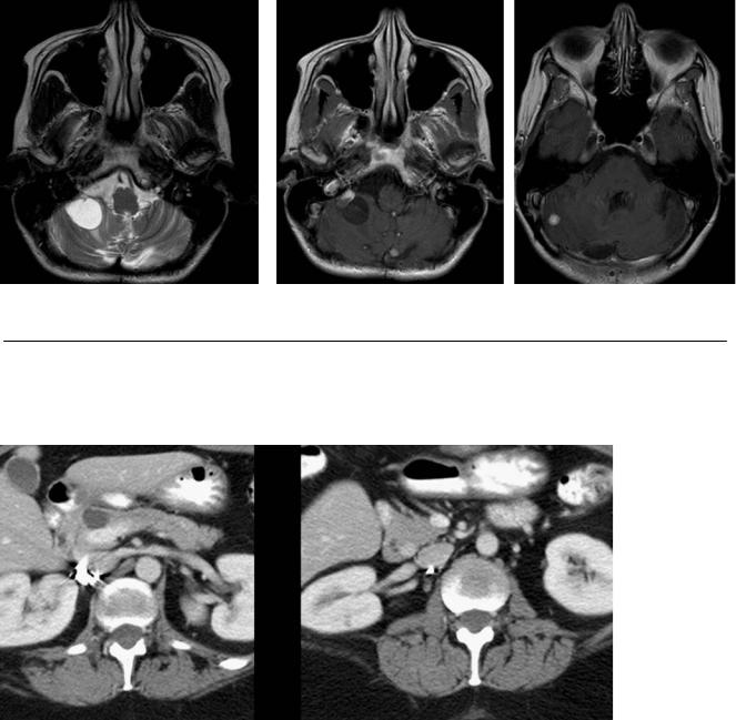
(A) Axial T2-weighted image (WI) of the posterior fossa shows a cystic lesion on the right cerebellar hemisphere (arrow). There is surrounding vasogenic edema. (B) On axial T1WI with contrast, the right cerebellar lesion shows a mural nodule (white arrow); the cystic component (black arrow) does not enhance. A second enhancing lesion (arrowhead) is noted near the midline. (C) Axial T1WI of the posterior fossa with contrast shows another enhancing lesion on the left cerebellar hemisphere (arrow). (D) Axial computed tomography (CT) scans of the abdomen with contrast show a simple pancreatic cyst (white arrow). A small enhancing lesion on the pancreatic head is also seen (black arrow).
■ Di erential Diagnosis
•Cerebellar hemangioblastoma: These are the most common primary posterior fossa tumors in adults. They appear as an intra-axial cystic mass with a well-defined mural nodule (75%). Of all hemangioblastomas, 4 to 20% are associated with Von Hippel-Lindau (VHL) disease. Eighty percent occur in the cerebellar hemisphere. They displace the 4th ventricle. The mural nodule shows avid enhancement. There are no calcifications.
•Pilocytic astrocytoma: These usually occur in children younger than 20 years of age. They appear as a cystic posterior fossa mass with an enhancing nodule. Twenty percent of them calcify. They are located in the cerebellar hemisphere and compress the 4th ventricle.
•Medulloblastoma: These are the most common posterior fossa tumors in children. They are highly malignant. They arise from the roof of the 4th ventricle, with an intraventricular location. They appear as a solid mass with cystic degeneration and would rarely present as a cyst with a mural nodule.
■ Other Imaging Findings
• CT: the nodule is isodense to brain.
¸Pearls & ˚ Pitfalls
¸Look for other lesions associated with VHL disease, such
as other hemangioblastomas, similar lesions in the retina, and renal cell carcinoma.
¸The most frequent posterior fossa mass in adults is metastasis.
¸VHL disease in percentages:
•Multiple brain hemangioblastomas: 45 to 70%
•Retinal hemangioblastomas: 50%
•Renal (70%) and pancreatic (50%) simple cysts
•Renal cell carcinoma: 30%
•Pheochromocytoma: 12%
•Islet cell tumor of the pancreas: 10%
■ Essential Facts
•Cerebellar hemangioblastomas are rare outside the posterior fossa.
•Up to 40% can be solid tumors.
•The nodule enhances intensely.
•The wall of the cyst rarely enhances.
•The nodule and cyst show high intensity on fluid-attenu- ated inversion recovery sequences.
57
Case 29
A B
C 
 D
D
■ Clinical Presentation
A 12-year-old boy with progressive headaches.