

Книги по МРТ КТ на английском языке / Neuro Imaging Redcases
.pdf98 |
RadCases.thieme.com |
RadCases Neuro Imaging |
■ Imaging Findings
A B C
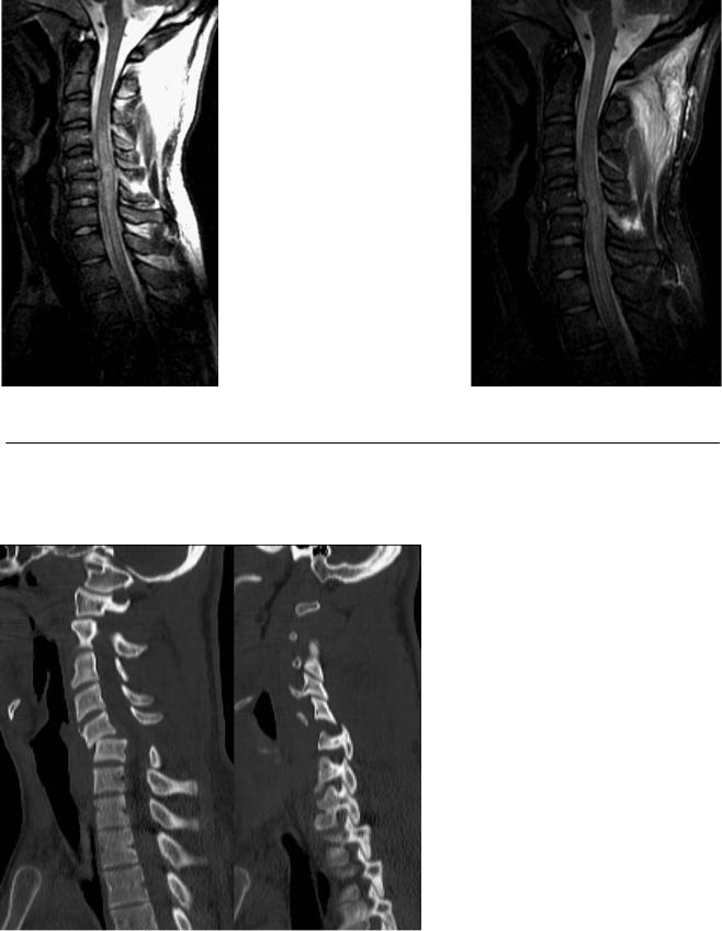
(A) Sagittal T2-weighted image (WI) shows subluxation and disk edema at C5-C6 with posterior osteophytes. Narrowing of the canal and edema of the cord (between arrowheads) are noted. (B) Sagittal T1WI shows subluxation and disk widening at C5-C6 (arrow). There are posterior osteophytes. (C) Note the central cord edema on the axial T2WI (arrow). No cord hemorrhage is demonstrated.
■ Di erential Diagnosis
•Cord contusion: Contusions tend to be severe in patients with degenerative changes. Magnetic resonance imaging (MRI) demonstrates increased cord signal and cord expansion.
•Wallerian degeneration of the cord: This develops in the subacute phase (about 1 month) after spinal cord injury. MRI reveals hyperintense signal on T2WIs in the white matter tracts above (dorsal columns) and below (lateral columns) the level of the injury.
•Cord infarction: This presents as the acute onset of myelopathy and is often heralded by sudden and severe back pain, which may radiate caudally. MRI shows a central cord lesion on the axial T2WIs. Causes include arteritis, embolism, aortic dissection, aortic surgery, decompression sickness, abdominal surgery, vertebral fracture, and disk herniation.
■Essential Facts
•Cord contusions are classified clinically as complete lesions (loss of motor and sensory function at the injured level and distally) or incomplete lesions (central cord syndrome, anterior cord syndrome, Brown-Séquard syndrome, conus medullaris syndrome, cauda equina syndrome).
•Trauma to the spinal cord involves either true cord injury or compression with secondary cord damage.
•Acute MRI findings:
•Transection
•Intramedullary hemorrhage that is usually associated with a clinically complete and irreversible spinal cord injury
•Contusion
•Edema
•Ligamentous injuries, muscular lesions, facet joint dislocations, and bone marrow edema
•Chronic MRI findings:
•Syringomyelia: cystic degeneration of the injured spinal cord at or near the site of trauma
•Progressive posttraumatic myelomalacic myelopathy, which may occur at 2 months and up to 30 years following injury
•Spinal cord atrophy
■ Other Imaging Findings
•Although plain radiography still has a role in spinal cord trauma, particularly cervical spinal cord trauma, it has largely been replaced by multislice computed tomography.
•The multislice coronal and sagittal reconstructions often reveal more than plain radiographs do, and fractures and joint dislocations can be detected accurately.
¸Pearls & ˚ Pitfalls
¸Spinal cord injury without radiographic abnormality
(SCIWORA) is well-known in the pediatric population.
¸It is a consequence of the inherent elasticity of the vertebral column in infants and young children, which makes the pediatric spine exceedingly vulnerable to deforming
forces.
˚In the chronic stage of cervical injuries, intramedullary cysts should be di erentiated from expected “static” myelomalacia, which has a less well-defined border and is not progressive.
99
Case 50
A
■ Clinical Presentation
A 7-month-old child with a history of vomiting. Retinal hemorrhages were found at the clinical examination.
Further Work-up
B 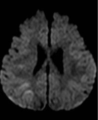 C
C 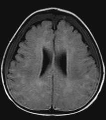
 D
D

100 RadCases.thieme.com |
RadCases Neuro Imaging |
|
|
■ Imaging Findings |
|
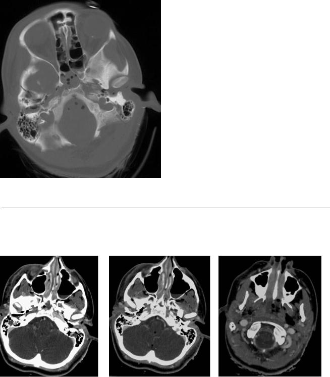
A 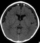 B
B 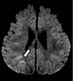 C
C 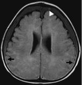
 D
D
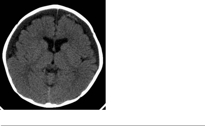
(A) Computed tomography (CT) of the brain demonstrates subdural collections in both frontal convexities, with mixed attenuation (asterisks). (B) Di usionweighted magnetic resonance imaging (MRI) shows restricted di usion in the right parietal subcortical white matter (arrow). (C) Axial fluid-attenuated inversion recovery image shows hematocrit e ect, with blood elements layering in the dependant portion of the collection (arrows) and focal areas of high signal within the subdural space anteriorly, consistent with acute blood (arrowhead). (D) Note the presence of retinal hemorrhages in the posterior pole of the left globe (arrow).
■ Di erential Diagnosis
•Nonaccidental head injury (NAHI): NAHI should be suspected in an infant or young child who presents with no history of injury; with a discrepancy between the explanation and nature of the lesions; with injuries of di erent ages or multiple injuries; with associated retinal hemorrhages; or with a change or inconsistency in the history, delayed medical care, repeated injuries, or evidence of overall poor care.
•Benign external hydrocephalus with spontaneous hemorrhage: Prominent subarachnoid spaces in infants are secondary to insu cient resorption of cerebrospinal fluid (CSF) as a result of immature arachnoid villi. These should be of the same density/intensity as CSF on CT and MRI. The condition predisposes infants to subdural hematoma (SDH), which may be spontaneous or associated with trauma of any type.
•Subdural hygroma: This is a CSF collection secondary to traumatic arachnoid tears. Attenuation and signal intensity are similar to those of CSF. Blood vessels crossing through the extra-axial collection will help di erentiate subdural hygromas from chronic SDHs.
■Essential Facts
•NAHI is the leading cause of morbidity and mortality in abused children.
•NAHI occurs in ~12% of physically abused children.
•It accounts for 80% of deaths from head injury in children younger than 2 years of age.
•Clinical presentation: the triad of subdural hemorrhage, retinal hemorrhage, and encephalopathy (i.e., di use axonal injury) occurs in the context of an inappropriate or inconsistent history and is commonly accompanied by other apparently inflicted injuries.
•Spectrum of injuries:
•Skull fracture (multiple or complex fractures, bilateral fractures, and fractures that cross suture lines or involve more than one bone are more suspicious)
•SDH
•Shearing injury
•Cerebral contusions
•Brain swelling
•Cerebral infarction (caused by shaking, strangulation, smothering)
•Chronic injuries (chronic SDH, communicating hydrocephalus, atrophy, or encephalomalacia)
■ Other Imaging Findings
•Imaging evaluation includes CT, a radiographic or radionuclide skeletal survey, MRI, and serial imaging in some cases.
•Nonhead injuries associated with non-accidental trauma include rib fracture, metaphyseal fracture, vertebral compression fracture, and small-bowel hematoma and laceration.
¸Pearls & ˚ Pitfalls
¸Skull fractures are not rare in household trauma but
rarely present with significant underlying brain injury (with the exception of epidural hematoma).
¸Soft-tissue swelling may be absent, even in acute skull fracture; thus, a lack of swelling should not be construed
as evidence that the fracture is subacute.
˚Retinal hemorrhages have been reported with accidental trauma, resuscitation, increased intracranial pressure, increased venous pressure, subarachnoid hemorrhage, sepsis, coagulopathy, certain metabolic disorders, systemic hypertension, and other conditions, and are therefore not specific for NAHI.
101
Case 51
A
■ Clinical Presentation
A 36-year-old woman injured in a motor vehicle collision.
Further Work-up
B C D

102 RadCases.thieme.com |
RadCases Neuro Imaging |
|
|
■ Imaging Findings |
|
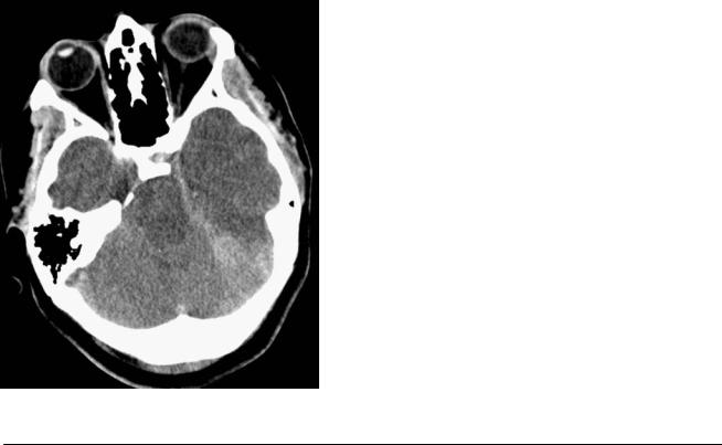
A B C D
(A) Computed tomography (CT) of the brain with bone windows shows a complex fracture of the skull base involving the sphenoid and temporal bones (black arrows) and compromise of the anterolateral wall of the right carotid canal (white arrow). (B) Axial image from a CT angiogram (CTA) of the brain shows absent opacification of the petrous right internal carotid artery (ICA) secondary to traumatic vascular injury (arrow). (C) Axial image from a CTA of the brain shows absent opacification of the petrous right ICA secondary to traumatic vascular injury (arrow). Pneumocephalus and neck emphysema are also noted. (D) Axial CTA at the level of C1 shows absent flow in the right ICA secondary to traumatic vascular injury (arrow). Pneumocephalus and neck emphysema are also noted.
■ Di erential Diagnosis
•Skull base fracture with arterial transection: Direct injury to the vessel is caused by the displaced bone fragments. Symptoms of stroke may be masked by the brain injuries.
•Extracranial carotid dissection: This is a posttraumatic intimal tear that commonly a ects the distal ICA. Some tears probably heal spontaneously, but others lead to dissection for a variable distance, with or without pseudoaneurysm formation, and some thrombose.
•Embolic occlusion of the ICA: The appearance of the vessels would be similar, but this diagnosis is less likely in the setting of skull base fractures.
■Essential Facts
•Because of the complex anatomic relationships of the skull base, fractures may damage critical neighboring structures, including cranial nerves, the ICA, and the cavernous sinus.
•Complications:
•Vascular injuries: carotid-cavernous fistula, pseudoaneurysm, dissection, sinus thrombosis
•Cerebrospinal fluid (CSF) fistula and infection
•Cranial nerve injury
•Central nervous system injury: contusions, extra-axial hemorrhages, brainstem infarctions, and posterior pituitary dysfunction in the form of diabetes insipidus
■ Other Imaging Findings
•CTA is used to identify potentially treatable vascular injuries.
•For the localization of CSF fistulas, high-resolution CT with or without cisternography, heavily T2-weighted coronal magnetic resonance (MR) imaging, gadoliniumenhanced MR cisternography, or intrathecal radioisotope studies have been proposed.
¸Pearls & ˚ Pitfalls
¸Frontal sinus fractures may lead to cosmetic deformity,
CSF fistula, chronic meningitis, and mucocele. Sinus cranialization may be necessary to prevent CSF leak and meningitis.
¸Temporal bone fractures may cause damage of the facial nerve, labyrinthine fistula, conductive or sensorineural
hearing loss, CSF leak, or cephalocele.
˚Air in the cavernous sinuses may be the result of embolism from intravenous access and does not always indicate basilar skull fracture.
103
Case 52
A  B
B
■ Clinical Presentation
A 26-year-old who was involved in a car accident and is now comatose.
Further Work-up
C 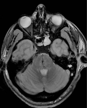
 D
D
104 RadCases.thieme.com |
RadCases Neuro Imaging |
|
|
■ Imaging Findings |
|
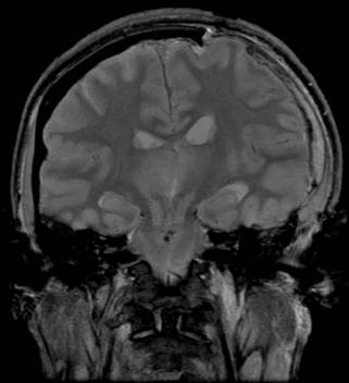
A B C D
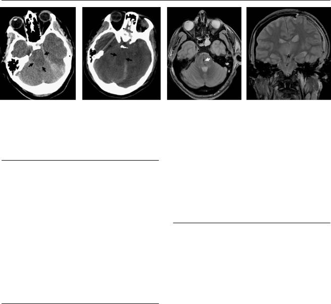
(A) Axial computed tomography (CT) of the brain shows signs of cerebral edema, with low attenuation of the brain and loss of distinction between gray and white matter. E acement of the basal cisterns (arrows) and deformity of the midbrain are evident. (B) Axial CT demonstrates e acement of the perimesencephalic cistern (arrows) and deformity of the midbrain, with reduced transverse diameter and elongation in the anteroposterior direction. Note a subdural hematoma over the left tentorium. (C) Axial gradient-echo T2*-weighted image (WI) reveals petechial hemorrhages in the central pons (arrow).
(D) Coronal T2WI shows downward displacement of the thalami and midbrain (asterisk). The pontine hemorrhages are again demonstrated.
■ Di erential Diagnosis
•Duret hemorrhages: Duret hemorrhages are brainstem hemorrhages that develop as a consequence of downward trans-tentorial herniation. Magnetic resonance imaging (MRI) findings of descending trans-tentorial herniation include lower position of the brainstem, e acement of the basal cisterns, flattening of the pons against the clivus, and inferoposterior displacement of the quadrigeminal plate.
•Interpeduncular subarachnoid hemorrhage: This is usually an early occurrence in trauma patients and can be mistaken for parenchymal hemorrhage.
•Shearing injury: This is characterized by multiple small hemorrhages in the deep white matter, including the corpus callosum and brainstem. In the pons, it may appear identical to and coexist with Duret hemorrhages.
■Essential Facts
•Descending trans-tentorial herniation consists of caudal descent of brain tissue through the tentorial incisura and occurs mainly in response to mass e ect in the frontal, parietal, and occipital lobes.
•The lower brainstem is made less mobile by the upper cervical dentate ligaments. Longitudinal compression of the upper brainstem against the unyielding medulla leads to buckling of the brainstem and further compression of the midbrain tegmentum.
•Stretching and shearing of the perforating branches of the basilar artery occur, resulting in ischemia and hemorrhage within the brainstem, called Duret hemorrhage.
•The clinical consequences of progressive central herniation are oculomotor paresis, progressive alteration of consciousness, decerebrate rigidity, coma, and death.
¸Pearls & ˚ Pitfalls
¸Downward descent of the brain may be secondary to
intracranial hypotension, which is caused by either an iatrogenic or a spontaneous cerebrospinal fluid leak.
¸There may be pachymeningeal venous engorgement and edema, downward descent of the brain, and descending
central trans-tentorial or tonsillar herniation.
˚Di use cerebral edema results in symmetric abnormalities on CT and MRI that may be overlooked.
˚The distortion of the brainstem caused by downward trans-tentorial herniation may mimic a Chiari type II malformation.
105
Case 53
A B  C
C
■ Clinical Presentation
A 21-year-old woman who fell on her head during gymnastics practice.
Further Work-up
D
106 RadCases.thieme.com |
RadCases Neuro Imaging |
|
|
■ Imaging Findings |
|
A B C 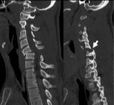 D
D
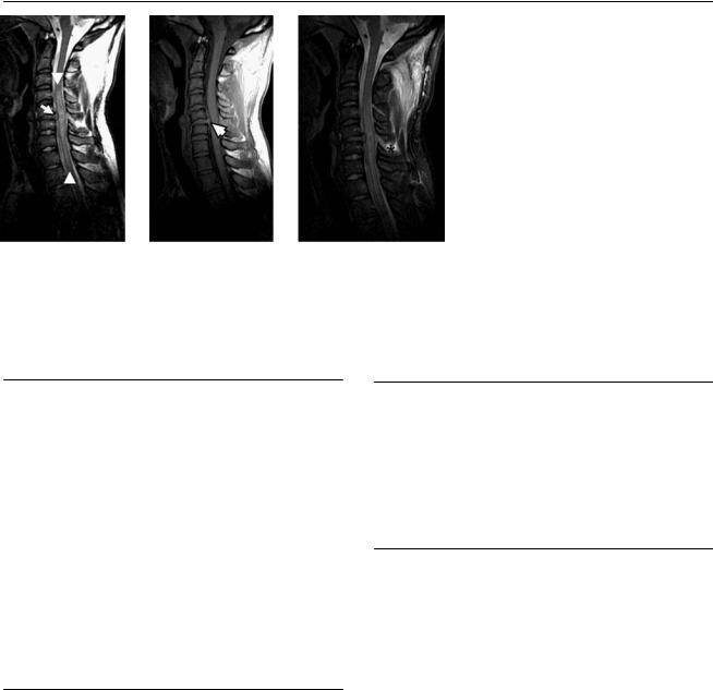
(A) Sagittal T2-weighted image (WI) demonstrates a disk protrusion (arrow) at C5-C6 impinging on the spinal cord, which demonstrates significant edema (between arrowheads). (B) Sagittal T1WI shows widening of the intervertebral space posteriorly (arrowhead). (C) Fat-suppressed sagittal T2WI shows disruption of the C5-C6 interspinous ligament with widening and edema (asterisk). There is also edema of the dorsal paraspinal soft tissues in the upper cervical region. (D) Sagittal and parasagittal reformatted images of a computed tomography (CT) scan obtained 12 hours earlier, before traction, show anterior translation of C5 on C6 secondary to facet dislocation (arrow), which was bilateral.
■ Di erential Diagnosis
•Traumatic disk herniation and cord contusion: Traumatic injuries can be suspected when the disk has a high signal on T2WIs. Also, concomitant traumatic vertebral body injuries and/or ligamentous injuries may be suggestive of traumatic disk lesions.
•Cord contusion secondary to chronic degenerative disk disease: In some cases, it may not be possible to determine if the disk herniation was present before the injury or is posttraumatic in nature. Patients with degenerative spinal stenosis are at higher risk for cord contusion.
•Epidural hematoma: The signal intensity of an acute epidural hematoma may be similar to that of the herniated disk material. The shape of the herniated disk and its connection with the native disk may help to di erentiate them.
■ Essential Facts
•Traumatic disk herniation is commonly caused by distraction and shearing in sudden extension, but it also occurs in flexion injuries.
•Cervical disk herniations are seen in 54 to 80% of patients who have joint facet dislocation.
•High signal intensity seen horizontally through a disk (involving the nucleus and annulus on a T2WI) is highly suggestive of disk and annulus disruption.
■ Other Imaging Findings
•CT will assess the bone and the articular alignment but is insensitive for the evaluation of soft-tissue injuries in the spine.
•Magnetic resonance imaging is the modality of choice for the evaluation of cord injuries and disruption of the disks and ligaments.
¸Pearls & ˚ Pitfalls
¸The integrity of the diskoligamental complex (DLC) is
thought to be directly proportional to spinal stability after cervical spine trauma.
¸The components of the DLC are the intervertebral disk, anterior and posterior longitudinal ligaments, ligamenta flava, interspinous and supraspinous ligaments, and facet
capsules.
˚Traumatic and nontraumatic disk herniations can be similar in appearance.
107
Case 54
A
■ Clinical Presentation
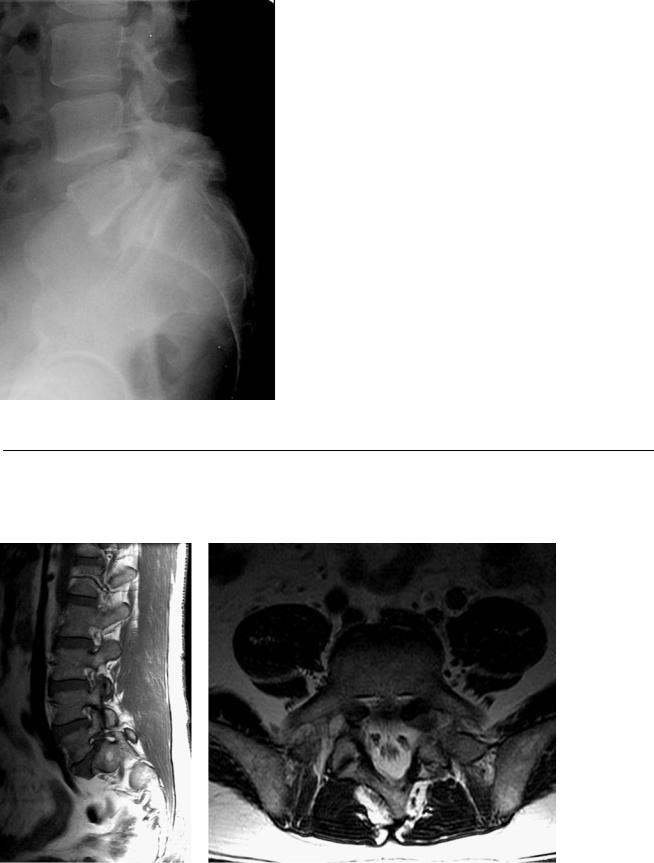
A 58-year-old man with left lumbar radiculopathy.
Further Work-up
B C