

Книги по МРТ КТ на английском языке / Neuro Imaging Redcases
.pdf158 RadCases.thieme.com |
RadCases Neuro Imaging |
|
|
■ Imaging Findings |
|
A B 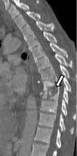 C
C 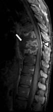 D
D
(A) Lateral radiograph of the thoracic spine shows irregular end plates and narrowed disk spaces of the midthoracic vertebrae (arrows). (B) Sagittal reformatted computed tomography (CT) of the thoracic spine without contrast shows hypodense destructive changes of a vertebra (arrow). There is a prevertebral collection (asterisk). (C) Sagittal T1and T2-weighted images (WIs) of the thoracic spine show low T1 and high T2 signal of three adjacent vertebral bodies (asterisks). There is an anterior well-defined fluid collection with a thin capsule adjacent to these segments (arrows). (D) Fat-saturated sagittal T1WI of the thoracic spine with contrast shows enhancement of the three thoracic segments and rim enhancement of the fluid collection (arrow). There is a small area of posterior epidural enhancement (arrowhead).
■ Di erential Diagnosis
•Tuberculous spondylitis: Also known as Pott disease, tuberculous spondylitis has a predilection for the upper lumbar and lower thoracic spine. More than one vertebral body is usually involved. The disk is compromised late in the disease and may appear collapsed. Anterior wedging leads to gibbous deformity. There is usually early paravertebral abscess formation with a thin wall. Subligamentous spread of the infection a ects three or more levels. Softtissue calcifications may be present.
•Pyogenic spondylodiskitis: This is bacterial infection of the disk and vertebral body. It is more frequent in the lower lumbar or cervical spine. Pyogenic spondylodiskitis has a thick, irregular abscess wall. There is involvement of the intervertebral disk and the two adjacent vertebral bodies. Erosion of the end plates is typically seen.
•Spinal metastasis: Vertebral metastases involve multiple vertebral bodies, including the posterior elements. The disks are not a ected. Contrast enhancement is variable.
■Essential Facts
•The upper lumbar spine and lower thoracic spine are the most frequent sites for skeletal involvement of tuberculosis (TB).
•The disease starts in the anterior body adjacent to the end plate and progresses toward the posterior body.
•TB initially shows hematogenous seeding to the anterior vertebral body and then spreads to distant vertebral bodies through the anterior longitudinal ligament.
■ Other Imaging Findings
•CT shows destruction of the end plates and soft-tissue calcification.
•Magnetic resonance imaging shows low T1 signal intensity in the a ected body and increased T2 signal in the a ected body, abscess, and disk. There is enhancement of the vertebral body and abscess.
¸Pearls & ˚ Pitfalls
¸It is essential to evaluate for cord compression resulting
from epidural extension of the abscess.
¸ In a patient with vertebral body lesions and soft-tissue abscess, look for pulmonary findings of TB.
˚Systemic coccidioidomycosis can present with miliary disease and vertebral osteomyelitis, mimicking TB.

159
Case 80
A
■ Clinical Presentation
A 36-year-old woman with a history of heroin use presenting with di use headache, fever, and seizures.
Further Work-up
B
C 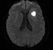 D
D
160 |
RadCases.thieme.com |
|
RadCases Neuro Imaging |
|
|
■ Imaging Findings |
|
|
|
A |
B |
C |
|
D |
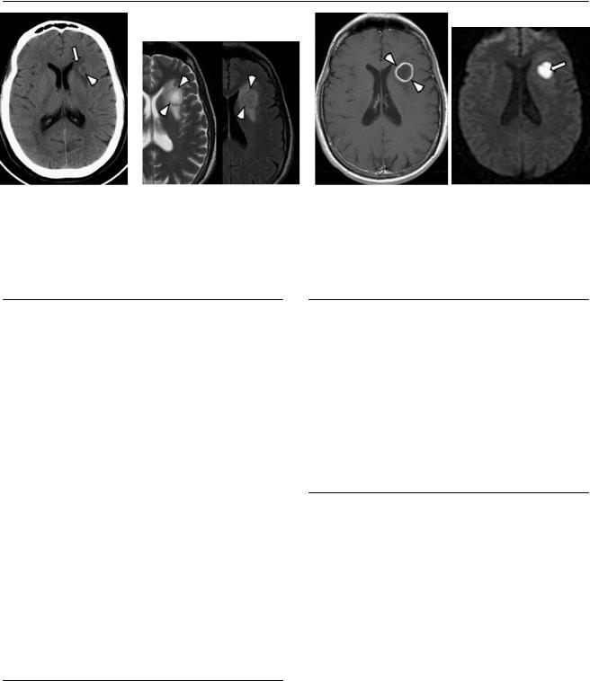
(A) Axial computed tomography (CT) without contrast shows a ring isodense to the gray matter (arrowhead) in the left frontal lobe. The lesion, located at the gray–white matter junction, has a hypodense center and is surrounded by vasogenic edema (arrow). (B) Axial T2-weighted image (WI) and fluid-attenu- ated inversion recovery image show the left frontal lesion with a rim of low intensity (arrowheads) and peripheral vasogenic edema. (C) Axial T1WI with contrast shows a round, well-defined, ring-enhancing lesion in the left frontal lobe (arrowheads). The inner and outer margins are smooth. (D) Di usion-WI shows a “lightbulb” sign of the left frontal lesion (arrow).
■ Di erential Diagnosis
•Cerebral abscess: This is the organized, late phase of a brain infection. A bacterial etiology is the most common. The gray–white matter junction in the territory of the middle cerebral artery is the most frequent location because of hematogenous spread. A ring-enhancing lesion is described as having smooth inner and outer margins. There is surrounding edema. The lesion presents as high signal on di usion-WIs with a dark apparent di usion coe cient (ADC) map.
•Metastasis: A hematogenous metastasis involves the cortical or subcortical white matter and occurs more frequently in the territory of the middle cerebral artery. The lesion demonstrates surrounding vasogenic edema, no restricted di usion, and nodular or ring enhancement. Hemorrhage may be present. A known primary tumor increases the likelihood of this diagnosis.
•Glioblastoma multiforme: This is the most common of the primary intracranial neoplasms. The usual presentation is thick, irregular ring enhancement with a serrated inner margin. There is a necrotic center with surrounding
edema. The corpus callosum can be a ected. Glioblastoma multiforme can have increased signal on di usion-WIs but usually has no restriction on the ADC map.
■Essential Facts
•Bacterial infection of the brain evolves from cerebritis to capsule formation.
•Abscesses form when a capsule of granulation tissue, reinforced by a collagen wall, confines the infected area of brain and pus.
•Pathogens may enter the brain by hematogenous spread or direct extension of an infection (from the sinuses, mastoid processes, or ears). They may also be introduced following penetrating injury or surgery.
■ Other Imaging Findings
•CT: The capsule appears as an isodense rim around a hypodense center with surrounding hypodense edema. The capsule enhances with contrast.
•Magnetic resonance imaging:
•T2WIs: the rim of an abscess is hypointense; this finding is the first to disappear after treatment.
•Di usion-WIs: there is restricted di usion with restriction on ADC maps.
•Spectroscopy shows increased levels of acetate, alanine and other amino acids, and lactate.
¸Pearls & ˚ Pitfalls
¸In the early phases of transition between cerebritis and
an organized abscess, the margins of the capsule can be poorly defined.
¸Highly cellular tumors with areas of necrosis may show restricted di usion that a ects the solid (enhancing) component of the tumor, whereas in abscesses, the re-
striction involves the pus-filled (nonenhancing) cavity.
˚Extension to the corpus callosum is rare in abscesses.
˚An abscess can rupture through the ependymal lining of the ventricle and cause ventriculitis.
˚Always look for additional lesions.
161
Case 81
A
■ Clinical Presentation
A 35-year-old drug addict with bacterial endocarditis presenting with the sudden onset of severe, worsening headache.
Further Work-up
B 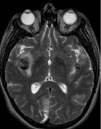 C
C 
 D
D

162 RadCases.thieme.com |
RadCases Neuro Imaging |
|
|
■ Imaging Findings |
|
A  B
B 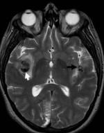 C
C 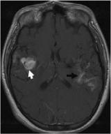
 D
D
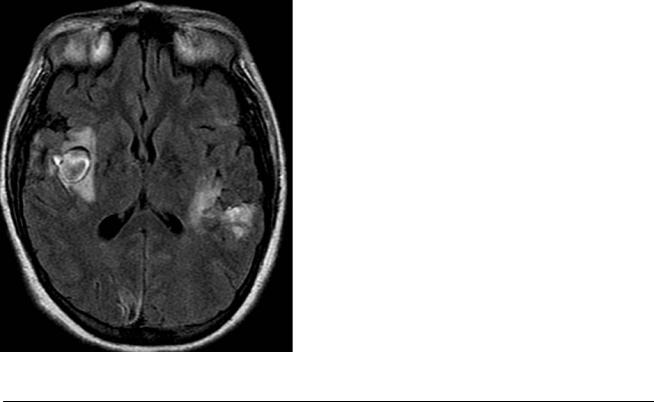
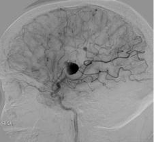
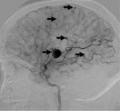
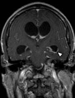
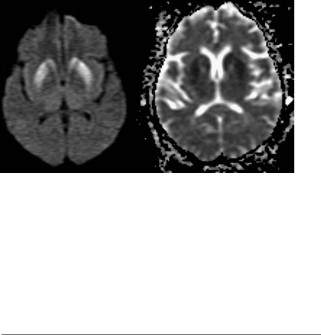
(A) Axial fluid-attenuated inversion recovery image shows a round lesion in the deep sylvian fissure on the right (black arrow) surrounded by vasogenic edema. The lesion has a well-defined capsule of low intensity. Additional hyperintensities are seen in the perisylvian cortex on the left and in the right occipital lobe (white arrows). (B) Axial T2-weighted image (WI) shows a hypointense round lesion in the deep sylvian fissure on the right (white arrow) and a smaller contralateral lesion (black arrow). (C) Axial T1WI with contrast shows di use enhancement of the right sylvian lesion (white arrow). There is cortical enhancement in the cortex surrounding the left sylvian fissure (black arrow) and in the left occipital lobe. (D) Lateral digital subtraction angiography (DSA) of the head in a late arterial phase of the right internal carotid artery injection shows multiple aneurysms throughout the middle cerebral artery (MCA); most are in the distal branches (arrows).
■ Di erential Diagnosis
•Mycotic aneurysm: Mycotic aneurysms account for 5% of intracranial aneurysms and are of infectious etiology. Multiple aneurysms are usually present bilaterally, involving the distal segments and unrelated to the branching sites. They can be fusiform. Rapid morphologic changes over time are not uncommon. Mycotic aneurysms are associated with nearby arterial occlusion or stenosis.
•Saccular aneurysm: Saccular aneurysms have a “berry” shape. They are located at branching points in the vessels of the circle of Willis. They are unusual in peripheral branches. They can be multiple and are more frequent in females. Ninety percent occur in the anterior circulation.
•Noninfectious fusiform aneurysm: These are rare and tend to be large. They form spontaneously and are not related to the bifurcation of vessels. They are not multiple or bilateral.
■Essential Facts
•Vessel wall necrosis after bacterial or fungal embolization results in the formation of multiple intracranial infectious aneurysms.
•Bacterial endocarditis is the most common cause.
•Mycotic aneurysms involve distal branches, typically in the distribution of the MCA.
•They can bleed, and subarachnoid hemorrhage or parenchymal hematoma may develop.
•They can regress after the treatment of endocarditis.
•Endovascular treatment has been proposed for large mycotic aneurysms because of the likelihood of rupture.
■ Other Imaging Findings
•Computed tomography (CT) is the initial study of choice if intracranial bleeding is suspected.
•Magnetic resonance (MR) imaging is good for detecting parenchymal hemorrhage and ischemia; however, it can miss small lesions.
•DSA is the imaging gold standard for the detection of the multiple infectious aneurysms; MR angiography and CT angiography are less sensitive.
¸Pearls & ˚ Pitfalls
¸The mortality rates are 32% for bacterial and 90% for
fungal infectious aneurysms.
¸ The gold standard for the diagnosis is histologic confirmation.
˚Mycotic aneurysms can be round and di cult to differentiate from “berry” aneurysms based solely on their morphology.
˚A history of endocarditis and a positive blood culture are part of the clinical criteria to confirm the diagnosis.

163
Case 82
A 
 B
B
C D
■ Clinical Presentation
A 38-year-old man with severe headache and fever.

164 RadCases.thieme.com |
RadCases Neuro Imaging |
|
|
■ Imaging Findings |
|
A 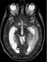 B
B  C
C 
 D
D
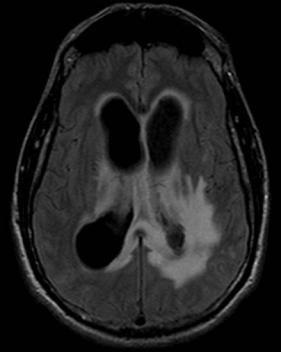
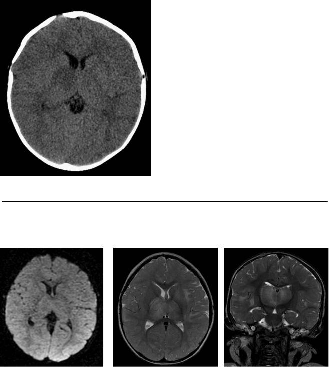
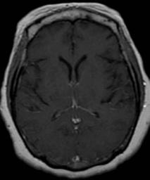
(A) Axial T2-weighted image (WI) shows dilated lateral ventricles with a septum in the right occipital horn (arrow). (B) Axial fluid-attenuated inversion recovery (FLAIR) image shows hyperintense signal around the ventricles (arrowheads) that is more prominent around the right atrium (arrow). (C) Axial T1WI of the brain with contrast shows enhancement at the ependymal lining (arrows). There is an enhancing nodule (arrowhead) in the left thalamic region. (D) Coronal T1WI with contrast shows enhancement of the ventricular wall (arrowhead). Note the dilatation of the lateral ventricles.
■ Di erential Diagnosis
•Ventriculitis: Ventriculitis is inflammation of the ependymal lining. Pyogenic infection is the most common cause. If there are pus collections within the ventricles, imaging studies will show bacterial debris, hydrocephalus, symmetric enhancement of the ventricular walls, and restricted di usion. Seventy-five percent of cases are associated with meningitis.
•Ventricular extension of tumor: Primary tumors (usually glioblastoma multiforme) extending to the ventricles will cause T2 signal abnormality in the periventricular region and ependymal enhancement. The enhancement is typically asymmetric. No ventricular debris should be present.
•Lymphoma: Ventricular extension of an intracranial lymphoma is one of the characteristics that allows clinicians to di erentiate lymphoma from toxoplasmosis in patients infected with human immunodeficiency virus. Imaging will show nodular ependymal enhancement, basal ganglia lesions, and ring-enhancing subcortical lesions. There may be meningeal thickening of the basal cisterns. Isolated ventricular disease without parenchymal disease is extremely rare.
■ Other Imaging Findings
•Computed tomography shows ventriculomegaly and a fluid level in the ventricle.
•Magnetic resonance imaging: Debris may be hyperintense on T1WIs, with periventricular low signal on T1 and increased signal on T2/FLAIR sequences. The debris and parenchymal abscesses show restricted di usion. Enhancement of ependymal, meningeal, and parenchymal lesions can be seen.
¸Pearls & ˚ Pitfalls
¸The most common causes of ventriculitis include ven-
tricular catheter infection and ruptured abscess.
¸ FLAIR images are superior to postcontrast T1WIs for detecting ventricular wall abnormalities.
˚In trauma patients, ventricular hemorrhage can show increased FLAIR signal in the ependymal lining with obstructive hydrocephalus.
■ Essential Facts
•Ventriculitis has a high mortality rate (> 50%).
•Ventricular debris is the most common imaging finding in pyogenic ventriculitis.
•Hydrocephalus develops very rapidly.
165
Case 83
A
■ Clinical Presentation
A 1-year-old with altered mental status and seizures following a viral illness.
Further Work-up
B C D

166 RadCases.thieme.com |
RadCases Neuro Imaging |
|
|
■ Imaging Findings |
|
A 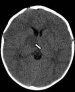 B
B 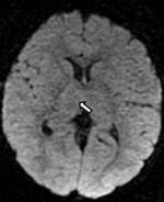 C
C 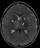
 D
D
(A) Axial computed tomography (CT) shows low attenuation in the right thalamus and posterior limb of the internal capsule (arrow). (B) Axial di usionweighted image (WI) fails to show restriction in the right thalamus (arrow). (C) Axial T2WI demonstrates increased signal intensity in the thalami, internal capsules (arrows), lenticular nucleus on the left (asterisk), and subcortical white matter bilaterally. (D) Coronal T2WI shows enlargement and increased signal in the thalami. There is also increased signal in the subcortical white matter bilaterally (arrows).
■ Di erential Diagnosis
•Acute disseminated encephalomyelitis (ADEM): ADEM is a monophasic demyelinating disease of the central nervous system that presents with numerous hyperintense white and gray matter lesions on T2WIs. It has an asymmetric distribution and is poorly marginated. Contrast enhancement is infrequent.
•Multiple sclerosis (MS): MS presents with multiple, wellmarginated lesions that have a predilection for the periventricular regions and corpus callosum. When the cord is involved, partial myelopathy is present.
•Viral encephalitis: This causes the acute onset of a febrile illness and neurologic signs (seizures, alteration of consciousness, meningeal irritation). On magnetic resonance imaging (MRI), there is a variable appearance on di usionWIs and T2WIs. Di erent viruses prefer specific areas: herpes encephalitis favors the medial temporal lobes, Japanese encephalitis is found in the gray matter, and enterovirus 71 favors the rhombencephalon. CT and MRI may show complications such as hemorrhage, hydrocephalus, and herniation.
■Essential Facts
•ADEM presents as multiple gray and white matter lesions of the brain and spinal cord.
•A prior infectious episode or vaccination triggers the inflammatory response.
•Young and adolescent children are most a ected.
•The severity of the clinical symptoms is variable; these include seizures, coma, behavioral disturbances, headaches, vomiting, fever, drowsiness, and/or focal neurologic signs.
•Selective involvement of the cerebral cortex may be the only manifestation of ADEM.
•Infratentorial lesions occur in more than 50% of cases (brainstem, middle cerebellar peduncles, and cerebellar white matter).
•ADEM has a favorable prognosis.
■ Other Imaging Findings
•CT findings are frequently normal.
•MRI: T2WIs and fluid-attenuated inversion recovery images show multiple, asymmetrically distributed, poorly marginated hyperintense areas involving the white matter and the deep gray matter nuclei (i.e., thalamus and basal ganglia). Contrast enhancement is infrequent. When present, it typically involves the vast majority of lesions simultaneously.
¸Pearls & ˚ Pitfalls
¸Restricted di usion does not imply irreversible damage
in ADEM.
¸ Approximately 30% of patients who have ADEM also have lesions in their spinal cord.
˚The neuroradiologic findings of ADEM are not specific and typically do not allow ADEM to be di erentiated from MS or encephalitis.
167
Case 84
A
B 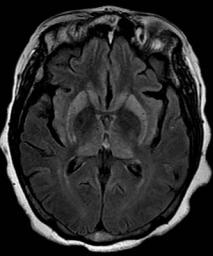 C
C 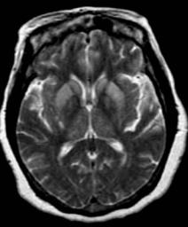
 D
D
■ Clinical Presentation
A 45-year-old woman with rapidly progressive dementia.