

Книги по МРТ КТ на английском языке / Neuro Imaging Redcases
.pdf38 |
RadCases.thieme.com |
RadCases Neuro Imaging |
|
■ Imaging Findings |
|
A B C D
(A) Axial computed tomography (CT) scan of the lumbar spine demonstrates a bony spur in the spinal canal (arrow). (B) Axial T2-weighted image (WI) of the lumbar spine shows two hemicords (arrows) inside the thecal sac. (C) Axial T2-weighted magnetic resonance imaging (MRI) of the lumbar spine demonstrates the septum (arrow) dividing the spinal cord. (D) Sagittal T2WI of the lumbar spine shows a tethered cord. The cystlike lesion (arrow) is where the cord splits (arrowhead).
■ Di erential Diagnosis
•Diastematomyelia (split cord malformation): Diastematomyelia is a focal or complete split of the spinal cord. It is the consequence of a split notochord. It is associated with congenital scoliosis (80% of cases), tethered cord syndrome (75% of cases), and spinal bony anomalies (50% of cases). Intersegmental laminar fusion is pathognomonic.
•Meningocele manqué: Meningocele manqué is characterized by dysraphic dorsal tethered bands (atretic neural tissue) adherent to dura. The following may be seen: diastematomyelia, filum lipoma, dermoid cysts, and neuroenteric cysts. These are di cult to see on imaging studies.
•Tethered cord syndrome: Tethered cord syndrome is characterized by a low-lying conus medullaris (below L2; some authors say below L3). Traction of the spinal cord is secondary to closed spinal dysraphism, diastematomyelia, spinal lipoma, tight filum terminale, caudal regression syndrome, scar tissue after meningocele repair, thickened filum terminale, and hydromyelia, and it is associated with progressive neurologic deterioration.
■Essential Facts
•There are two types of diastematomyelia:
•Type 1 (with septum): 25%
◦A bony septum results in separate dural tubes, each containing a hemicord.
◦The usual location is the thoracic or lumbar spine.
◦Scoliosis is present.
◦Hydromyelia is common.
•Type 2 (without septum): 75%
◦There is no bony spur; a fibrous septum may be present.
◦Only one thecal sac contains both hemicords.
■ Other Imaging Findings
•Plain radiographs are limited but can detect scoliosis and vertebral anomalies.
•CT can demonstrate the bony spur and hemivertebrae.
•Myelo-CT is a helpful tool to demonstrate the dural sac and hemicords.
•MRI:
•Axial and coronal views are best to demonstrate the split cord. This is di cult to see on sagittal images.
•T2 is best to demonstrate hydrosyringomyelia.
•It is limited in the evaluation of a bony spur.
•It can demonstrate a dermal sinus.
¸Pearls & ˚ Pitfalls
¸Cutaneous birthmarks (hemangiomas, dyschromic
patches, and hairy tufts) overlie the defect in 50% of cases.
¸Females are more often a ected than males.
¸Intersegmental laminar fusion is pathognomonic.
¸Image the spine of a patient whose skin marks are com-
patible with the cutaneous stigmata of diastematomyelia.
˚Diastematomyelia is very hard to see in sagittal views.
˚Evaluation for a bony spur is limited in MRI.
˚Meningocele manqué is hardly ever diagnosed with imaging (it is diagnosed surgically).

39
Case 20
A
■ Clinical Presentation
A patient with urinary incontinence and a subcutaneous mass in the sacral region.
Further Work-up
B 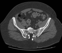 C
C 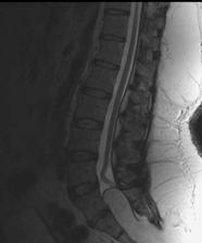 D
D
40 |
RadCases.thieme.com |
|
RadCases Neuro Imaging |
|
|
■ Imaging Findings |
|
|
|
A |
B |
C |
|
D |
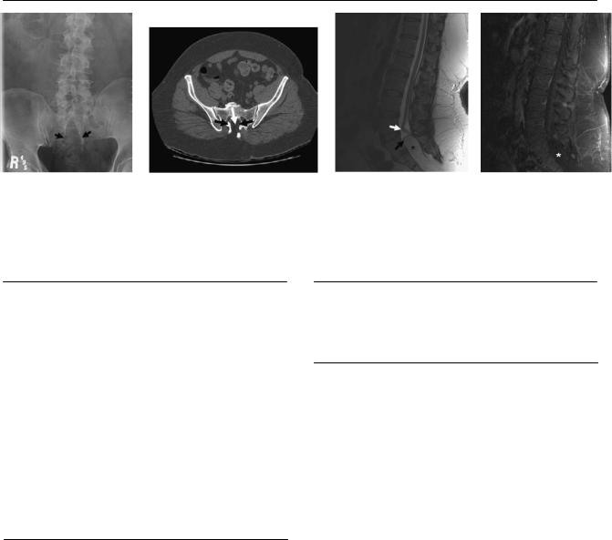
(A) Anteroposterior radiograph of the lumbar spine shows dysraphism of the sacrum (arrows). (B) Axial computed tomography (CT) scan of the pelvis without contrast demonstrates the lack of fusion of the posterior elements of the sacrum (black arrows). Fat corresponding to the lipoma occupies the defect (white arrow). (C) Sagittal T2-weighted image (WI) of the lumbar spine shows the conus descending to L4 (white arrow). The lipoma extends into the spinal canal (asterisk). The black arrow points to the placode-lipoma interface. (D) Sagittal T1WI of the lumbar spine with fat saturation and contrast; note the fat suppression of the lipoma (asterisk).
■ Di erential Diagnosis
•Lipomyelocele and lipomyelomeningocele: Lipomyeloceles and lipomyelomeningoceles are large subcutaneous lipomas with intraspinal extension through a wide spina bifida. Tethered cord is present in all cases. Skin abnormalities can occur in 50% of the cases: hypertrichosis, dimple, dermal sinus tract, and capillary hemangioma.
•Myelomeningocele (dorsal): This is a less extensive spinal dysraphism. Dermal stigmata or a mass can be present, but no lipoma is associated. Myelomeningocele is not always associated with tethered cord.
•Sacrococcygeal teratoma: This is a complex pelvic mass in newborns characterized by fat, calcifications in 60% of cases, debris, skin appendages, and enhancement of the solid portions.
■Essential Facts
•This is an abnormality of primary neurulation.
•The junction between fat and meninges is intact in lipomyelocele and lipomyelomeningocele.
•Lipomyelocele, also known as lipomyeloschisis (75% of cases): the placode-lipoma interface is inside the spinal canal.
•Lipomyelomeningocele (25% of cases): the placode-lipoma interface is outside the spinal canal in a meningocele.
•Plain radiography demonstrates sacral dysraphism.
•On CT, fat occupies the spinal canal, and bony defects are identified.
•T2WIs can detect the placode-lipoma interface; fat-satu- rated sequences confirm the lipoma.
•There is no enhancement.
■ Other Imaging Findings
•Obstetric ultrasound can show posterior dysraphism and fat in the lipomatous portion.
¸Pearls & ˚ Pitfalls
¸Because of its origin, the lipomatous portion of the lesion
is extradural in location.
¸The lipoma is asymmetric in almost half of the cases.
¸If a dorsal myelomeningocele is associated with a teth-
ered cord, look for a lipoma.
˚Meningoceles are associated in only 25% of cases (lipomyelomeningocele).
41
Case 21
A
■ Clinical Presentation
A 25-year-old woman presenting with headaches and papilledema.
Further Work-up
B C D
42 |
RadCases.thieme.com |
RadCases Neuro Imaging |
|
■ Imaging Findings |
|
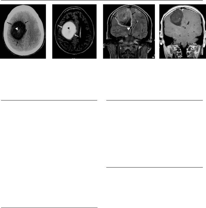
A B C D
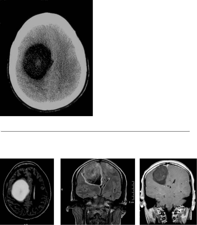
(A) Computed tomography (CT) scan without contrast shows a round, hypoattenuated lesion in the right frontal lobe (arrows). The lesion is located in the subcortical white matter and demonstrates a coarse central calcification (arrowhead). (B) Axial T2-weighted image (WI) shows a hyperintense subcortical mass (arrows) in the right frontal lobe; no vasogenic edema is evident. An area of low signal is seen in the center of the mass (arrowhead), representing the calcification evident on the CT scan. (C) Coronal fluid-attenuated inversion recovery image shows a mass in the right frontal lobe with heterogeneous signal (arrows). There is e acement of the right frontal horn (arrowhead) and displacement of the midline to the left. (D) Coronal T1WI with contrast shows mild enhancement of the right frontal mass.
■ Di erential Diagnosis
•Oligodendroglioma: This is a tumor of glial origin presenting as a mass in the cortex or subcortical white matter with coarse calcification (20–90% of cases) and occasional cystic degeneration or hemorrhage. Most do not enhance, but there may be subtle enhancement in 20%. They are more frequently supratentorial in location (90% of cases). Vasogenic edema is uncommon.
•Low-grade astrocytoma: In low-grade astrocytoma, the location is more central, and the cortex is rarely involved. Calcification is infrequent. All the other characteristics are similar to those of oligodendroglioma.
•Dysembryoplastic neuroepithelial tumor (DNET): DNET is a benign, well-marginated lesion. DNET has a cortical location but is more frequently in the temporal lobe. It has a “bubbly” appearance on magnetic resonance imaging (MRI), variable enhancement (20%), and calcification
(25%). It presents in patients younger than 20 years of age, usually with seizures.
■Essential Facts
•Calcification is associated with higher-grade tumors.
•Calcification and vasogenic edema are rare in children.
•Low-grade tumors (World Health Organization [WHO] grades I and II) are called oligodendrogliomas.
•High-grade oligodendrogliomas (WHO grade III) are called anaplastic oligodendrogliomas.
■ Other Imaging Findings
•CT: Sixty percent have low attenuation and 23% are isodense to the parenchyma. DNETs may not be detected on CT.
•MRI:
•Better definition of tumor extent
•Typically heterogeneous signal
•T1WIs: hypointense to gray matter
•T2WIs: hyperintense to gray matter
•Di usion-WIs: higher-grade tumors tend to have lower apparent di usion coe cient values.
¸Pearls & ˚ Pitfalls
¸Tumors with 1p and 19q deletions are more likely to
calcify and have ill-defined margins; even though these tumors tend to be more aggressive, they also respond better to chemotherapy.
¸The more aggressive variant—anaplastic oligodendroglioma (WHO grade III)—is commonly associated with these deletions.
¸Consider DNET if a cortically based tumor in the tempo-
ral lobe presents in a young patient.
˚Oligodendrogliomas are the most common intracranial tumors that have calcifications; however, only 20% calcify.

43
Case 22
A 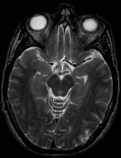
 B
B
C 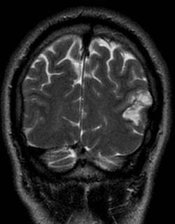
 D
D
■ Clinical Presentation
A 27-year-old with a history of seizures.

44 |
RadCases.thieme.com |
|
RadCases Neuro Imaging |
|
|
■ Imaging Findings |
|
|
|
A |
B |
C |
|
D |
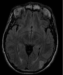
(A) Axial T2-weighted image (WI) of the brain shows a lesion in the left parietal lobe (arrow) with a small, cystic, “bubbly” appearance. (B) Axial fluid-attenu- ated inversion recovery (FLAIR) image demonstrates increased cortical signal in the left parietal lobe (arrow) without surrounding edema. (C) Coronal T2WI demonstrates the cortically based, “bubbly” lesion (arrows) in the left parietal lobe. (D) Axial T1WI with contrast shows no enhancement in the left parietal mass (arrow).
■ Di erential Diagnosis
•Dysembryoplastic neuroepithelial tumor (DNET): DNETs are benign, well-demarcated cortical lesions. They present as partial seizures in patients younger than 20 years. They occur more frequently in the temporal lobe (60%) or frontal lobe (30%). Cortical dysplasia is often noted adjacent to the lesion. Calcification is rare (20%). DNET appears as a multilobulated “bubbly” lesion with high signal on T2WIs and low signal on T1WIs. There is no surrounding vasogenic edema. Thirty percent show enhancement.
•Ganglioglioma: Ganglioglioma is also a mixed neuronal glial cell tumor. This is a cystic mass and does not have the multilobulated appearance of DNET. Ganglioglioma presents as a mural enhancing nodule (40%), has a temporal lobe distribution, and is calcified in 30% of cases. It can scallop the inner table of the skull. There is no associated vasogenic edema.
•Neuroepithelial cyst: Neuroepithelial cysts are thin-walled and can be unior multilocular. They have no enhancement. The intraventricular location is more common. They do not have a bright rim on FLAIR images.
■Essential Facts
•Initially described in 1988
•Mixed neuronal glial cell tumor
•Located in the temporal lobe, usually near the amygdala of the hippocampus
•Inner scalloping of the skull
■ Other Imaging Findings
•Computed tomography (CT): Low density. CT can detect the calcifications, even though they are rare. CT allows the evaluation of inner table scalloping.
•Magnetic resonance imaging: faint enhancement (20%) and a bright rim are seen on FLAIR images.
¸Pearls & ˚ Pitfalls
¸Look for a FLAIR rim.
¸The multilobulated morphology helps in the di erential
diagnosis.
¸ Exclude DNET from your di erential list in adult patients.
˚On CT, DNET can resemble a stroke; however, it does not evolve to secondary atrophy.
45
Case 23
A 
 B
B
C D
■ Clinical Presentation
A 17-year-old girl with a history of seizures.
46 |
RadCases.thieme.com |
|
RadCases Neuro Imaging |
|
|
■ Imaging Findings |
|
|
|
A |
B |
C |
|
D |
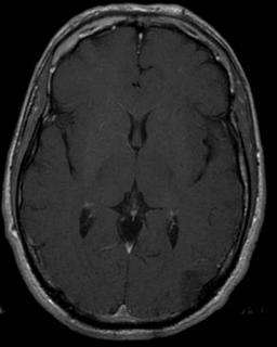
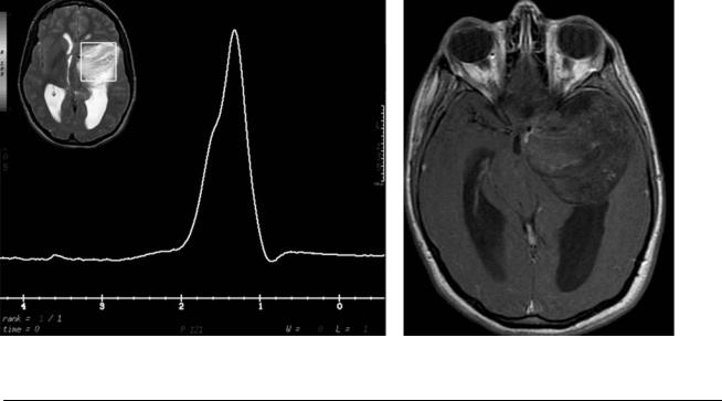
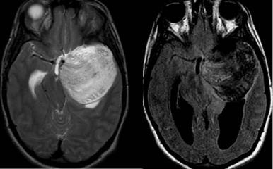
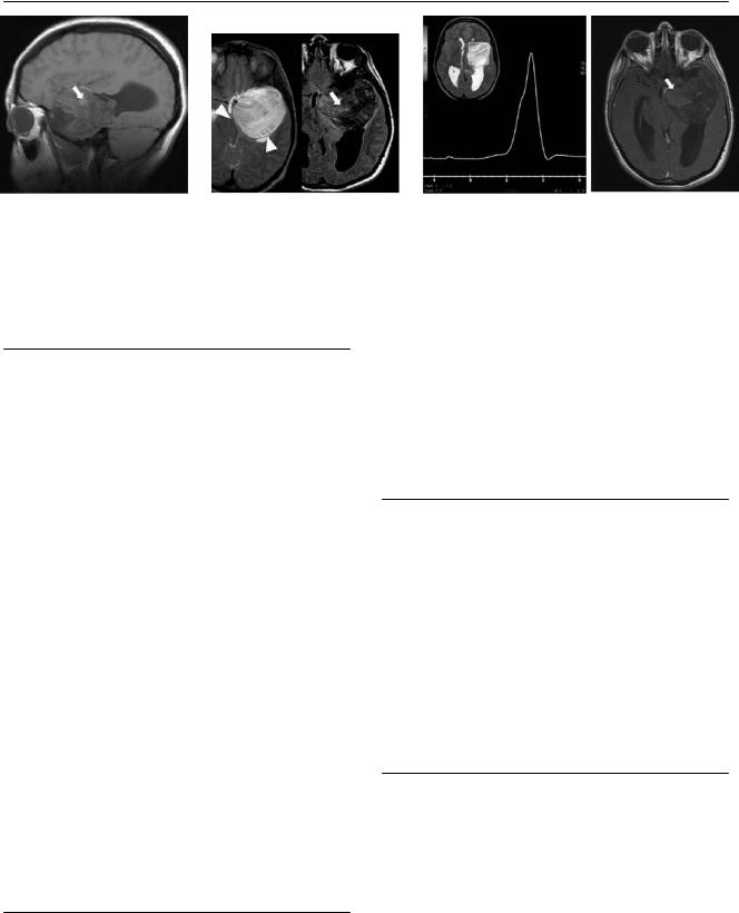
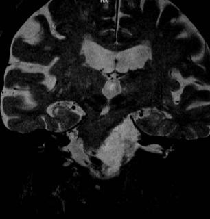
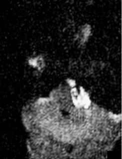
(A) Sagittal T1-weighted image (WI) shows a mass in the anterior aspect of the temporal fossa with areas of T1 hyperintensity (arrow). The mass is compressing the adjacent parenchyma. (B) Axial T2-weighted and fluid-attenuated inversion recovery (FLAIR) images demonstrate heterogeneous signal in the left temporal fossa mass. The mass shows areas of increased signal on the FLAIR image (arrow). The location of the lesion is extra-axial. It compresses the temporal lobe and cerebellar peduncle (arrowheads), displacing vessels without encasing them. (C) Magnetic resonance (MR) spectroscopy shows a high lipid peak with a broad base from –0.9 to –1.3 parts per million (ppm). (D) Axial T1WI with contrast shows no enhancement of the mass; the hyperintense regions (arrow) were bright in the nonenhanced sequences (related to fat).
■ Di erential Diagnosis
•Nonruptured dermoid cyst: Dermoid cysts are ectodermal inclusion cysts that contain squamous and glandular tissue. The tissue causes the cysts to grow, increasing the likelihood of ruptures. They account for fewer than 5% of intracranial tumors and are most frequent in the midline. A supratentorial location is more common than an infratentorial location. If a cyst ruptures, chemical meningitis develops with secondary vasospasm and stroke; the contents can be intraventricular. They have a well-defined capsule that is thicker than the capsule in epidermoid cysts.
•Epidermoid cyst: Epidermoid cysts are also ectodermal inclusion cysts, although they contain only squamous epithelium. The signal characteristics are similar to those of cerebrospinal fluid (CSF), except for slight hyperintensity and heterogeneity on FLAIR images (“dirty CSF appearance”). They are five to nine times more common than dermoid cysts. They are less common in pediatric patients. The most frequent locations are around the cerebellopontine angle and the ventricles. They typically show restricted di usion and no contrast enhancement.
•Craniopharyngioma: Craniopharyngiomas are benign dysontogenetic epithelial masses that originate at the epithelium of the Rathke pouch. They are the most common pediatric tumors in the suprasellar region. They have mixed solid and cystic components with calcification. Most of them enhance (90%). Calcifications can be seen on computed tomography or gradient-echo T2*WIs. Cyst contents are typically hyperintense on FLAIR images.
■Essential Facts
•The sellar, parasellar, and frontonasal regions are common locations for nonruptured dermoid cysts.
•Dermoid cysts contain ectodermal derivatives such as squamous epithelium and dermal appendages such as hair follicles, sebaceous glands, and apocrine glands, which produce keratin and oils.
•The meninges enhance when rupture and chemical meningitis have occurred; otherwise, no enhancement is noted.
■ Other Imaging Findings
•Computed tomography:
•Round, lobulated mass with low attenuation (cholesterol content)
•Capsular calcification in 20%
•Can remodel the adjacent skull
•MR imaging: hyperintense on T1; both hypoand hyperintense regions common on T2
•If ruptured: typical appearance of fat-fluid levels in the frontal horns; di use, hyperintense droplets in the subarachnoid or intraventricular spaces on T1WIs; extensive pial enhancement on T1WIs with contrast
•Broad lipid peak on MR spectroscopy
¸Pearls & ˚ Pitfalls
¸Both dermoid and epidermoid cysts are ectodermal in-
clusion cysts; the di erence is that dermoid cysts contain squamous epithelium and skin appendages, whereas epidermoid cysts are lined only by squamous epithelium.
¸Ruptured and unruptured dermoid cysts di er in appearance.
¸Enhancement is rarely seen and is usually secondary to chemical meningitis.

47
Case 24
A 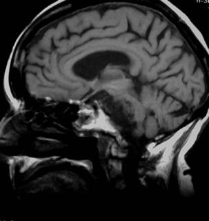
 B
B
C 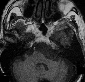
 D
D
■ Clinical Presentation
A 35-year-old man with progressive left-sided hearing loss.