

Книги по МРТ КТ на английском языке / Neuro Imaging Redcases
.pdf8 |
RadCases.thieme.com |
|
RadCases Neuro Imaging |
|
|
■ Imaging Findings |
|
|
|
A |
B |
C |
|
D |
(A) Axial computed tomography (CT) scan of the head shows an absence of the anterior fibers of the corpus callosum (arrow) and a dilated lateral ventricle on the right (asterisk). (B) Axial T1-weighted magnetic resonance (MR) image of the brain shows an absence of the anterior fibers of the corpus callosum (arrow) and a dilated lateral ventricle on the right (asterisk). (C) Coronal T1-weighted image (WI) of the brain after intravenous contrast shows absence of the corpus callosum with a high-riding 3rd ventricle (arrow). Note the “bullhorns” appearance of the lateral horns (asterisks). Thick bands of white matter superior to the lateral ventricles are Probst fibers (arrowheads). (D) Sagittal T1WI of the brain shows absence of the corpus callosum. Note the lack of a cingulate gyrus, the vertically oriented sulci (arrow), and the colpocephaly of the right lateral ventricle (asterisk).
■ Di erential Diagnosis
•Dysgenesis of the corpus callosum (CC): This is suggested by partial or complete absence of the CC.
•Lobar holoprosencephaly: Partial agenesis of the CC can be seen in lobar holoprosencephaly. Other findings include fusion of anterior horns and lateral ventricles and fusion of the anterior fornix. There is an absence of septum pellucidum. This entity is associated with midline facial defects. There is no thalamic fusion (seen in more severe forms of holoprosencephaly).
•Schizencephaly: This is a migration disorder that can be associated with partial dysplasia of the CC. The main findings include hemispheric cerebrospinal fluid–filled clefts through which the ventricles communicate with the subpial space. The clefts are lined by gray matter. Unilateral schizencephaly is more frequent than bilateral defects.
■Essential Facts
•Bundles of Probst: tracks of white matter that run parallel to the ventricle as an alternative to the CC
•High-riding 3rd ventricle
•Absent cingulate sulcus with radially oriented fissures
•Crescent-shaped lateral horns; small (“bullhorns”) appearance of the frontal horns
•Enlarged occipital horns (colpocephaly)
•Absence of the septum pellucidum or severe widening of the cavum septum pellucidum
•The precursors of the CC develop between 8 and 20 weeks. Many di erent genes control the movement of axons across the midline, and numerous mutations can result in callosal defects.
•The CC develops from the anterior genu to the posterior splenium. The rostrum is the last portion to develop.
•Associated with other central nervous system anomalies: anomalies of cortical development, lipoma or interhemispheric cyst, Dandy-Walker complex, Chiari type II malformation, encephalocele (midfacial anomalies), and holoprosencephaly
•Sagittal and coronal MR imaging of the brain is the best diagnostic tool for this condition.
■ Other Imaging Findings
•Fetal ultrasound after week 20 can detect severe callosal defects.
•Partial callosal agenesis can be hard to detect on ultrasound.
¸Pearls & ˚ Pitfalls
¸Lipomas of the midline associated with agenesis of the CC
calcify in 10% of cases and are seen on CT.
¸ Callosal agenesis is the most common finding in fetal alcohol syndrome.
˚In patients with an enlarged 3rd ventricle secondary to hydrocephalus, evaluation of the CC is limited because of the thinning and superior displacement of the fibers.
Wait until the ventricles are decompressed before undertaking further assessment.
˚When all the segments of the CC are present but small, the condition is considered to be callosal hypoplasia.

9
Case 5
A 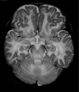
 B
B
C
■ Clinical Presentation
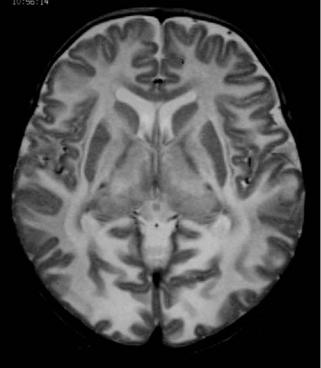
An infant presenting with marked hypotonia, macrocephaly, and seizures.
10 |
RadCases.thieme.com |
RadCases Neuro Imaging |
|
■ Imaging Findings |
|
A B C
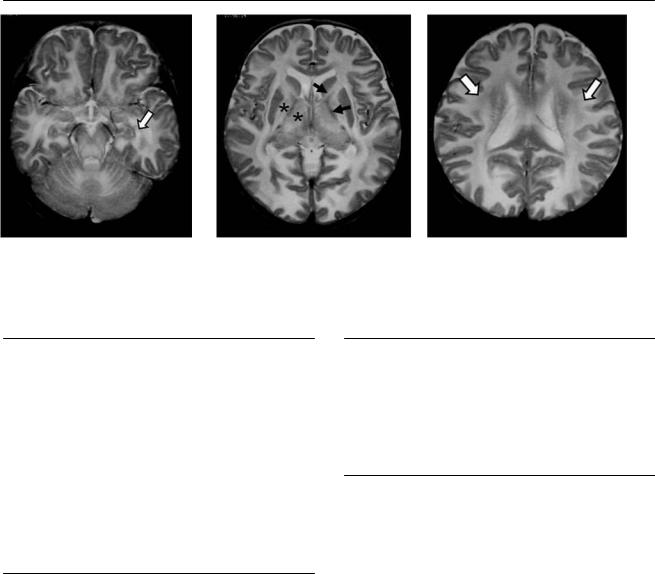
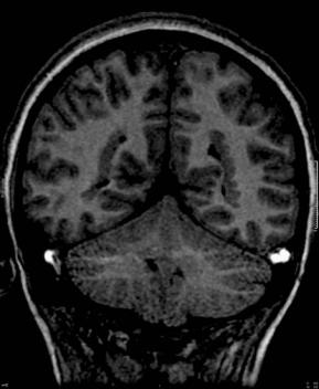
(A) Axial T2-weighted magnetic resonance (MR) image of the brain reveals an increased white matter T2 signal (arrow). (B) There is relative sparing of the internal capsule (arrows). Abnormal T2 signal is demonstrated in the thalami (asterisks) and globus pallidus. (C) Note the di use increase in the white matter T2 signal (arrows).
■ Di erential Diagnosis
•Canavan disease: This involves predominantly the subcortical U fibers. The occipital lobes are more involved than the frontal and parietal lobes. The thalami and basal ganglia are a ected in severe cases.
•Alexander disease: This also presents with macrocephaly but mostly involves frontal white matter. The a ected areas may show enhancement.
•Pelizaeus-Merzbacher disease, connatal type: This presents with a nearly total lack of myelination. The brainstem, diencephalon, cerebellum, and subcortical white matter may demonstrate myelin preservation.
■Essential Facts
•Autosomal-recessive condition, most common in Ashkenazi Jews
•Deficiency of aspartoacylase
•Hypotonia, macrocephaly, and seizures in newborn period
•Spasticity, optic atrophy, and intellectual failure as disease progresses
•Death usually by second year of life
•Computed tomography: di use low attenuation in cerebral and cerebellar white matter
•MR imaging: White matter has high T2 and low T1 signal. The thalami and basal ganglia can be a ected.
•The peripheral white matter is preferentially a ected early in the course of the disease.
•In severe cases, there may be an extensive lack of myelination with relative sparing of the internal capsules.
■ Other Imaging Findings
•MR spectroscopy may reveal an increased N-acetylaspar- tate (NAA) peak. Because of the deficiency in aspartoacylase, NAA accumulates in the mitochondria and impairs myelin synthesis.
•Myoinositol elevation due to gliosis may also be present.
¸Pearls & ˚ Pitfalls
¸As opposed to Canavan disease, Krabbe disease and
metachromatic leukodystrophy involve the deep white matter early, and the peripheral white matter is a ected as the disease progresses.
˚In infants born prematurely, myelination may be delayed for the corrected gestational age, mimicking leukodystrophy. Adequate clinical correlation and follow-up imaging are helpful to make the distinction.
11
Case 6
A B
C 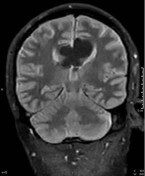 D
D
■ Clinical Presentation
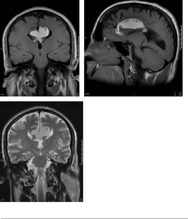
A 30-year-old man with a history of headaches.

12 |
RadCases.thieme.com |
|
RadCases Neuro Imaging |
|
|
■ Imaging Findings |
|
|
|
A |
B |
C |
|
D |
(A) Coronal T1-weighted magnetic resonance imaging (MRI) of the brain demonstrates a high-signal lesion in the midline (asterisk) at the site of the corpus callosum. The lesion shows ventricular extension (arrow). (B) Sagittal T1-weighted MRI of the brain demonstrates a high-signal lesion in the midline (asterisk) that shows ventricular extension (arrow). Note the hypoplasia of the corpus callosum (arrowhead). (C) Coronal T2-weighted MRI of the brain demonstrates a high-signal lesion in the midline (asterisk) at the site of the corpus callosum. The lesion shows ventricular extension (arrow). (D) Coronal T2-weighted fat-satu- rated MRI of the brain demonstrates loss of signal in the midline lesion (asterisk), which was bright before fat saturation.
■ Di erential Diagnosis
•Intracranial lipoma: Intracranial lipomas are fatty, extraaxial, pia-based lesions. Calcifications are common in lesions with an interhemispheric location. They may encase vessels. Pericallosal lipomas are associated with choroidal extension. Fat suppression can aid the diagnosis.
•Dermoid cyst: Dermoid cysts are usually located near the parasellar or frontal region at the midline. They are well circumscribed and contain fat. Twenty percent have capsular calcification (lipomas at this location rarely calcify). Rupture of a dermoid cyst causes chemical meningitis and leptomeningeal enhancement (lipomas typically do not rupture).
•Epidermoid cyst (ectodermal inclusion cyst): Epidermoid cysts are lobulated. They are unusual at the midline. Fifty percent occur at the cerebellopontine angle (CPA), 17% are located at the 4th ventricle, and 10% are parasellar. On T1and T2-weighted images, they are isointense to cerebrospinal fluid. They have a dirty fluid-attenuated inversion recovery signal. These cysts restrict on di usion (this feature is diagnostic).
■Essential Facts
•Poor di erentiation of the embryonic meninx primitiva (forms leptomeninges and cisterns), then di erentiation into fat
•Locations in order of frequency: interhemispheric (40–45%), suprasellar-infundibular (25%), quadrigeminal cistern (25%), and CPA (14%)
•Associated with other malformations
•When associated with dysgenesis of the corpus callosum (interhemispheric), described as tubulonodular type
•Cephaloceles
■ Other Imaging Findings
•Computed tomography can identify calcifications.
•Lipomas are suppressed on fat-saturated MRI.
•Magnetic resonance spectroscopy will show a high lipid peak.
¸Pearls & ˚ Pitfalls
¸There are two types of pericallosal lipomas: bulky (tu-
bulonodular), which are associated with agenesis of the corpus callosum, and ribbon-like (curvilinear).
˚Other entities with a high T1 signal:
•Hemorrhage
◦Early subacute (3–7 days): intracellular methemoglobin
◦Late subacute (7–14 days): extracellular methemoglobin
•Lesions with a high protein content
•Melanin
•Lipids
•Minerals (manganesium)
•Slow flow
13
Case 7
A
B C
■ Clinical Presentation
A newborn with a posterior neck mass diagnosed on prenatal ultrasound.

14 |
RadCases.thieme.com |
RadCases Neuro Imaging |
■ Imaging Findings
A 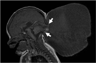 B C
B C
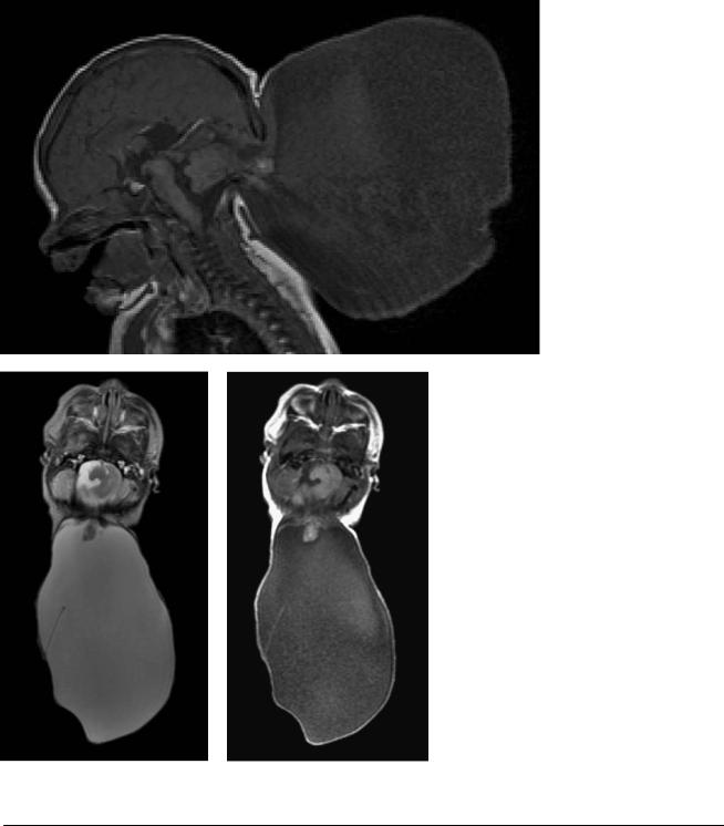
(A) Sagittal T1-weighted image of the brain shows a posterior skull defect (arrows) through which cerebellar tissue and cerebrospinal fluid are protruding. (B) Axial T2WI of the brain demonstrates elongation of the cerebellum, which protrudes toward the neck (arrow) and is surrounded by a large cystic lesion. Note a thin septum within the cyst (arrowhead). (C) Axial T1WI of the brain demonstrates elongation of the cerebellum, which protrudes toward the neck (arrow) and is surrounded by a large cystic lesion.
■ Di erential Diagnosis
•Occipital meningoencephalocele: In occipital meningoencephalocele, there is posterior traction of the cerebellum. The cystic lesion communicates with the subarachnoid space of the posterior fossa.
•Chiari type III malformation: Chiari type III malformation has the same features as a Chiari type II malformation, but with an occipital cephalocele instead of a myelomeningocele. This patient’s brainstem is not elongated.
•Cystic hygroma: Although hygromas may be unilocular, they involve the neck, oral cavity, face, or airway as well as surrounding normal muscles. No skull defect is present.
■ Essential Facts
•Occipital cephaloceles originate between the foramen magnum and the lambda and have variable contents. The brain within them is usually dysplastic, gliotic cerebellum.
•Associated anomalies include Chiari type II and III malformations, Dandy-Walker complex, cerebellar dysplasias, diastematomyelia, and Klippel-Feil syndrome.
■ Other Imaging Findings
•Ultrasound may detect occipital meningoencephaloceles early in pregnancy.
•Computed tomography demonstrates the bony defects.
¸Pearls & ˚ Pitfalls
¸For the purposes of surgical planning, it is important to
identify the position of the venous sinuses and the presence of associated brain anomalies.
˚Occipital cephaloceles without brainstem anomalies are often mislabeled as Chiari type III malformations.
15
Case 8
A B
C D
■ Clinical Presentation
A 2-year-old girl presenting with developmental delay and seizures.

16 |
RadCases.thieme.com |
RadCases Neuro Imaging |
|
■ Imaging Findings |
|
A B C D
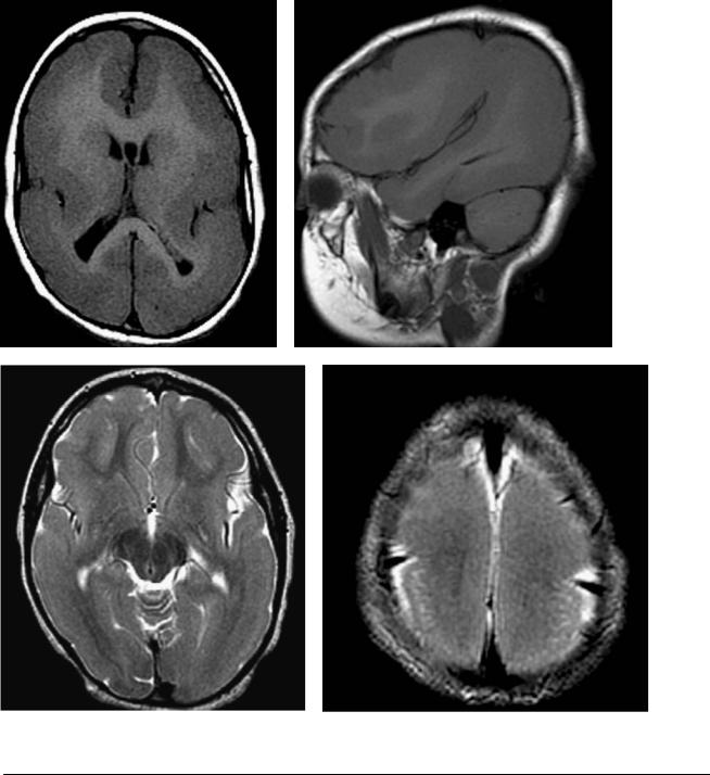
(A) Axial T1-weighted image (WI) shows severely decreased sulcation of the brain with a thick cortex (arrows). (B) Sagittal T1WI shows a thick cortical layer with severely decreased sulcation. (C) Axial T2WI shows severely decreased sulcation of the brain with a thick cortex (arrows).
(D) Axial T2WI shows severely decreased sulcation of the brain with a thick cortex (arrows).
■ Di erential Diagnosis
•Type I (classic) lissencephaly: Type I lissencephaly is a migration disorder of the gray matter, with the formation of a smooth, four-layered cortex (a normal cortex has six layers). Type I lissencephaly may be isolated or part of a syndrome, most frequently Miller-Dieker syndrome.
•Type II lissencephaly (cobblestone complex): Type II lissencephaly has a disorganized, unlayered cortex and no ribbons of band heterotopia. Neurons migrate past the cortical plate to the subpial space. No subependymal heterotopia should be present. Type II lissencephaly is associated with congenital muscular dystrophy (Walker-Warburg syndrome). Ocular defects are common.
•Cytomegalovirus (CMV) infection: Cortical gyral abnormalities, including lissencephaly, can be seen in CMV infections. Other findings include microcephaly, cerebral periventricular and parenchymal calcifications, and cerebellar hypoplasia.
■ Essential Facts
•Type I lissencephaly is part of the agyria/pachygyria spectrum (hourglass configuration of brain).
•In the usual presentation, the outer cortex exhibits ribbons of band heterotopia separated by white matter.
•Subependymal heterotopia is commonly seen.
•There is no abnormal enhancement.
•Fetal ultrasound can detect severe cases of subependymal heterotopia.
■ Other Imaging Findings
•Di usion tensor tracking has shown a band of anisotropic di usion related to the dysplastic 4th cortical layer and
a disconnection between the cortex and the deep white matter.
¸Pearls & ˚ Pitfalls
¸Always look for other sites of heterotopic cortex. Some
enhancement has been described at the dense cellular layer (dysplastic cortical layer). Remember that a smooth cortex can be seen in normal fetuses until up to 26 weeks of gestation.
˚Multiple migration disorders can appear similar to lissencephaly.
˚Knowledge of the gestational age is important to avoid overcalling agyria.
˚Only magnetic resonance imaging late in pregnancy can detect lissencephaly in utero.

17
Case 9
A 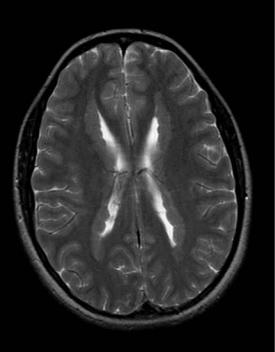
 B
B
■ Clinical Presentation
A patient undergoing a work-up for epilepsy.