

Книги по МРТ КТ на английском языке / Neuro Imaging Redcases
.pdf18 |
RadCases.thieme.com |
RadCases Neuro Imaging |
■ Imaging Findings
A B
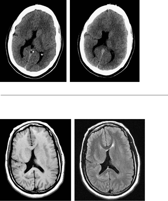
(A) Axial T2-weighted image (WI) of the brain demonstrates multiple nodules, with signal intensity similar to that of the cerebral cortex, lining the lateral ventricles (arrows). There is a serrated appearance of the inner margin of the ventricles. (B) Coronal T1WI confirms the presence of nodules along the ventricular walls. The nodules have signal intensity like that of the cortex (arrows).
■ Di erential Diagnosis
•Subependymal heterotopia: Nodules of gray matter along the ventricular surface are suggestive of subependymal heterotopia. This may appear exophytic, extending to the ventricle.
•Subependymal nodules of tuberous sclerosis: These are irregularly shaped and often calcified. They are not isointense to cortex. They may enhance.
•Band heterotopia: Band heterotopia appears as homogeneous bands of gray matter between the lateral ventricles and cerebral cortex, separated from both by layers of normal-appearing white matter.
■ Essential Facts
•Heterotopia: arrest of the radial migration of neurons, leading to ectopic neurons in areas other than the cortex
•Three types
•Subependymal
◦Normal development; seizures
◦Smooth, ovoid masses isointense to gray matter in all sequences
•Focal subcortical
◦Motor and intellectual disturbance, epilepsy
◦Large heterogeneous mass, isointense to cortical gray matter
◦The a ected hemisphere is small with thin cortex and shallow sulci.
◦Associated hypogenesis of the corpus callosum and dysplastic basal ganglia
•Band (laminar) heterotopia
•Considered a mild form of classic lissencephaly
•Variable developmental delay and seizures
•Female preponderance (90%)
•Homogeneous bands of gray matter between the lateral ventricles and cerebral cortex, separated from both by a layer of normal-appearing white matter
•May have foci of high T2 signal in the white matter
■Other Imaging Findings
•On positron emission tomography with fluorodeoxyglucose, band heterotopia has glucose uptake similar to or greater than that of normal cortex. This contrasts with the hypometabolism found in cortical dysplasias.
¸Pearls & ˚ Pitfalls
¸Lesions may not be apparent in infants and do not
become noticeable until myelination has reached an advanced stage.
˚The caudate tail along the lateral wall of the lateral ventricles should not be misinterpreted as ectopic gray matter.
19
Case 10
A B
■ Clinical Presentation
A patient with a history of developmental delay and seizures.
Further Work-up
C D
20 |
RadCases.thieme.com |
|
RadCases Neuro Imaging |
|
|
■ Imaging Findings |
|
|
|
A |
B |
C |
D |
|
|
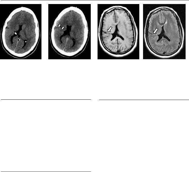
(A) Axial computed tomography scan of the brain demonstrates a small outpouching of the lateral wall of the right lateral ventricle, lined |
|||
|
by gray matter (arrow). A deep sulcus is noted in the subjacent cortex. (B) |
A contiguous slice demonstrates a cleft with cerebrospinal flu- |
||
|
id (CSF) attenuation through which the lateral ventricle communicates with the subarachnoid space. The defect is lined by gray matter (arrows). |
|||
|
(C) Axial T1-weighted image of the brain show a CSF-filled cleft, lined by gray matter, between the subarachnoid space and the lateral wall of the right |
|||
|
lateral ventricle (arrow). (D) Axial fluid-attenuated inversion recovery image of the brain shows a CSF-filled cleft, lined by gray matter, between the |
|||
|
subarachnoid space and the lateral wall of the right lateral ventricle (arrow). |
|
|
|
■ Di erential Diagnosis
•Closed-lip schizencephaly: Closed-lip schizencephaly presents with a CSF cleft, lined by gray matter, that extends from the ependymal surface of the brain to the pia, with apposition of the cleft walls.
•Focal transmantle dysplasia: Focal transmantle dysplasia presents with abnormal cells extending from the cortex to the ventricular surface, seen as a curvilinear focus of hyperintensity relative to the mature white matter. No CSF-filled cleft is present.
•Band heterotopia: The abnormally positioned bands of gray matter are parallel in band heterotopia, not perpendicular to the ventricular walls, and are not surrounded by CSF.
¸Pearls & ˚ Pitfalls
¸A thin pial membrane covers the cerebral defect in
schizencephaly. It separates the cleft from the subarachnoid space.
˚A cleft with closed lips in the imaging plane may be missed. At least two planes are necessary to assess patients with seizure or developmental delay.
■ Essential Facts
•Schizencephaly is an anomaly of neuronal migration in which a CSF-filled cleft is lined by gray matter. It extends from the ventricular surface (ependyma) to the periphery (pial surface) of the brain.
•Types
•Closed-lip (type I) schizencephaly: cleft walls in apposition, with a ventricular dimple
•Open-lip (type II) schizencephaly: cleft walls separated (more common)
•Polymicrogyric and nonpolymicrogyric cleft linings, cystlike diverticula and membranous structures, and subependymal heterotopia can be found at the cleft
•Concomitant anomalies are polymicrogyria outside the cleft, white matter volume loss, septal and optic anomalies, callosal anomalies, and hippocampal anomalies.
21
Case 11
A
■ Clinical Presentation
A newborn infant with feeding problems and seizures. The baby has a cleft lip.
Further Work-up
B C D
22 |
RadCases.thieme.com |
|
RadCases Neuro Imaging |
|
|
■ Imaging Findings |
|
|
|
A |
B |
C |
|
D |
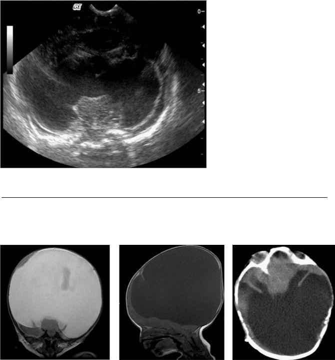
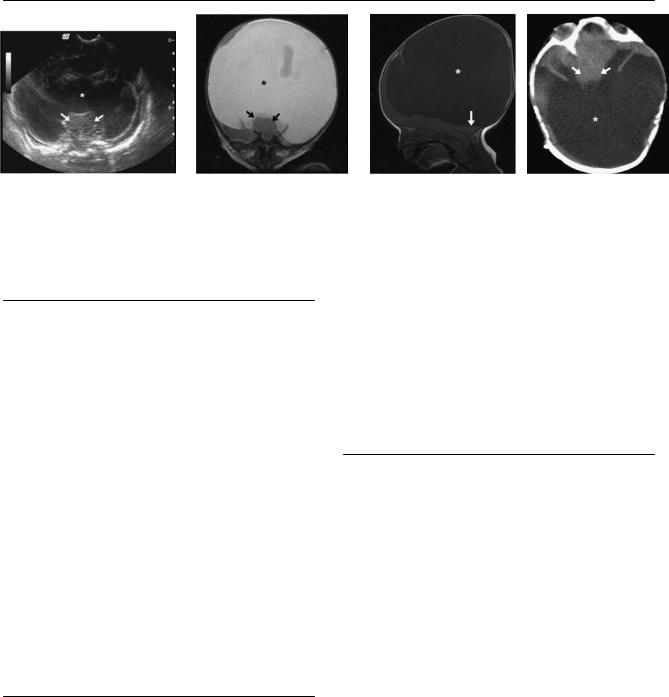
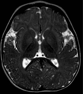
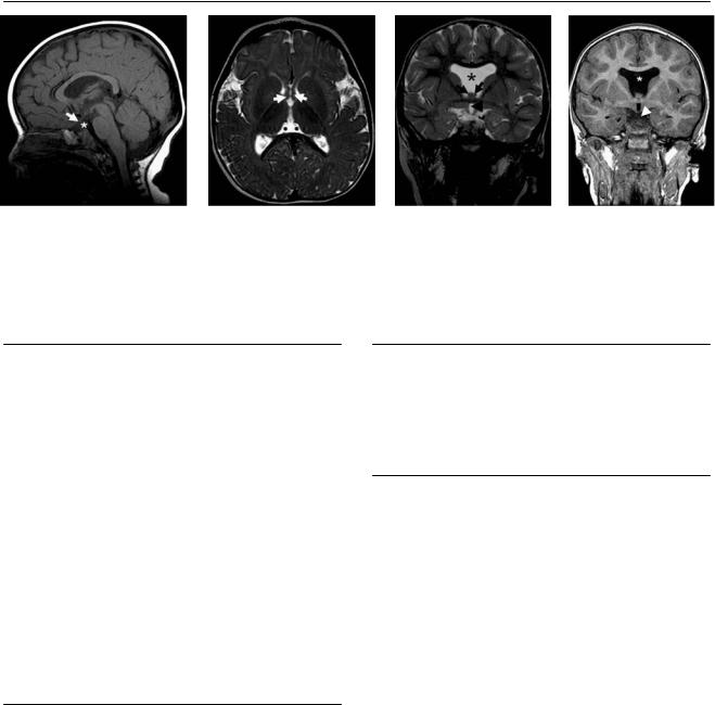
(A) Transfontanellar ultrasound of the brain in a coronal view at the level of the thalami shows a prominent monoventricle (asterisk) and thalamic fusion (arrows). (B) Coronal T2-weighted magnetic resonance image of the brain at the level of the thalami shows a monoventricle, seen as a prominent supratentorial cyst (asterisk), and fusion of the thalami (arrows). Note the absence of a cerebral falx. (C) Sagittal T1-weighted image of the brain shows a large supratentorial cyst (asterisk); note the hypoplasia of the posterior fossa structures (arrow). (D) Axial computed tomography scan of the brain without contrast shows fusion of the thalami (arrows) and a large posterior cyst-monoventricle (asterisk).
■ Di erential Diagnosis
•Alobar holoprosencephaly: Alobar holoprosencephaly is a congenital malformation of the forebrain. The corpus callosum is absent. There is a dorsal cyst with hydrocephalus. Macrocephaly can arise from high pressure. The anterior brain is compressed anteriorly, and the thalami are fused.
•Hydranencephaly: Intrauterine destruction of the cerebral hemispheres secondary to occlusion of the internal carotid arteries is called hydranencephaly. The thalami are not fused as in holoprosencephaly. The cerebral falx is intact. Macrocrania is also noted. The posterior fossa structures are intact, and there is absence of a cortical mantle.
•Severe (obstructive) hydrocephalus: There is increased cerebrospinal fluid formation versus insu cient resorption in severe hydrocephalus. Macrocrania can develop if severe hydrocephalus occurs early in life, when the sutures are open. The compressed brain has a cortical mantle appearance (characteristic) not seen in holoprosencephaly. No thalamic fusion is present. The site of the obstruction should be located. There is a high T2 signal in the periventricular white matter, which is not seen in holoprosencephaly or hydranencephaly.
■Essential Facts
•Alobar holoprosencephaly is the most severe form of a defect in dorsoventral patterning and cleavage of the forebrain.
•The hypothalamus and caudate nuclei are the most severely a ected structures.
•Azygos anterior cerebral artery can be present.
•Complex facial malformations are associated with this condition:
•Cleft lip/palate
•Hypotelorism
•Arhinencephaly
•Cyclopia
•Three types of appearance have been described for the remaining brain: “cup,” “pancake,” and “ball.”
¸Pearls & ˚ Pitfalls
¸“The face predicts the brain”: if facial malformations are
present, the brain must be studied.
¸Absence of the falx and thalamic fusion are the most relevant imaging findings.
¸Facial changes are not associated with hydranencephaly
or severe hydrocephalus.
˚Macrocephaly can be seen in the three entities in the differential diagnosis.

23
Case 12
A 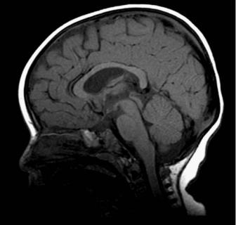
 B
B
C 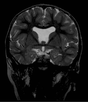
 D
D
■ Clinical Presentation
A 20-year-old man with a history of bilaterally decreased eyesight and hypopituitarism.
24 |
RadCases.thieme.com |
RadCases Neuro Imaging |
|
■ Imaging Findings |
|
A B C D
(A) A T1-weighted sagittal image of the brain shows a small optic chiasm (arrow) and absence of the pituitary stalk (asterisk). (B) A T2-weighted axial image of the brain shows two separate anterior horns of the fornix (arrows). (C) A T2-weighted sagittal image of the brain shows absence of the septum pellucidum (asterisk). Note the lack of fusion of the fornix (arrows) and the hypoplastic sella, with no evidence of a pituitary gland or pituitary stalk. Hypoplasia of the optic nerves is seen (arrowhead). (D) A T2-weighted sagittal image of the brain shows absence of the septum pellucidum (asterisk). Note the hypoplastic sella, with no evidence of a pituitary gland or pituitary stalk. Hypoplasia of the optic chiasm is seen (arrowhead).
■ Di erential Diagnosis
•Septo-optic dysplasia (de Morsier syndrome): Septo-optic dysplasia is considered the mildest form of holoprosencephaly. It is caused by dysgenesis of the septum pellucidum. The following may be present: optic nerve hypoplasia, hypothalamic and pituitary dysfunction (60% of cases), schizencephaly (50% of cases), thin corpus callosum, and square frontal horns.
•Lobar holoprosencephaly: A fused anterior fornix is the main feature in the brain that di erentiates lobar holoprosencephaly from septo-optic dysplasia. There is an absence of the corpus callosum. This diagnosis is associated with midline defects, which are rarely seen in septo-optic dysplasia. The optic nerves and hypothalamus are spared.
•Partial agenesis of the corpus callosum: Here, absence of the septum pellucidum or severe widening of the cavum septum pellucidum can be seen. The optic nerves and hypothalamus are intact.
■Essential Facts
•Two distinct forms of septo-optic dysplasia are described:
•Associated with schizencephaly: usually, a remnant of the septum pellucidum is present.
•White matter hypoplasia, including optic radiations. There is no schizencephaly. Hypopituitarism may be present.
•Magnetic resonance imaging:
•Best diagnostic tool
•Thin slices through the optic nerves are helpful.
■ Other Imaging Findings
•Computed tomography can detect absence of the septum pellucidum but has less contrast for visualizing migrational disorders. Evaluation of the optic tract is limited.
•It is di cult to identify findings on prenatal ultrasound.
¸Pearls & ˚ Pitfalls
¸The anterior fusion of the fornix helps to di erentiate
lobar holoprosencephaly from septo-optic dysplasia. ¸ Always look for schizencephaly.
˚If dysgenesis or absence of the septum pellucidum is detected, investigate further to exclude septo-optic dysplasia or lobar holoprosencephaly.
25
Case 13
A B
C D
■ Clinical Presentation
A 40-year-old man with progressive weakness of both upper extremities for 1 year.

26 |
RadCases.thieme.com |
|
RadCases Neuro Imaging |
|
|
■ Imaging Findings |
|
|
|
A |
B |
C |
|
D |
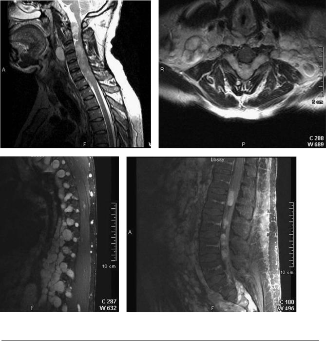
(A) Sagittal T2-weighted image (WI) of the cervical spine demonstrates a widened spinal canal with an intradural, extraspinal mass that compresses the spinal cord posteriorly (arrowhead). A soft-tissue mass in the prevertebral soft tissues shows a high T2 signal (arrow). (B) Axial T2WI of the spine at the level of C5 shows anterior compression of the spinal cord (black arrow) by a hyperintense, lobulated mass that occupies the spinal canal and extends through the neural foramina to the paraspinal soft tissues (arrowheads). Note the widening of the neural foramina (white arrows). (C) Parasagittal fat-saturated T1WI of the thoracic spine with gadolinium shows enhancing masses (arrowheads) at multiple levels that widen the neural foramina and extend to the paraspinal soft tissues (arrows). (D) Sagittal fat-saturated T1WI shows multiple oval, enhancing masses in the spinal canal (arrowheads).
■ Di erential Diagnosis
•Spinal neurofibromatosis type 1 (NF1; also known as von Recklinghausen disease): Scoliosis is seen in 71% of cases. Plexiform neurofibromas are pathognomonic for NF1; they can have a dumbbell appearance if located in a neural foramen, which they can enlarge. They typically enhance.
•Spinal meningioma: Spinal meningioma appears as an enhancing mass in the spinal canal. If an extradural mass is located in the lateral and posterior aspects of the spinal canal, it is likely a meningioma, not a neurofibroma. Meningiomas do not usually extend through the neural foramen. A“dural tail” is seen in one-third of cases and is not present in neurofibromas.
•Traumatic lateral meningocele: Meningocele is secondary to traction on nerve root sleeves. There is usually associated traumatic injury. Look for avulsion of the nerve roots. A meningocele is a fluid-filled cavity that does not enhance. It is not associated with a soft-tissue mass or dural ectasia.
■Essential Facts
•NF1 is a chromosome 17 abnormality.
•Spinal NF1 is associated with dural ectasia, lateral meningoceles, and posterior scalloping of the vertebral bodies.
•NF1 is also associated with the following:
•Plexiform neurofibromas
•Optic gliomas (30%)
•Osseous dysplasias
•Skin findings (café au lait spots, freckling, skin neurofibromas)
■ Other Imaging Findings
•Plain radiographs are helpful in the evaluation of spinal curvature deformities, expanded neural foramen, and scalloping.
•Computed tomography (CT): Neurofibromas are hypodense, enhancing masses. (Hypoattenuation may be related to lipid content.)
•Magnetic resonance imaging:
•Focal areas of signal intensity (FASI)
◦Hyperintense T1 and T2 lesions (presumed hamartomas)
◦Basal ganglia, thalami, cerebellum, and subcortical white matter
◦No mass e ect
◦No enhancement
•Optic glioma
◦Optic nerve most frequently involved
◦Variable enhancement
◦Buphthalmos
•Spectroscopy can di erentiate FASI from normal brain and tumors.
¸Pearls & ˚ Pitfalls
¸Skeletal dysplasias are the most common spinal manifes-
tation of NF1.
¸Plexiform neurofibromas can show low attenuation on noncontrasted CT scans because of the lipid content.
¸Suspect NF1 in patients with dural ectasia and lateral meningoceles.
¸Always study the entire neuraxis in NF1.
˚Use contrast to di erentiate plexiform neurofibromas from lateral meningoceles.

27
Case 14
A B
C
■ Clinical Presentation
A 22-year-old man presenting with hearing loss and tinnitus.