

Книги по МРТ КТ на английском языке / Neuro Imaging Redcases
.pdf178 RadCases.thieme.com |
RadCases Neuro Imaging |
|
|
■ Imaging Findings |
|
A 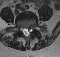 B
B 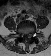 C D
C D
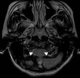
(A) Axial T1-weighted image (WI) at the level of L4-L5 demonstrates an oval lesion in the left lateral recess with a rim of low signal and central heterogeneous high signal (asterisk). The left L5 nerve root cannot be separated from the mass. (B) Axial T1WI shows heterogeneous high signal in the left lateral recess lesion, which abuts the facet joint (arrow). (C) Sagittal T2WI shows the close relationship of the lesion with the facet joint (arrow). There is no contact with the intervertebral disk. (D) Sagittal T1WI shows the close relationship of the lesion with the facet joint (arrow). There is no contact with the intervertebral disk.
■ Di erential Diagnosis
•Facet synovial cyst: Facet synovial cysts frequently occupy the lateral recess and may extend to central spinal canal. Cysts can contain synovial serous fluid, more gelatinous material, air, or blood. They are continuous with the facet joint but may abut the disk as well.
•Disk herniation: This causes a focal contour abnormality along the posterior disk margin that displaces the epidural fat and sometimes the dural sac and nerve roots. The herniation is usually contiguous with the rest of the disk, but free fragments are possible.
•Meningioma: This is usually an intradural extramedullary mass. It appears isointense to the spinal cord on both T1WIs and T2WIs but may be hypointense on T1WIs and hyperintense on T2WIs. Homogeneous intense enhancement is present.
■ Essential Facts
•Facet joints are frequently involved in spondyloarthrosis.
•The typical imaging findings are joint space narrowing, subchondral sclerosis, cysts, osteophytosis, ligament thickening, intra-articular vacuum, and joint fluid.
•Severe facet osteoarthritis can cause lateral recess and neural foramen stenosis.
•Facet joint degeneration is complicated by synovial cysts, which originate from the joint and can keep or lose their connection with the joint.
■ Other Imaging Findings
•Plain films can show degenerative changes; however, the anatomic complexity of this region requires computed tomography (CT) or magnetic resonance imaging (MRI) for a complete evaluation of the degenerative process.
•CT is more accurate for identifying bony abnormalities, but MRI more clearly shows the neural structures and soft tissues.
¸Pearls & ˚ Pitfalls
¸The role of facet joints in back pain is often di cult to
assess because the symptoms can be unspecific and imaging findings of degeneration are common.
¸In selected cases, nerve block or steroid and anesthetic injections of the facet joint are useful for diagnostic and
therapeutic purposes.
˚Spondylolysis may result in heterogeneous signal in the vicinity of the facets and should not be mistaken for synovial cyst formation.
179
Case 90
A B
C 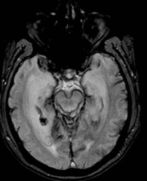
 D
D
■ Clinical Presentation
A 57-year-old man with three prior episodes of intraventricular hemorrhage and secondary hydrocephalus, now presenting with bilateral hearing loss and ataxia.
180 |
RadCases.thieme.com |
|
RadCases Neuro Imaging |
|
|
■ Imaging Findings |
|
|
|
A |
B |
C |
|
D |
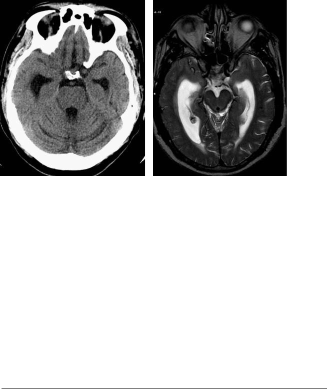
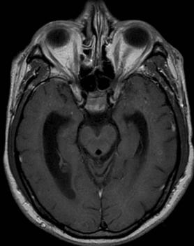
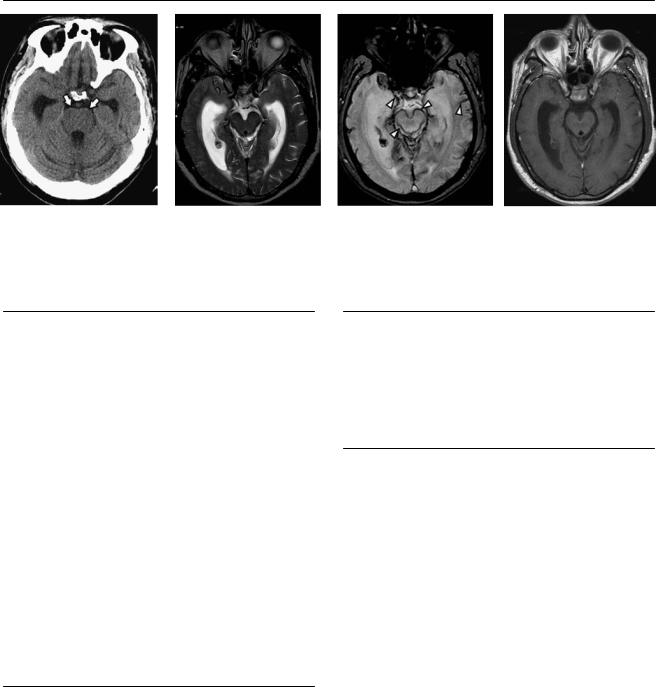
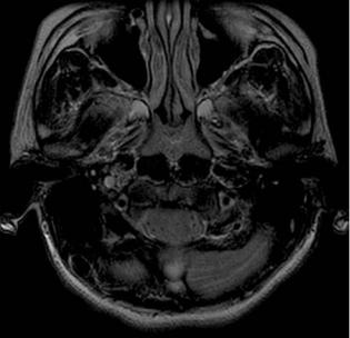
(A) Axial computed tomography without contrast shows a thin rim of high intensity on the anterior aspect of the pons (arrows). There is cerebellar atrophy with prominence of the sulci and hydrocephalus. (B) Axial fast spin-echo (FSE) T2-weighted image (WI) shows a rim of low intensity on the surface of the central nervous system. (C) Axial gradient-echo (GRE) T2*WI of the brain shows a thick rim of dark signal on the surface of the central nervous system (arrowheads). (D) Axial T1WI with contrast shows no abnormal enhancement.
■ Di erential Diagnosis
•Superficial siderosis: Deposits of hemosiderin on the surface of the central nervous system (CNS) occur in some patients after multiple episodes of intraventricular or subarachnoid hemorrhage. A rim of low intensity in the surface of the CNS is best seen on GRE T2*WIs. There is no enhancement.
•Leptomeningeal melanin: An increased amount of melanin in the leptomeninges can be an anatomic variant. It is seen as a thick, T2-hypointense rim on the surface of the medulla oblongata. GRE T2*WIs do not show these findings. Evaluation of the degree of skin pigmentation (individuals with heavier skin pigmentation demonstrate denser leptomeningeal pigmentation) and the absence of any other pathologic leptomeningeal characteristics may aid in the di erentiation of this entity from ironor melanincontaining di use leptomeningeal diseases.
•Magnetic resonance artifacts: T1 or T2 shortening of the brain surface is seen as a rim of hypointensity of variable thickness on the surface of the brain. This is evidenced only in certain sequences.
■Essential Facts
•Superficial siderosis is an uncommon condition.
•Reactive gliosis and demyelination a ect the cerebellum and cranial nerves I through VIII.
•A minimal rim of increased density will be present on the CNS surface, with cerebral and cerebellar atrophy.
■ Other Imaging Findings
•GRE T2*WIs show a hemosiderin rim on the surface of the CNS because of the susceptibility artifact of hemosiderin.
•FSE T2WIs show a dark rim on the CNS surface.
•Typically, no enhancement is seen.
¸Pearls & ˚ Pitfalls
¸Many times, superficial siderosis is an incidental finding,
manifesting only if gliosis is present.
¸ A history of repeated intracranial hemorrhage is the usual scenario.
˚Always ask for GRE T2*WIs if superficial siderosis is suspected because the sensitivity of the other imaging sequences for detecting this entity is poor.
˚In half of cases, the cause of siderosis is not identified.

181
Case 91
A 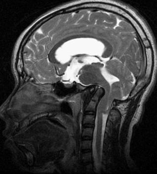
 B
B
C 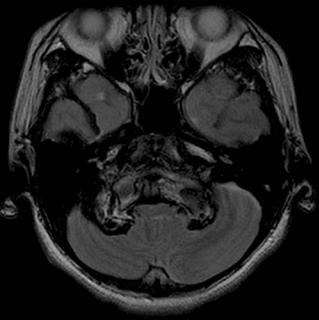
 D
D
■ Clinical Presentation
A 17-year-old with a history of hydrocephalus.

182 RadCases.thieme.com |
RadCases Neuro Imaging |
|
|
■ Imaging Findings |
|
A 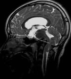 B
B  C
C 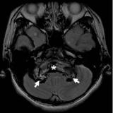
 D
D
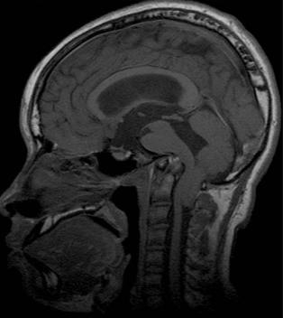
(A) Sagittal T2-weighted image (WI) of the brain: The basal angle measures 176 degrees. This angle is subtended by the junction of the nasion-tuberculum and tuberculum-basion tangents. Average is 134 to 135 degrees, minimum is 121 degrees, and maximum is 148 to 149 degrees. Consider platybasia if the angle is greater than 150 degrees. (B) Sagittal T1WI of the brain: The Chamberlain line extends from the posterior margin of the hard palate to the opisthion (posterior margin of the foramen magnum). It is abnormal if the tip of the odontoid projects more than 5 mm above the Chamberlain line. (C) Axial T2WI at the level of the foramen magnum demonstrates the tip of the dens (asterisk) at the same level as the occipital condyles (arrows). Note the compressive e ect on the medulla oblongata. (D) Axial T2WI again demonstrates the compressive e ect on the medulla oblongata and on the cerebellar tonsils (arrowheads), which are low-lying.
■ Di erential Diagnosis
•Basilar invagination and platybasia: This is a developmental abnormality in which the vertebral column is situated in an abnormally high position because of decreased height of the skull base. The odontoid tip lies more than 5 mm above the Chamberlain line (posterior margin of hard palate to opisthion); the anterior arch of C1 typically lies below. The tip of the odontoid is located more than 7 mm above the McGregor line (posterior margin of hard palate to the lowest point of the occipital squamosal surface); the anterior arch of C1 typically lies below. The base of the skull is congenitally flat.
•Chiari type I malformation: This is characterized by a caudal extension of the cerebellar tonsils below the foramen magnum. It may present in isolation or be associated with shortening of the clivus, basilar invagination, C1 assimilation, and fused cervical vertebrae (Klipper-Feil syndrome).
•Sagging brain: This is secondary to spinal hypotension and may present spontaneously or after spinal procedures; it may also be due to cerebrospinal fluid leak. The brainstem and 3rd ventricle “sag” inferiorly, and the pituitary gland is upwardly convex. If contrast is administered, dural thickening and enhancement are demonstrated.
■Essential Facts
•Basilar impression refers to the acquired form of basilar invagination, associated with conditions that soften the skull base, such as osteogenesis imperfecta, hyperparathyroidism, rickets, and Paget disease.
•Platybasia refers to flattening of the skull base. It may be present in patients with basilar invagination and basilar impression, but these conditions may exist in the absence of platybasia.
•Other abnormalities of the craniovertebral junction may coexist:
•Basiocciput hypoplasia (short clivus)
•Occipital condyle hypoplasia
•Atlanto-occipital nonsegmentation (i.e., assimilation or occipitalization of the atlas)
•Atlas anomalies
•Axis anomalies (os odontoideum, aplasia, or hypoplasia of the dens)
■ Other Imaging Findings
•Plain radiographs are inaccurate for measurements because of the superimposition of osseous structures.
•Computed tomography provides optimal visualization of the osseous structures.
•Magnetic resonance imaging (MRI) also permits assessment of the surrounding soft-tissue structures and cervicomedullary junction.
¸Pearls & ˚ Pitfalls
¸Numerous syndromes may involve the craniovertebral
junction and cause atlantoaxial instability:
•Klippel-Feil syndrome
•Down syndrome
•Connective tissue disorders, such as Ehlers-Danlos syndrome
•Inflammatory conditions such as rheumatoid arthritis and oropharyngeal infections
•Achondroplasia
•Mucopolysaccharidoses
•Osteogenesis imperfecta
˚In the assessment for platybasia, measurement of the basal angles with MRI yields lower values than those previously reported with traditional radiography.

183
Case 92
A 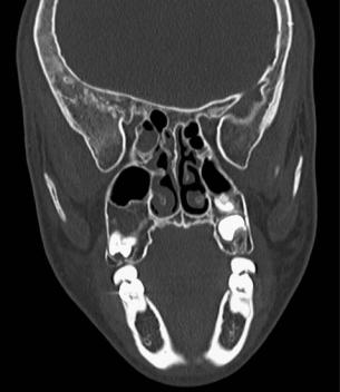
 B
B
■ Clinical Presentation
A 36-year-old woman with long-standing cosmetic deformity of the face.
Further Work-up
C 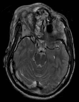
 D
D

184 RadCases.thieme.com |
RadCases Neuro Imaging |
|
|
■ Imaging Findings |
|
A  B
B 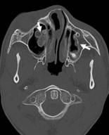 C
C 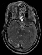
 D
D
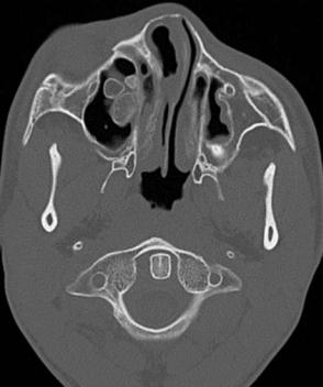
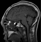
(A) Reformatted coronal computed tomography (CT) of the face shows replacement and enlargement of the bone marrow in the greater sphenoid wing and maxillary bones (asterisks), predominantly on the right, with a ground glass attenuation. Note the narrowing of the right superior orbital fissure (arrow) in comparison with the left. (B) Axial CT shows proliferation of abnormal bone marrow within the right maxillary sinus and lateral wall of the left maxillary sinus (arrows). Asymmetry of the face is evident. (C) Axial T2-weighted image (WI) through the orbital roof shows heterogeneous signal of the abnormal bone marrow. Note the elongation of the right optic nerve (arrows) and proptosis, caused by the enlarged orbital walls. (D) Sagittal T1WI shows that the areas of marrow replacement display relatively low signal (arrow). Areas of high T1 signal within the ethmoid complex may represent secretions or secondary retention of mucus (arrowhead). Note the magnetic susceptibility artifact in the region of the nasion, secondary to prior surgery.
■ Di erential Diagnosis
•Fibrous dysplasia (FD): FD presents with deformity of the face secondary to enlargement of multiple bones and abnormal attenuation of the bone. The skull base foramina may be distorted.
•Paget disease (PD): PD is a chronic progressive disease of unknown etiology in which initial destruction of bone is followed by a reparative process. The radiographic changes are progressive and include osteoporosis circumscripta, enlargement of the skull, thickening of the outer table, and finally complete sclerosis with loss of distinction between the diploë and calvarial bone tables.
•Chronic invasive fungal sinusitis: Inhaled fungal organisms invade the mucosa, submucosa, blood vessels, and bony walls of the paranasal sinuses. On CT, a hyperattenuating soft-tissue collection within one or more of the paranasal sinuses is present. It may be masslike, with destruction of the sinus walls and extension beyond the confines of the sinus. On magnetic resonance imaging (MRI), there may be decreased signal intensity on T1WIs and markedly decreased signal intensity on T2WIs.
■Essential Facts
•FD is a benign bone condition of unknown etiology in which normal bone is replaced by an abnormal proliferation of fibroconnective tissue.
•The three types are monostotic, polyostotic, and polyostotic with cutaneous and endocrine abnormalities (McCune-Albright syndrome).
•Craniofacial FD may a ect the base of the skull, orbits, maxillae, and mandibles; symptoms result from the mass e ect exerted by the lesion; cranial neuropathies may result from involvement of the foramina.
•The radiographic appearance includes intramedullary lesions that distort and expand the bone, mixed radiolucent and radiopaque lesions, “ground-glass” attenuation, and narrowing of the skull base foramina.
■ Other Imaging Findings
•CT shows patterns similar to those visible on plain radiography and assesses the compromise of the skull base foramina.
•MRI shows variable signal intensity, usually intermediate signal on T1WIs and hypointense signal on T2WIs.
•Scintigraphy demonstrates increased uptake of the radioisotope. It may be useful for the detection of subtle pathologic fractures.
¸Pearls & ˚ Pitfalls
¸Tips to di erentiate FD from PD:
•“Ground-glass” bone matrix is typical of FD but is not seen in PD.
•FD tends to a ect cranial bones asymmetrically, whereas the cranium is symmetric in PD.
•The paranasal sinuses are not usually involved in PD.
•The cortical tables are thick in PD (especially the inner table) but tend to be thin in FD.
˚Sites of high signal intensity on T2WIs in the frontoethmoid area should raise the possibility of a coexistent mucocele.
185
Case 93
A
B C
■ Clinical Presentation
A 30-week premature infant, now 3 months of age, presenting with increased head circumference.
Further Work-up
D
186 |
RadCases.thieme.com |
|
RadCases Neuro Imaging |
|
|
■ Imaging Findings |
|
|
|
A |
B |
C |
|
D |
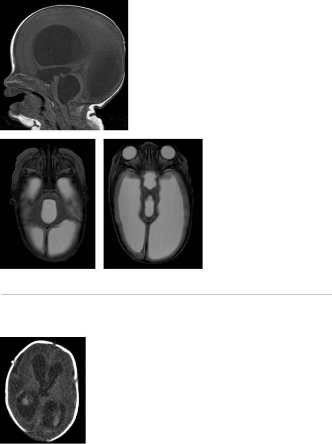
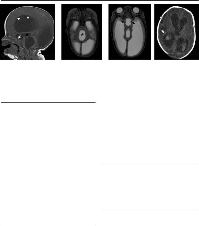
(A) Sagittal T1-weighted image (WI) shows dilatation of the entire ventricular system. Note the elongation and thinning of the corpus callosum (arrowheads), descent of the floor of the 3rd ventricle, which is obliterating the suprasellar cistern and abutting the pituitary gland (white arrow), and enlargement of the aqueduct (black arrow) and 4th ventricle. (B) Axial T2WI shows that the anteroposterior diameter of the 4th ventricle (asterisk) is significantly increased, resulting in flattening of the dorsal surface of the pons. (C) Axial T2WI shows that the dilated 3rd ventricle occupies the suprasellar cistern. The hypothalamic structures are splayed (arrows). (D) Computed tomography of the brain obtained 3 months earlier, when the patient was 2 weeks old. Intraventricular hemorrhage and mild ventricular dilatation are evident. Note the parenchymal hemorrhage in the right periventricular white matter (arrow).
■ Di erential Diagnosis
•Communicating hydrocephalus: Enlargement of the ventricular system and obliteration of the extra-axial spaces. There may be a history of prior intraventricular hemorrhage or meningitis.
•Hydranencephaly: The cerebral hemispheres are replaced by cerebrospinal fluid (CSF), with preservation of the thalami, inferior aspect of the frontal lobes, and inferior and medial aspect of the temporal lobes. Atrophy of the brainstem may be present. The cerebellum is usually normal. The key to distinguishing hydrocephalus from hydranencephaly is the presence of a thin rim of residual cerebral cortical tissue in hydrocephalus that is not present in hydranencephaly. Vascular, infectious, and genetic causes have been implicated.
•Severe brain atrophy: Enlargement of the ventricular system and prominent extra-axial spaces, secondary to loss of brain volume, are characteristic. Elongation of the corpus callosum, descent of the 3rd ventricular floor, ballooning of the 3rd ventricle, and lateral displacement of the hypothalamic structures are indicative of increased pressure within the ventricles and are absent in passive ventricular enlargement.
■Essential Facts
•Hydrocephalus is defined as ventricular dilatation and increased pressure resulting from an imbalance between CSF production and absorption.
•It is associated with the following:
•Ventricular enlargement
•E acement of the extra-axial spaces
•Dilatation of the 3rd ventricle, downward displacement of 3rd ventricular floor, and splaying of the hypothalamic nuclei
•Transependymal flow of CSF
•Communicating hydrocephalus:
•The CSF flow blockage is outside the ventricular system (e.g., basal cisterns or parasagittal arachnoid villi).
•Causes include meningitis, subarachnoid hemorrhage, meningeal carcinomatosis, and abnormal skull base.
•Noncommunicating hydrocephalus:
•There is intraventricular obstruction (at or proximal to the 4th ventricular outlets).
•The dilatation of the segments of the ventricular system is proximal to the obstruction.
■ Other Imaging Findings
•CSF flow voids are accentuated on magnetic resonance imaging (MRI).
•Signal abnormalities can be found within the corpus callosum after ventricular shunting and are probably caused by compression of the corpus callosum against the falx before ventricular decompression.
¸Pearls & ˚ Pitfalls
¸Rapid-sequence MRI has been used for the nonemergent
imaging evaluation of pediatric hydrocephalus.
¸MRI yields reliable visualization of the ventricular catheter and o ers superior anatomic detail while limiting
exposure to radiation.
˚Periventricular edema due to transependymal CSF flow or hydrostatic stasis from elevated intraventricular pressure may be evident as blurred or ill-defined ventricular margins. This finding favors acute or subacute or progressive hydrocephalus; however, in an infant, the normally high water content of the immature white matter may obscure the edema.
187
Case 94
A B
C 
 D
D
■ Clinical Presentation
A 72-year-old with gait disturbance and memory loss.