

Книги по МРТ КТ на английском языке / Neuro Imaging Redcases
.pdf28 |
RadCases.thieme.com |
RadCases Neuro Imaging |
|
■ Imaging Findings |
|
A B C
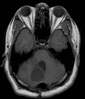
(A) Axial postcontrast image of the posterior fossa demonstrates bilateral enhancing masses in the cerebellopontine angle (CPA) cistern. On the left side, an intracanalicular component is noted (arrow). Two additional enhancing masses with central areas of fluid signal (asterisk) near the torcular and in the right cerebellum showed dural attachment in the tentorium on other images. (B) Coronal postcontrast image confirms the presence of CPA masses with an intracanalicular component and shows an additional enhancing lesion in the left parafalcine region with broad attachment to the dura (arrow). (C) Axial postcontrast image reveals another tiny dura-based mass in the left frontal convexity (arrow).
■ Di erential Diagnosis
•Neurofibromatosis type 2 (NF2): Bilateral vestibular schwannomas are a defining feature of this disease. Additionally, multiple meningiomas are identified.
•Von Hippel-Lindau syndrome: This presents with multiple retinal and cerebellar hemangioblastomas. The lesions are parenchymal tumors, unlike the masses in the present case.
•Dural metastasis: Dural metastases are similar in appearance to meningiomas, but they are unusual in the CPA.
■Essential Facts
•A defect in the NF2 gene on chromosome 22q11 is inherited in an autosomal-dominant fashion.
•Intracranial lesions include vestibular schwannomas (in 95% of adults, they occur in the internal auditory canal or porus acusticus); multiple meningiomas; and calcifications of the choroid plexus, cerebellar cortex, and occasionally cerebral cortex.
•Spinal lesions include cord ependymomas, multiple schwannomas of exiting nerve roots, and meningiomas.
■ Other Imaging Findings
•Plain radiographs are helpful in the evaluation of spinal curvature deformities, an expanded neural foramen, and scalloping.
•On computed tomography, neurofibromas appear as hypodense, enhancing masses. (Hypoattenuation may be related to lipid content.)
•Magnetic resonance imaging is excellent for the evaluation of spinal deformities and masses.
¸Pearls & ˚ Pitfalls
¸Cutaneous manifestations are less frequent in NF2 than
in NF1; therefore, the patients are older at the time of diagnosis.
¸Multiple masses along the exiting spinal nerve roots can be features of both NF1 and NF2. In NF2, these are usually
schwannomas.
˚Multiple tumors in a young patient should always raise suspicion for phakomatosis.
29
Case 15
A
■ Clinical Presentation
A 19-year-old man with a history of seizures presenting with progressive mental status changes.
Further Work-up
B C D
30 |
RadCases.thieme.com |
RadCases Neuro Imaging |
|
■ Imaging Findings |
|
A B C D
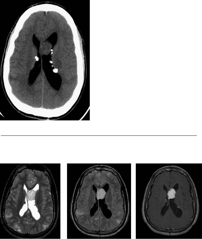
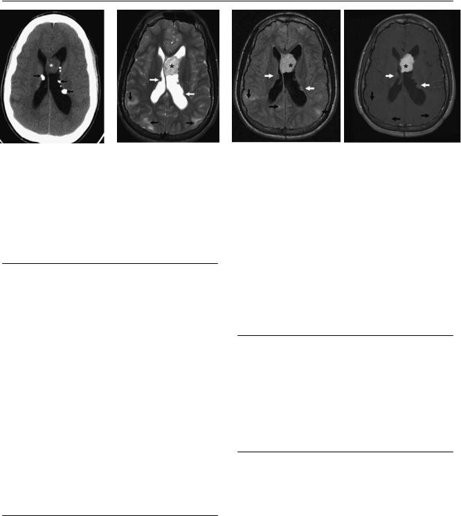
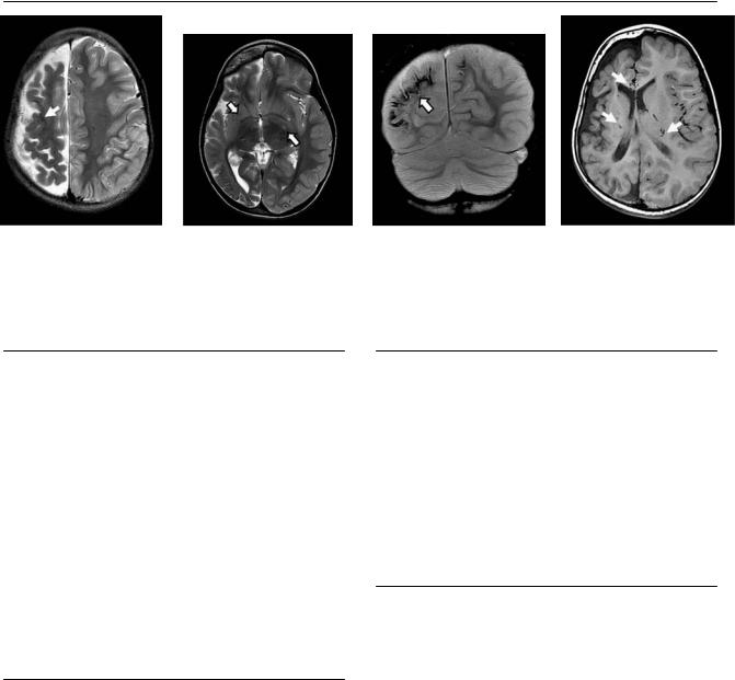
(A) Axial computed tomography (CT) scan of the head without contrast shows multiple subependymal calcified nodules (arrows). A mass is seen in the left lateral ventricle adjacent to the septum pellucidum (asterisk). Note the dilatation of the left ventricle due to obstruction of the foramen of Monro on that side. (B) Axial T2-weighted image (WI) of the brain shows multiple subependymal nodules (white arrows). A giant cell astrocytoma with heterogeneous signal (asterisk) is deforming the septum pellucidum. Multiple subcortical and cortical tubers are seen as areas of hyperintensity (black arrows). (C) Fluid-attenuated inversion recovery (FLAIR) image of the brain shows multiple subependymal nodules (white arrows). A giant cell astrocytoma with heterogeneous signal (asterisk) is deforming the septum pellucidum. Multiple subcortical and cortical tubers are seen as areas of hyperintensity (black arrows). (D) Axial T1-weighted postcontrast magnetic resonance imaging (MRI) of the brain shows multiple enhancing subependymal nodules (white arrows). An enhancing giant cell astrocytoma (asterisk) is displacing the septum pellucidum on the right. Multiple subcortical and cortical tubers do not show enhancement (black arrows).
■ Di erential Diagnosis
•Tuberous sclerosis complex (Bourneville-Pringle syndrome): In this diagnosis, look for cortical and subcortical tubers, subependymal nodules, subependymal giant cell astrocytomas, and white matter radial migration lines (heterotopic glia).
•Central neurocytoma: Central neurocytoma is in the differential diagnosis for a frontal intraventricular mass. This tumor consists of a well-circumscribed mass that is confined to the anterior portion of the lateral ventricles. No subependymal nodules or cortical tubers are present.
There are punctate calcifications within the mass. Multiple small cysts can be seen. There is mild to moderate contrast enhancement. Central neurocytomas rarely extend outside the ventricles.
•Congenital cytomegalovirus infection: Periventricular and parenchymal calcifications are seen in congenital cytomegalovirus infection, as are microcephaly, cortical gyral abnormalities (agyria and thin cortex), hippocampal dysplasia, and cerebellar hypoplasia.
■Essential Facts
•Tuberous sclerosis complex is a phakomatosis caused by abnormal di erentiation of the cells of the germinal matrix and is associated with migrational neuronal arrest.
•Cortical and subcortical tubers are dysmorphic neurons. Ninety percent are cerebral and 10% are cerebellar. Only 3% enhance.
•Subependymal nodules are seen in the walls of the lateral ventricles. They calcify and may enhance. Malignant degeneration to giant cell astrocytoma occurs in 10 to 15%.
•Microcephaly is usually present as a consequence of brain volume loss.
•Tuberous sclerosis is associated with rhabdomyoma of the heart, angiomyolipomas of the kidneys (50%), drusen, and cystic lesions.
■ Other Imaging Findings
•Computed tomography can demonstrate calcification in the subependymal nodules and ventriculomegaly.
•On MRI, cortical and subcortical tubers appear as areas of high signal on T2WIs and FLAIR images.
•Positron emission tomography can identify epileptogenic tubers (tubers that are disproportionately hypometabolic for their size).
¸Pearls & ˚ Pitfalls
¸Tubers are multiple in 95% of patients.
¸Ninety percent of tubers are located in the frontal lobes.
˚In newborns, the nodular subependymal lesions appear hyperintense on T1WIs and hypointense on T2WIs (the reverse of what is seen in adults) because of the lack of myelination.

31
Case 16
A 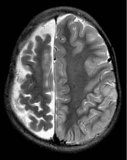
 B
B
C 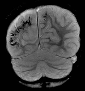
 D
D
■ Clinical Presentation
A 17-year-old girl with seizures, hemiparesis, and homonymous hemianopsia.
32 |
RadCases.thieme.com |
|
RadCases Neuro Imaging |
|
|
■ Imaging Findings |
|
|
|
A |
B |
C |
|
D |
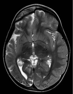
(A) Axial T2-weighted image (WI) of the brain demonstrates atrophy of the right cerebral hemisphere and linear areas of decreased signal along the cortex (arrow). (B) Axial T2WI of the brain demonstrates atrophy of the right cerebral hemisphere. Flow voids of dilated ependymal veins are also noted (arrows).
(C) Coronal gradient-echo image reveals tram-track cortical calcifications in the atrophic right hemisphere (arrow). Less extensive changes are present in the left parietal cortex. (D) Note the dilated medullary veins, which appear as large flow voids on the axial T1WI (arrows).
■ Di erential Diagnosis
•Sturge-Weber syndrome (also known as encephalotrigeminal angiomatosis): There is atrophy of the a ected hemisphere with tram-track cortical calcifications. The calvarium is thickened.
•Cortical laminar necrosis from old hemispheric infarct: More significant volume loss and dilatation of the ventricles would be expected in this diagnosis. Dilatation of the medullary and subependymal veins is not a feature of cortical laminar necrosis.
•Superficial siderosis: Hemosiderin is deposited along the leptomeninges, subpial tissues, and subependyma of the ventricles as a result of chronic, recurrent subarachnoid hemorrhage. T2-weighted and gradient-echo images reveal hypointensity along the pial surface of the brain and spinal cord and along the ependyma of the ventricles.
■Essential Facts
•The postcapillary circulation of the brain and face is abnormal.
•A facial port wine stain and choroidal, scleral, or episcleral telangiectasia are characteristic.
•Abnormal venous drainage of the hemispheres results in the recruitment of collateral veins from the choroid plexus and medullary veins.
•Chronic cerebral ischemia leads to atrophy and calcification.
■ Other Imaging Findings
•On computed tomography (CT), tram-track calcification is unusual before 2 years of age.
•Magnetic resonance imaging shows contrast enhancement of the subarachnoid space, calcifications on T2and T2*WIs, enlargement of the choroid plexi, and T2 prolongation in the adjacent white matter.
•Functional neuroimaging with fluorodeoxyglucose positron emission tomography often demonstrates cortical hypometabolism extending beyond the apparent structural abnormalities in Sturge-Weber syndrome.
¸Pearls & ˚ Pitfalls
¸The Dyke-Davido -Masson sequence consists of unilat-
eral skull and sinus hypertrophy (mainly the frontal sinus), then atrophy of the mastoid air cells and ipsilateral cerebral hemisphere (in childhood). Causes are cerebral infarction (most often), infection, trauma, and SturgeWeber syndrome.
˚Tram-track calcifications of the brain may mask contrast enhancement on CT.
33
Case 17
A 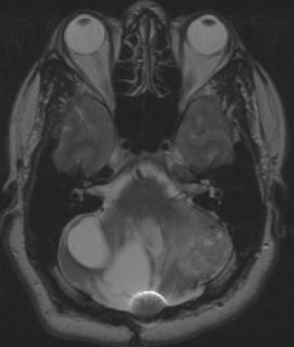
 B
B
C D
■ Clinical Presentation
A 35-year-old man with the history of a resected tumor in the cerebellum presenting with worsening dysmetria.
34 |
RadCases.thieme.com |
RadCases Neuro Imaging |
|
■ Imaging Findings |
|
A B C D
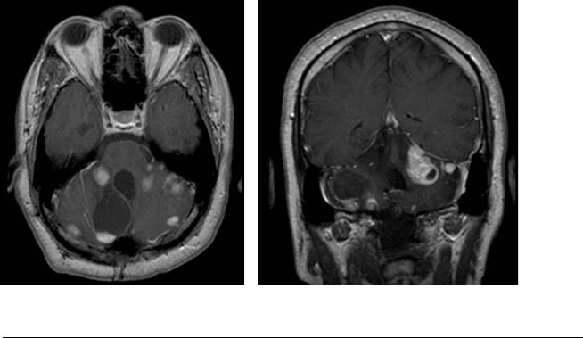
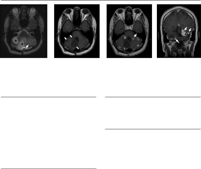
(A) Axial T2-weighted image (WI) demonstrates multiple cystic lesions (asterisks) in the cerebellum bilaterally and areas of encephalomalacia on the left side. Note the magnetic susceptibility artifact near the occipital bone resulting from prior surgery (arrow). (B) Axial T1WI of the brain without contrast showing well-defined cystic lesions with signal slightly higher than that of cerebrospinal fluid (arrows). (C) Axial postcontrast T1WI shows numerous enhancing solid lesions (arrow) and a mural nodule (asterisk) along the wall of one of the cerebellar cysts. (D) Coronal postcontrast T1WI shows numerous enhancing solid lesions (arrows) and a mural nodule (asterisk) along the wall of one of the cerebellar cysts.
■ Di erential Diagnosis
•Von Hippel-Lindau (VHL) disease (also known as hemangioblastomatosis): Multiple hemangioblastomas in the posterior fossa, which are cystic with a mural nodule, are characteristic of this diagnosis. No surrounding vasogenic edema is present.
•Metastatic disease: this presents as numerous mass lesions with surrounding edema and mass e ect.
•Multiple brain abscesses: These are characterized by multiple necrotic, thick, ring-enhancing lesions with significant edema and without a mural nodule. It is unusual for only the cerebellum to be involved.
■Essential Facts
•The VHL gene on chromosome 3 is inherited in an autoso- mal-dominant fashion.
•Central nervous system (CNS) neoplasms include hemangioblastomas in the cerebellum, brainstem, spinal cord, and retina as well as endolymphatic sac tumors.
•Non-CNS lesions include clear cell renal carcinoma, pheochromocytoma, pancreatic neuroendocrine tumors, and epididymal cystadenoma.
•Hemangioblastomas are solid, cystic, hemorrhagic, or mixed. They are frequently cystic with a mural enhancing nodule.
•Hemangioblastomas may have flow voids from feeding arteries.
■ Other Imaging Findings
•Hemangioblastomas are highly vascular lesions, demonstrating arterial blush on conventional angiography. There is no arteriovenous shunting.
¸Pearls & ˚ Pitfalls
¸Hemangioblastomas may occur sporadically; however, 20
to 38% of cerebellar hemangioblastomas are associated with VHL disease.
¸ Spinal hemangioblastomas are infrequent and highly suggestive of VHL disease.
˚Spinal cord hemangioblastomas may be mistaken for drop metastases from a cerebellar hemangioblastoma.
35
Case 18
A 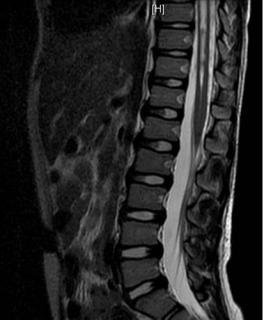
 B
B
C D
■ Clinical Presentation
A 7-year-old boy with neck paresthesias and hand tingling.
36 |
RadCases.thieme.com |
|
RadCases Neuro Imaging |
|
|
■ Imaging Findings |
|
|
|
A |
B |
C |
|
D |
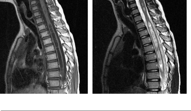
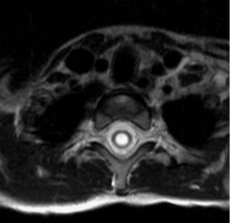
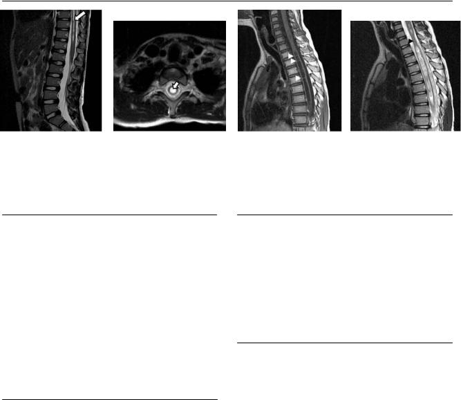
(A) Sagittal T2-weighted image (WI) of the lower spine demonstrates a well-defined central area of fluid signal within the cord, which tapers toward the conus medullaris (arrow). (B) Axial T2WI in the cervical region shows a central fluid cavity within the cord with well-defined margins (arrow). There is mild cord expansion. (C) Sagittal T1WI shows extension of the cystic cavity to the cervical cord and multiple septa (arrowheads).
(D) Sagittal T2WI of the upper spine demonstrates expansion of the cord in the area that contains the fluid cavity, with a waist in the region where the cord is normal (arrowhead).
■ Di erential Diagnosis
•Syringohydromyelia: This is characterized by a longitudinally oriented cavity within the spinal cord, with cerebrospinal fluid (CSF) signal on all the sequences. There is no solid component or enhancement.
•Myelomalacia: Myelomalacia has moderately increased T2 signal and moderately reduced T1 signal relative to the cord, poorly defined margins, and no CSF flow artifacts.
•Cystic astrocytoma: Cystic astrocytoma is an eccentrically located lesion with low T1 signal and high T2 signal relative to the cord. It has a cystic component in 25 to 38% of cases. If the cystic component is lined with tumor cells, it usually demonstrates peripheral wall enhancement, unlike a secondary syrinx.
■Essential Facts
•A longitudinally oriented, fluid-filled cavity originates in the spinal cord tissue or in the central canal.
•Distinguishing between hydromyelia (dilatation of the central canal) and syringomyelia (the cavity is lateral to the central canal) is frequently not possible.
•Causes may be congenital, tumor-related (astrocytoma, glioma, ependymoma), trauma-related, or infective/inflammatory.
■ Other Imaging Findings
•Magnetic resonance imaging shows single or multiple cystic intramedullary collections with CSF signal on all sequences, tapered cranial and caudal ends, and expansion of the cord. The lesion may have a beaded appearance.
•Atypical signal: The T1 signal is high when the lesion contains infected or proteinaceous material. An artifactual low T2 signal is due to CSF pulsation.
¸Pearls & ˚ Pitfalls
¸When a syrinx is initially found on imaging, the full
craniocaudal extent needs to be assessed. If no obvious cause is identified, gadolinium-enhanced images must be obtained in an attempt to detect any underlying tumor.
¸Syrinx cavities that are secondary to Chiari malformations, foramen magnum stenosis, or tumor may resolve
after blockage of the CSF circulation is resolved.
˚Gibbs or truncation artifacts are bright or dark lines that are seen parallel and adjacent to the borders of abrupt changes in intensity, such as the transition from bright CSF to dark spinal cord on a T2WI. In the spinal cord, this artifact can simulate a small syrinx to the unaware.
˚Ventriculus terminalis is a nonenhancing, ovoid, nonseptate cystic structure localized within a normally positioned conus; it is seen frequently in patients younger than 5 years of age.
37
Case 19
A B
C  D
D
■ Clinical Presentation
A 40-year-old woman with progressive leg weakness that has worsened in the last year.