

Книги по МРТ КТ на английском языке / Neuro Imaging Redcases
.pdfAcknowledgments
I have to thank so many people for helping in the preparation of this book. To Santiago Restrepo for introducing us to this exciting project. To my coauthor Eliana for her support and endless drives to the island to complete cases. To all my mentors who had the patience to guide me to this point: Miguel Ruiz, Leonidas Borrero, Ramon Figueroa, Rafael Rojas, and Leonard Swischuk. I thank Dr. Faustino Guinto, who created the amazing teaching file of the University of Texas Medical Branch (UTMB) neuroradiology and taught me so many things, and Gregory Chaljub and Greg Katzman for their support.
To my colleagues in neuroradiology at UTMB, who contributed to the cases and supported me during the preparation of the material. Special thanks to all the neuroradiology fellows and residents who have inspired me on a daily basis and were the fuel to make this project a reality. Mul-
I have immense gratitude for my partner Roy Riascos for inviting me on this stimulating journey. This project would have not been possible without all of the help from my mentors and supportive colleagues, or without the forgiveness and patience of my loving family. Two of my professors departed, but the drive and wisdom that they gave me remained. God bless Dr. Edwin D. Cacayorin and Dr. Joel Yeakley. I thank Leo Hochhauser, Clark Sitton, Emilio Supsupin, and Alex Simonetta, my benevolent colleagues, who have been strength-
tiple medical students helped to arrange our neuroradiology teaching file, and without their e ort this book would not be possible.
Some of the cases were obtained from the Medical College of Georgia, Charity Hospital in New Orleans, and The Children’s Hospital in New Orleans—my thanks to everybody who contributed in our search for cases. This project would have not been possible without the support of my family, who has lived through the long hours of preparation of the material. To my parents Lucy and Roy for helping me become who I am.
To Tim Hiscock and Adriana di Giorgio at Thieme Medical Publishers for their everlasting patience.
Thank you.
—Roy Riascos
ening our department, contributing to the teaching file, and continuously encouraging me with this endeavor. I thank my Colombian teachers, Anibal Morillo, Maria Isabel Mantilla, and Sonia Bermudez, who opened up my mind to this fascinating world of neuroradiology. And of course, my beloved parents, Juan and Ester, who taught me what can be found at the other end of persistence.
—Eliana Bonfante
1
Case 1
A
B C
■ Clinical Presentation
A 14-year-old girl presenting with occipital headaches during Valsalva maneuver and anesthesia in both arms.
2 |
RadCases.thieme.com |
RadCases Neuro Imaging |
|
■ Imaging Findings |
|
A B
|
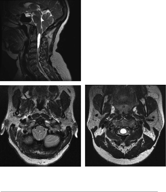
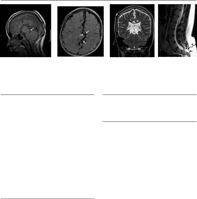
(A) Midsagittal T2-weighted image (WI) at the foramen magnum demonstrates a 12-mm de- |
|
scent of the tip of the cerebellar tonsils (arrow) below a line between the basion and the opis- |
|
thion. A cervical syrinx is also noted. (B) Axial T2WI at the foramen magnum demonstrates |
|
ectopia of the cerebellar tonsils and e acement of the subarachnoid space. (C) Axial T2WI of |
C |
the cervical spine shows a syrinx (arrow). |
■ Di erential Diagnosis
•Chiari type I malformation: This patient has the typical peglike pointed tonsils displaced into the upper cervical canal with associated syringomyelia.
•Chiari type II malformation: Chiari type II malformation presents with deformity of the brainstem and enlargement of the foramen magnum. It is associated with myelomeningocele.
•Spinal hypotension syndrome: This is characterized by downward displacement of the cerebellar tonsils secondary to low pressure within the spinal canal. The brainstem and 3rd ventricle “sag” inferiorly, and the pituitary gland is upwardly convex. If contrast is administered, dural thickening and enhancement are demonstrated.
•Associated anomalies are shortening of the clivus, basilar invagination, C1 assimilation, and fused cervical vertebrae (Klipper-Feil syndrome).
•Cerebrospinal fluid flow studies may be useful if the imaging findings or clinical symptoms are not typical.
¸Pearls & ˚ Pitfalls
¸On axial images, visualization of the cerebellar tonsils at
the level of the dens is indicative of ectopia.
˚Patients with long-standing compensated hydrocephalus may present with descent of the cerebellar tonsils and symptoms similar to those of patients with Chiari type I malformation.
■ Essential Facts
•Chiari type I malformation is defined as caudal extension of the cerebellar tonsils 5 mm below the foramen magnum.
•Syringomyelia is seen in one-half of the patients.
•It is not associated with other neurulation malformations.

3
Case 2
A B
C 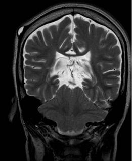
 D
D
■ Clinical Presentation
A 25-year-old patient initially presenting with lower extremity paralysis, respiratory distress, and impaired swallowing.
4 |
RadCases.thieme.com |
|
RadCases Neuro Imaging |
|
|
■ Imaging Findings |
|
|
|
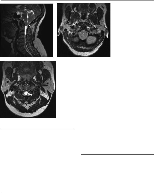
A |
B |
C |
|
D |
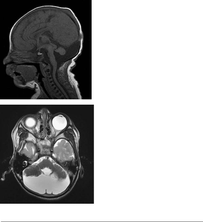
(A) Sagittal T1-weighted image (WI) of the brain shows inferior protrusion of the cerebellar vermis through a widened foramen magnum (asterisk). Note the tectal beaking (arrow) and dysgenesis of the corpus callosum (arrowhead). The 4th ventricle is e aced. (B) Axial fluid-attenuated inversion recovery image of the brain demonstrates an interdigitation of the sulci of the brain in the midline (arrows) secondary to hypoplasia of the cerebral falx. (C) Coronal T2WI of the brain shows a towering cerebellum and inferior tilting of the cerebellar tonsils through the foramen magnum (white arrows) along with the vermis (asterisk). (D) Sagittal T2WI of the spine shows posterior dysraphism of the sacrum (arrows) with an associated sinus tract (arrowhead).
■ Di erential Diagnosis
These three pathologic conditions are associated with crowding of the foramen magnum:
•Chiari type II malformation: Chiari type II malformations are complex developmental disorders of the posterior brain that can be associated with closure defects of the neural tube, myelomeningocele, and dysgenesis of the corpus callosum.
•Chiari type III malformation: Chiari type III malformations have the same characteristics as Chiari type II malformations but are associated with encephalocele instead of myelomeningocele.
•Medulloblastoma: Medulloblastomas are malignant primitive neuroectodermal tumors of the posterior fossa that arise from the roof of the 4th ventricle. They can be similar to Chiari type II malformations, as they e ace the 4th ventricle and can be isointense to the cerebellum on T1-weight- ed noncontrasted images. Contrast helps to di erentiate them, as more than 90% of medulloblastomas enhance.
■ Other Imaging Findings
•Magnetic resonance imaging is the imaging modality of choice for the characterization of Chiari malformations.
¸Pearls & ˚ Pitfalls
¸Myelomeningocele and dysgenesis of the corpus callo-
sum are seen in almost all cases of Chiari type II malformation.
˚A complete evaluation of the spine is needed when posterior fossa defects are detected, as posterior dysraphism may not be clinically evident.
˚Early detection and surgical treatment of a Chiari type II malformation will reduce the risk for permanent neurologic defect.
■ Essential Facts
•Chiari type II malformations are associated with defects of the following structures:
•Skull: lacunar skull; concave clivus; low-riding torcula; wide foramen magnum and upper cervical canal
•Dura: fenestrated falx causing interdigitating sulci; heartshaped incisura; hypoplastic cerebellar tentorium
•Posterior fossa: towering cerebellum; downward vermian displacement; slitlike 4th ventricle; tectal beaking; wrapping of cerebellum around brainstem; medullary kink
•Other common associated defects: myelomeningocele (90%); dysgenesis of the corpus callosum (85%); hydrocephalus (75%); syringomyelia (50%); aqueductal stenosis (50%); holoprosencephaly; tethered cord
5
Case 3
A 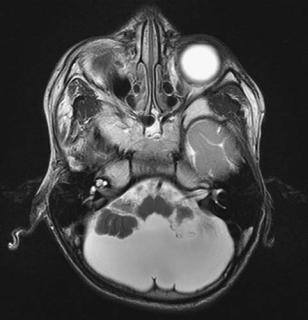 B
B
C
■ Clinical Presentation
A 4-year-old boy with developmental delay and seizures.
6 |
RadCases.thieme.com |
RadCases Neuro Imaging |
|
■ Imaging Findings |
|
A B C
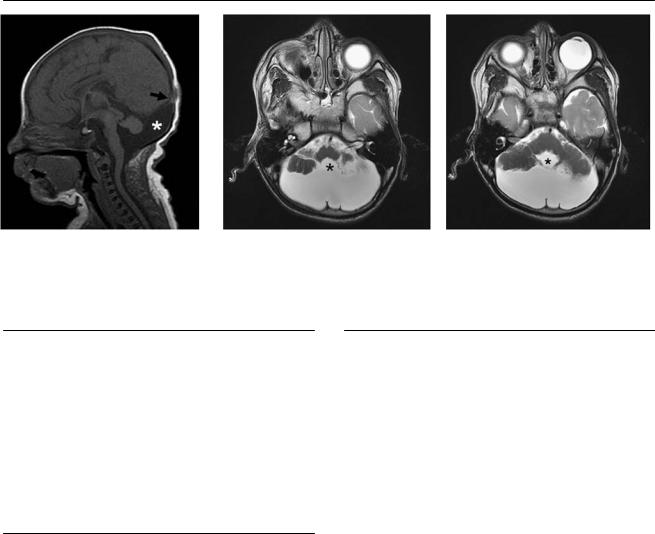
(A) Sagittal T1-weighted image (WI) of the brain demonstrates a large posterior fossa and an elevated insertion of the tentorium (arrow) with respect to the expected position. A cyst (asterisk) that communicates with the 4th ventricle fills the posterior fossa. (B,C) Axial T2WIs of the brain show cystic dilatation of the 4th ventricle (asterisks), which is ballooning between hypoplastic cerebellar hemispheres. The cerebellar vermis is absent.
■ Di erential Diagnosis
•Dandy-Walker continuum: The high insertion of the tentorium and cystic dilatation of the 4th ventricle are typical features of Dandy-Walker continuum. The degree of cerebellar hypogenesis may vary.
•Mega cisterna magna: Enlargement of the cisterna magna and posterior fossa without abnormality in the 4th ventricle or vermis is typical of this diagnosis.
•Arachnoid cyst: The 4th ventricle and vermis are normal but displaced by the cyst.
■Essential Facts
•An enlarged posterior fossa and a high position of the tentorium with upward displacement of the lateral sinuses and torcular Herophili are common in Dandy-Walker continuum.
•Varying degrees of cerebellar agenesis or hypogenesis are seen.
•As a result of cystic dilatation of the 4th ventricle, it fills nearly the entire posterior fossa.
•Cerebellar hypoplasia: hypogenesis of the cerebellar vermis and cystic dilatation of the 4th ventricle without posterior fossa enlargement were formerly known as Dandy-Walker variant.
•Associated anomalies, present in 70% of cases, include the following:
•Agenesis of the corpus callosum, migration anomalies, cephalocele, holoprosencephaly, hydrocephalus, and porencephaly, among others
¸Pearls & ˚ Pitfalls
¸Conditions associated with abnormal cerebellar size:
•Cerebellar atrophy: the cerebellum is small and the cerebellar fissures are enlarged.
•Cerebellar hypoplasia: the cerebellum is small, but the fissures are normal in size compared with the folia.
•Cerebellar dysplasia: disorganized development; an abnormal folial pattern ranging from absence of fissures to large folia with shallow fissures to foliation in abnormal directions or heterotopic nodules of gray matter may be present.
•Cerebellar hypogenesis: the superior portions of the cerebellum are formed, but the inferior portions are not; this term should not be used because it overlaps
with dysplasia.
˚An isolated 4th ventricle, which is the result of obstruction of both the aqueduct of Sylvius and the 4th ventricular outflow foramina, may give the appearance of a hypoplastic cerebellum. The lateral and 3rd ventricles
appear slightly enlarged or normal if there is a functioning ventriculoperitoneal shunt. The distal aqueduct may be enlarged.

7
Case 4
A
■ Clinical Presentation
A 12-year-old boy with slight cognitive defects.
Further Work-up
B  C D
C D