

Книги по МРТ КТ на английском языке / Neuro Imaging Redcases
.pdf58 |
RadCases.thieme.com |
|
RadCases Neuro Imaging |
|
|
■ Imaging Findings |
|
|
|
A |
B |
C |
|
D |
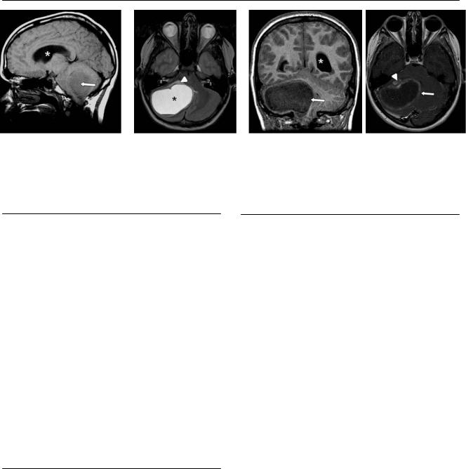
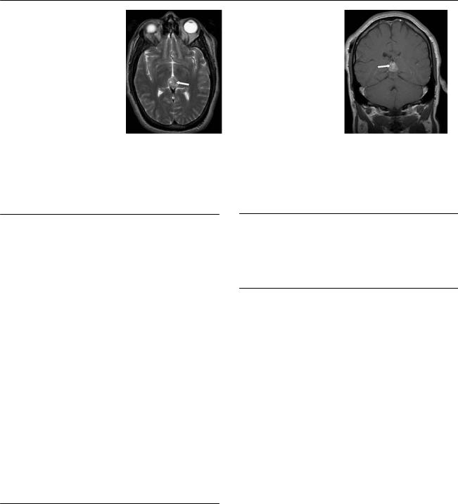
(A) Sagittal T1-weighted image (WI) shows noncommunicating hydrocephalus with dilated lateral and 3rd ventricles (asterisks). The 4th ventricle is not dilated (arrow), and the anatomy of the tectum is distorted (arrowhead). (B) Axial fluid-attenuated inversion recovery (FLAIR) image shows hyperintensity in the midbrain (arrow). The lateral and 3rd ventricles are dilated (asterisks). (C) Axial T1WI with contrast shows no enhancement in the midbrain (arrow). Hydrocephalus is seen (asterisks). (D) Axial FLAIR image shows normal size of the lateral ventricles (asterisks) after decompression through a shunt. The area of hyperintensity in the midbrain is again seen (arrow).
■ Di erential Diagnosis
•Tectal glioma: These are focal or infiltrative gliomas arising in the tectum. They a ect children, usually at 4 to 10 years of age. Early obstruction of the aqueduct can cause hydrocephalus. Calcification is common. Tectal gliomas can grow slowly or show no progression.
•Hamartoma in neurofibromatosis type 1 (NF1): The incidence of gliomas and hamartomas is increased in patients with NF1. There is initial growth and enhancement. It is impossible to di erentiate hamartoma from tectal glioma by imaging. There is spontaneous regression over time in NF1.
•Congenital aqueductal stenosis: This is the most frequent cause of hydrocephalus in the fetus or newborn. Macrocrania is common. There is no mass, FLAIR signal abnormality, or contrast enhancement.
■Essential Facts
•Acquired aqueductal stenosis is the most common manifestation at presentation.
•The pattern of enhancement is variable, but there may be no enhancement.
•Consider a glioma in a young patient with a prominent midbrain and hydrocephalus.
■ Other Imaging Findings
•Computed tomography:
•Calcifications
•Hydrocephalus
•Magnetic resonance imaging:
•T1: isointense
•T2: slightly hyperintense
•Contrast: variable enhancement
•FLAIR: best for delineating tumor
¸Pearls & ˚ Pitfalls
¸Tectal gliomas have a better prognosis if associated with
NF1.
¸ The more pronounced the enhancement at presentation, the worse the prognosis.
˚In patients with NF1, di erentiating a tectal glioma from a hamartoma may be di cult.
˚Conservative management, if possible, is urged for these patients.
59
Case 30
A B
C 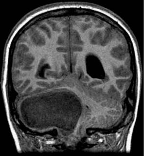
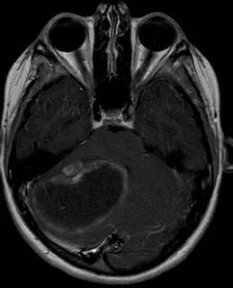 D
D
■ Clinical Presentation
A 13-year-old boy with a history of gait disturbance and vomiting.
60 |
RadCases.thieme.com |
RadCases Neuro Imaging |
|
■ Imaging Findings |
|
A B C D
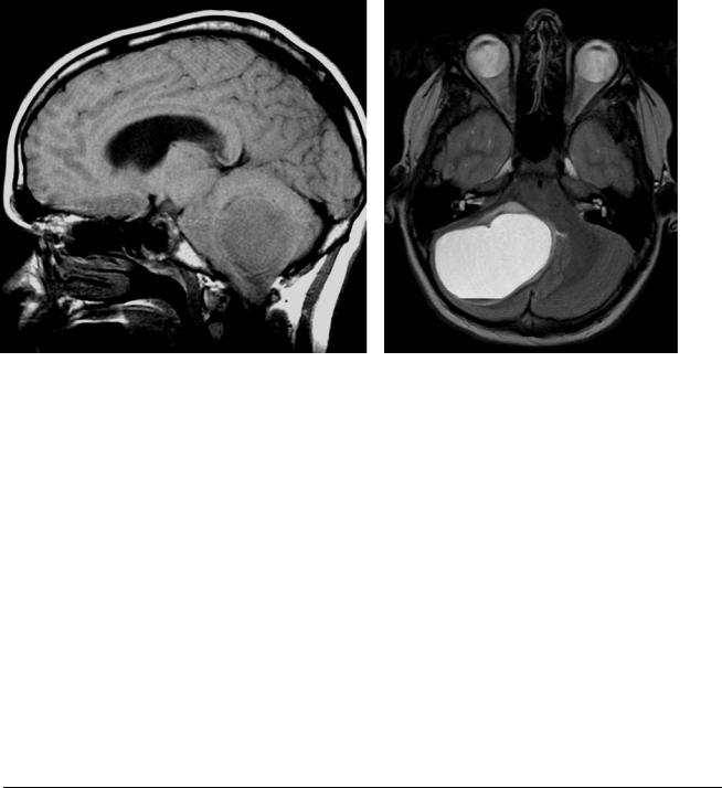
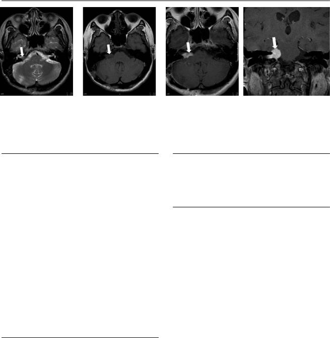
(A) Sagittal T1-weighted image (WI) shows an enlarged 4th ventricle. A mass (arrow) is e acing the 4th ventricle. The lateral ventricles are dilated (asterisk). (B) Axial T2WI shows a cystic mass centered on the right cerebellar hemisphere (asterisk). There is mild surrounding edema (arrowhead).
(C) Coronal T1WI shows the right cerebellar mass (arrow) and the dilated lateral ventricle (asterisk). (D) Axial T1WI with contrast shows peripheral enhancement of the mass (arrow); there is an anterior mural nodule (arrowhead).
■ Di erential Diagnosis
•Pilocytic astrocytoma: This is the most common pediatric brain tumor (85%). Patients younger than 20 years are a ected (75%), with a peak incidence in those 5 to 15 years of age. It appears as a cystic cerebellar mass with a mural nodule. There is low attenuation on computed tomography (CT). The most common location is in the cerebellar hemisphere. It deforms the 4th ventricle.
•Medulloblastoma: This is the most common posterior fossa tumor and the second most frequent brain tumor in children (after astrocytoma). It is rare in young adults. It is a highly malignant neuroepithelial tumor. Hyperattenuation on CT is typical. It almost always enhances. Medulloblastoma arises from the roof of the 4th ventricle. Fifty percent have cystic degeneration.
•Ependymoma: This is a midline cerebellar enhancing mass. Calcifications are frequent. It is hypoattenuated on CT. It typically extends through the foramen of Luschka into the subarachnoid space. It arises from the floor of the 4th ventricle.
■Essential Facts
•Ten percent of all childhood intracranial neoplasms are pilocytic astrocytomas.
•CT: the cyst is hypodense, the mural nodule is isodense; 20% show calcification.
•Magnetic resonance imaging:
•T1: low signal
•T2: high signal of cyst
•Contrast: enhancement of nodule; possible ring enhancement of cyst
•Fluid-attenuated inversion recovery: no suppression of cyst
¸Pearls & ˚ Pitfalls
¸Thirty-three percent of patients with a pilocytic astrocy-
toma involving the optic pathway have neurofibromatosis type 1.
¸ Patients with hypothalamic and optic nerve involvement have a worse prognosis.
˚When the optic nerves are involved, the enhancement is variable.
˚A location outside the 4th ventricle can help to di erentiate pilocytic astrocytoma from medulloblastoma.
˚Pilocytic astrocytoma is not a likely diagnosis in adults.
61
Case 31
A B
C 
 D
D
■ Clinical Presentation
A 43-year-old man with right-sided progressive hearing loss.
62 |
RadCases.thieme.com |
RadCases Neuro Imaging |
|
■ Imaging Findings |
|
A B C D
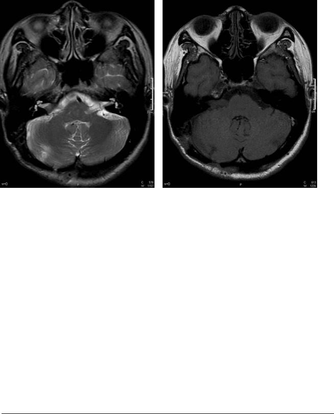
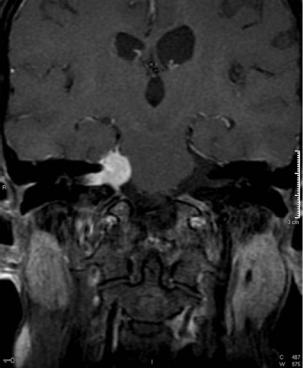
(A) Axial T2-weighted image (WI) shows expansion of the right internal auditory canal (IAC). A mass is e acing the cerebrospinal fluid (CSF) inside the canal (arrow). (B) Axial T1WI without contrast shows a mass with intensity similar to that of the brain (arrow) in the right IAC and cerebellopontine angle (CPA). (C) Axial T1WI with contrast shows intense enhancement of the mass (arrow). (D) Coronal T1WI with contrast shows the enhancing mass in the right CPA and IAC (arrow).
■ Di erential Diagnosis
•Vestibular schwannoma: Vestibular schwannomas present as a well-defined mass. They start in the IAC and extend to the CPA. They are the most common CPA mass (85%). They have di use enhancement and no dural tail. They expand the IAC, depending on their size. Microhemorrhage can be seen on T2*WIs.
•Meningioma: This is a well-defined, avidly enhancing mass with a dural tail. It starts in the CPA and rarely extends through the IAC. Meningiomas account for 10% of CPA tumors (second most common). No microhemorrhage is seen on T2*WIs.
•Epidermoid cyst: Epidermoid cysts are the third most common CPA masses. They are ectodermal inclusion cysts composed of epithelial cells that form a keratohyalin matrix as a consequence of desquamation. They engulf vessels and nerves. The cysts follow the CSF on T1 and T2WIs but show signal on fluid-attenuated inversion recovery images. They have a high signal on di usion-WIs secondary to T2 “shine through.” They do not have enhancement.
■ Other Imaging Findings
•Computed tomography shows an enlarged ICA (depending on size) with no calcification. The tumor is isodense to brain.
¸Pearls & ˚ Pitfalls
¸The vestibular nerve is more commonly a ected than the
cochlear nerve, hence the name vestibular schwannoma.
¸Malignant transformation is rare.
¸Vestibular schwannomas are associated with meningiomas in neurofibromatosis type 1.
¸Fifteen percent present with mural cysts.
˚A schwannoma can trap CSF and present with arachnoid cysts.
■ Essential Facts
•Consider if there is a unilateral enhancing mass in the CPA in adults.
•There is no extension to the 4th ventricle.
•Magnetic resonance imaging:
•T1: isointense to brain
•T2: heavy T2-weighted, high-resolution images good for demonstration
•T2*: shows microhemorrhage
•T1 with contrast: avid enhancement
•Di usion-WIs: low signal
63
Case 32
A
■ Clinical Presentation
A 64-year-old woman with progressive mental deterioration, nausea, and vomiting.
Further Work-up
B C D
64 |
RadCases.thieme.com |
RadCases Neuro Imaging |
|
■ Imaging Findings |
|
A B C D
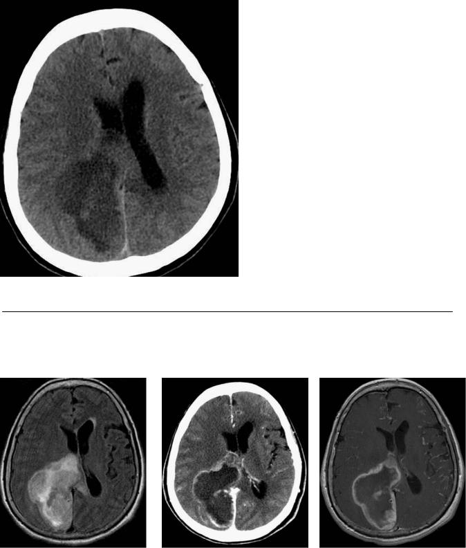
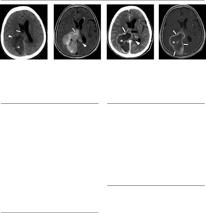
(A) Axial computed tomography (CT) scan of the head without contrast shows a low-density mass in the right occipital region (asterisk) with an isodense halo (arrowhead) and mass e ect that e aces the lateral ventricle (arrow). (B) Axial fluid-attenuated inversion recovery (FLAIR) image shows a heterogeneous mass (asterisk) with peripheral high signal that crosses the corpus callosum (arrowhead). The mass e aces the lateral ventricle (arrow). (C) Axial CT scan of the head with contrast shows irregular, thick peripheral enhancement (white arrow) and the low-density mass (asterisk). The edema crossing the corpus callosum is seen (arrowhead). The mass has subependymal extension to the lateral ventricle (black arrow).
(D) Axial T1-weighted image (WI) with contrast shows irregular enhancement (arrows) of the right occipital mass (asterisk).
■ Di erential Diagnosis
•Glioblastoma multiforme (GBM): GBM is a World Health Organization grade IV astrocytic tumor. It presents as a large, irregular heterogeneous mass with central necrosis and thick peripheral enhancement. It is typically located in the supratentorial white matter, involves the corpus callosum, and causes extensive vasogenic edema. It presents in patients 50 to 70 years old. Calcifications are extremely rare.
•Solitary metastasis: These typically are round masses located in the gray-white matter interface, demonstrate vasogenic edema, do not cross commissures, do not restrict di usion, and are usually multiple.
•Abscess: Abscesses are solitary or multiple ring-enhancing lesions with a cystic center. The rim is usually thinner than the rim of GBM. They show restricted di usion, which helps di erentiate them from GBM. They also are associated with vasogenic edema, do not cross commissures, and are located in the gray-white matter interface.
■Essential Facts
•Astrocytomas account for half of primary intracranial neoplasms. Of these, 50% are classified as GBM, making it the most common primary intracranial neoplasm.
•The mass e ect is extensive.
•If the mass crosses the corpus callosum, it has the typical “butterfly wing” pattern.
■ Other Imaging Findings
•CT:
•Isodense mass with central hypodensity
•No calcifications
•Magnetic resonance (MR) imaging:
•T2: heterogeneous mass; necrosis shows high signal
•FLAIR: high signal of periphery and center of mass; demonstrates vasogenic edema and early extension
•T1 with contrast: peripheral enhancement, usually thick, can be nodular
•MR spectroscopy: reversal of Hunter angle with decreased N-acetylaspartate and elevated choline peak; increased myoinositol level may be present
•Di usion-WIs: no restriction
¸Pearls & ˚ Pitfalls
¸GBM can extend through white matter tracts.
¸Patterns of metastatic spread may be through cerebrospi-
nal fluid, subependymal or hematogenous spread.
¸Tumor goes beyond area of enhancement.
¸Evaluate for mass e ect.
˚Associated with genetic syndromes:
•Turcot syndrome: GBM, colonic adenomatous polyposis, and medulloblastoma
•Increased incidence in neurofibromatosis type 1
˚Rarely, it can arise in the periphery of the brain, resembling a dura-based process.

65
Case 33
A 
 B
B
C 
 D
D
■ Clinical Presentation
A patient with precocious puberty and limited upward gaze.
66 |
RadCases.thieme.com |
RadCases Neuro Imaging |
■ Imaging Findings
A 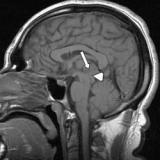 B C
B C 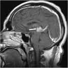 D
D
(A) Sagittal T1-weighted image (WI) shows a mass in the pineal region (arrow) compressing the tectum (arrowhead). There is a ventricular shunt in place to drain hydrocephalus from aqueductal compression. (B) Axial T2WI shows a heterogeneous mass in the pineal region (arrow). The mass has punctate areas of hypointensity (calcifications). (C) Sagittal T1WI with contrast shows di use enhancement of the mass (arrow). (D) Coronal T1WI with contrast shows the enhancing mass in the pineal region (arrow).
■ Di erential Diagnosis
•Germinoma: Germinomas account for two-thirds of pineal region masses. Eighty percent of germinomas occur in the pineal region, and the other 20% occur in the infundibular region. These lesions have a male predominance. They present as hyperdense lesions on computed tomography (CT). Germinomas manifest by the second to third decade of life. Pineal calcification in a child is highly suspicious for germ cell tumor. Germinomas engulf the central calcified gland. They have di use enhancement. They are malignant tumors.
•Pineoblastoma: These are malignant tumors that are less frequent than germinomas (15% of pineal masses).
Pineoblastomas arise from the pineal gland, displacing the calcifications superiorly. There is no gender predilection. Because they have no capsule, they can directly invade the brain or spread to the cerebrospinal fluid (CSF).
•Teratoma: These are the second most common tumors of the pineal region. Teratomas are well-circumscribed, multicystic masses that show fat signal. They can have ring enhancement or enhancement limited to the solid component. Calcifications are frequent. There is a male predilection.
■Essential Facts
•Germinomas are not encapsulated masses, which increases the likelihood of drop metastasis.
•Pineal germinomas tend to invade the posterior 3rd ventricle.
■ Other Imaging Findings
•Magnetic resonance imaging: isoor hyperintense to brain on T1, isointense on T2; shows enhancement
•CT: shows calcifications and increased attenuation
¸Pearls & ˚ Pitfalls
¸A CSF study is used to detect increased levels of α-feto-
protein, β-human chorionic gonadotropin, and placental alkaline phosphatase; these can indicate the presence of a germ cell tumor.
¸Germ cell tumors include germinomas, teratomas, endodermal sinus tumors, and embryonal carcinomas.
¸Germinoma is the most common pure germ cell tumor.
¸Most of the pineal tumors present with hydrocephalus
secondary to compression of the aqueduct.
˚Most solid masses in the pineal region are malignant germ cell tumors.
˚If the mass is multilocular with a lipid signal, favor teratoma (another germ cell tumor).
˚Most of the lesions in the pineal region enhance because there is no blood-brain barrier.
67
Case 34
A
■ Clinical Presentation
A 48-year-old man with left-sided motor weakness and vomiting.
Further Work-up
B
C  D
D