

Книги по МРТ КТ на английском языке / Neuro Imaging Redcases
.pdf78 |
RadCases.thieme.com |
|
RadCases Neuro Imaging |
|
|
■ Imaging Findings |
|
|
|
A |
B |
C |
|
D |
(A) Sagittal T1-weighted image (WI) of the cervical spine shows a low-signal lesion at the level of C3 (arrow). The lesion is extra-axial. (B) Sagittal T2WI of the cervical spine shows high signal of the mass. (C) Axial T2WI shows the mass (arrowhead) in the right aspect of the intradural space. The lesion compresses the spinal cord (arrow). (D) Axial fat-saturated T1WI of the cervical spine with contrast shows the “target” pattern of enhancement of the intradural, extramedullary mass (arrow). The lesion extends to the right neural foramen (arrowhead).
■ Di erential Diagnosis
•Spinal schwannoma: Spinal schwannomas comprise 80% of intradural extramedullary masses. They usually have an anterior and lateral location in the spinal canal. They extend to the neural foramen and enlarge it. They often compress the spinal cord. These lesions are typically solitary (unless they are related to neurofibromatosis type 2), demonstrate avid enhancement (which can have a “target” appearance), and do not calcify.
•Meningioma: Meningiomas are the second most common intradural extramedullary masses. They are localized in the posterior and lateral aspect of the spinal canal, more frequently at the thoracic level. They show intense enhancement and do not extend to the neural foramen. They may have a dural tail. Calcifications are rare.
•Neurofibroma: This presents as multiple masses exiting the neural foramina as part of neurofibromatosis type 1. Neurofibromas a ect multiple levels, enhance intensely, and expand the neural foramina. When a lesion is solitary, it is impossible to di erentiate it from a schwannoma.
■Essential Facts
•Schwannoma is the most common intradural extramedullary mass.
•Schwannoma can have fatty degeneration, cystic degeneration, or hemorrhage.
•Intensity is low on T1WIs and high on T2WIs in comparison with the spinal cord. There is a characteristic change if fatty or cystic degeneration is present.
•Enhancement is intense; the “target sign” (low central signal along with peripheral enhancement) may be seen.
■ Other Imaging Findings
•Schwannomas are isodense to the spinal cord on computed tomography.
¸Pearls & ˚ Pitfalls
¸Schwannoma is the most common intradural extramed-
ullary mass.
¸ The location is important: schwannomas tend to be anterior and lateral, whereas meningiomas are posterior.
˚ No bony erosions are seen with schwannomas.
79
Case 40
A
■ Clinical Presentation
A 40-year-old woman with severe headache and blurry vision.
Further Work-up
B C D
80 |
RadCases.thieme.com |
|
RadCases Neuro Imaging |
|
|
■ Imaging Findings |
|
|
|
A |
B |
C |
|
D |
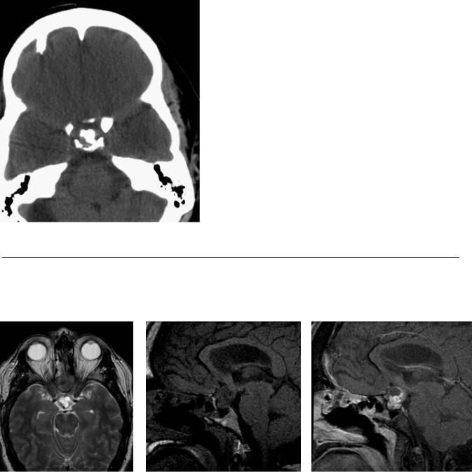
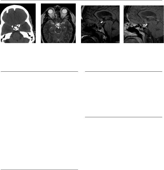
(A) Axial computed tomography scan of the brain shows irregular calcifications in the supraclinoid region (arrow). (B) On axial T2-weighted image (WI), the lesion has a multiloculated appearance. Note the dark posterior rim arising from calcification (arrow). (C) Sagittal T1WI shows sellar and suprasellar components of the mass, which is heterogeneous in signal (arrow). (D) Sagittal T1WI after gadolinium injection shows enhancement in the solid areas of the tumor (arrow) and nonenhancing cystic areas.
■ Di erential Diagnosis
•Craniopharyngioma: This is a suprasellar mass with a mixed solid and cystic appearance. It shows reticulated enhancement after the administration of gadolinium.
It has a mass e ect on the floor of the 3rd ventricle and hypothalamus.
•Rathke cleft cyst: This is a benign epithelium-lined cyst that is thought to be a remnant of the Rathke pouch. Seventy percent of Rathke cleft cysts involve both intrasellar and suprasellar regions. It is a cystic lesion, with a smooth margin and homogeneous signal intensity. It may have a high T1 signal with a thin rim or no enhancement.
•Pituitary adenoma: This is the most common of the lesions involving both the intrasellar and suprasellar regions. Pituitary adenomas arise from the pituitary gland and extend through the diaphragma sellae up toward the optic chiasm. They are isointense to gray matter and show homogeneously intense enhancement after gadolinium administration. Necrosis, cystic degeneration, and hemorrhage are not uncommon. Pituitary adenomas with suprasellar extension typically have a “figure of eight” or “snowman” appearance.
■ Other Imaging Findings
•Magnetic resonance (MR) imaging:
•Suprasellar (75%), suprasellar and infrasellar (20%), and infrasellar (5%) regions
•Partly solid, partly cystic, calcified mass lesion
•Reticular enhancement in the solid portion
•Superior extension compressing the 3rd ventricle
•MR angiography is useful to delineate the course of the vessels.
¸Pearls & ˚ Pitfalls
¸Lateral extension beyond the lateral wall of the internal
carotid artery may be seen in adenomas and craniopharyngiomas, but not in Rathke cleft cysts.
˚High T1 signal within the cystic component of a craniopharyngioma, adenoma, or Rathke cleft cyst may mimic pituitary apoplexy.
■ Essential Facts
•Craniopharyngiomas are epithelial neoplasms arising from squamous epithelial rests of the Rathke pouch.
•The clinical presentation includes visual disturbance, endocrine dysfunction, and raised intracranial pressure.
•The distribution by age is bimodal, with a peak incidence in children 5 to 14 years of age and again in adults 65 to 74 years of age.

81
Case 41
A
■ Clinical Presentation
A young woman presenting with loss of consciousness after being involved in a motor vehicle collision.
Further Work-up
B C D
82 |
RadCases.thieme.com |
|
RadCases Neuro Imaging |
|
|
■ Imaging Findings |
|
|
|
A |
B |
C |
|
D |
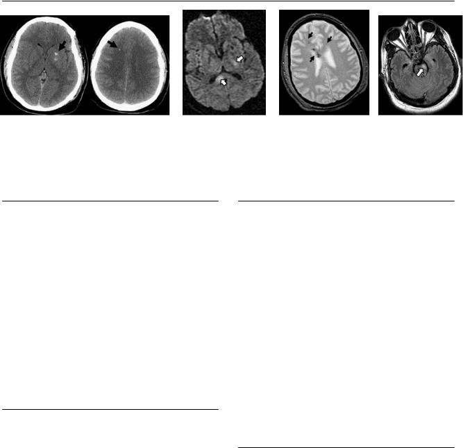
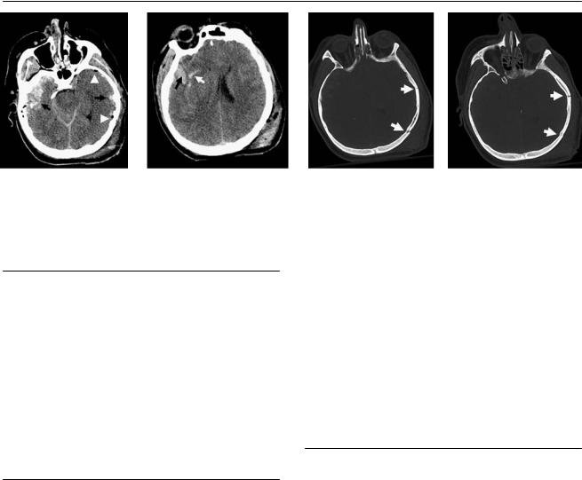
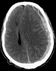
(A) Computed tomography (CT) scan of the brain demonstrates multiple punctate foci of hemorrhage in the left basal ganglia and right frontal deep white matter (arrows). (B) Di usion-weighted image (WI) shows restricted di usion in the left basal ganglia and in the splenium of the corpus callosum (arrows). (C) Petechial hemorrhage in the deep white matter and corpus callosum is evident in the gradient-echo (GRE) T2*WI (arrows). Multiple other foci were demonstrated in the brainstem and centrum semiovale. (D) Fluid-attenuated inversion recovery image shows increased signal in the left cerebral peduncle (arrow).
■ Di erential Diagnosis
•Di use axonal injury (DAI): This presents with significant impairment of mental status and relatively scarce CT abnormalities. It is characterized by multiple small hemorrhages in the deep white matter, including the corpus callosum and brainstem.
•Cerebral contusions: These are more frequent in the frontal and temporal lobes and are usually adjacent to the skull. Edema may precede hemorrhage by a few hours.
•Cerebral amyloid angiopathy: This presents as spontaneous cortical-subcortical intracranial hemorrhage in the normotensive elderly. The intracranial hemorrhage generally spares the deep white matter, basal ganglia, and brainstem. It may involve the cerebellum. Microhemorrhages are visualized on T2*-weighted GRE magnetic resonance (MR) imaging.
■Essential Facts
•DAI is a frequent cause of morbidity in patients with traumatic brain injuries, most commonly injuries resulting from high-speed motor vehicle accidents.
•The traumatic deceleration impact produces rotational forces that injure the areas where the density di erence is greatest.
•Two-thirds of DAI lesions occur at the gray-white matter junction. The corpus callosum, dorsolateral rostral brainstem, caudate nucleus, thalamus, tegmentum, and internal capsule are also a ected.
■ Other Imaging Findings
•CT:
•Normal CT scan at presentation in 50 to 80% of patients
•One or more areas of hemorrhage smaller than 2 cm in diameter in the cerebral hemispheres or adjacent to the 3rd ventricle
•Intraventricular hemorrhage
•Hemorrhage in the corpus callosum
•Brainstem hemorrhage
•MR imaging:
•Di usion-WIs: hyperintensity in areas of axonal injury
•GRE T2*WIs: petechial hemorrhages; can persist for many years after the injury
•T2WIs: multifocal areas of high signal at the white matter in the temporal or parietal corticomedullary junction, corpus callosum, dorsolateral rostral midbrain, and corona radiata
•MR spectroscopy may show di usely elevated lactate levels, even in tissues that appear normal on MR imaging; these correlate with a poor clinical outcome.
¸Pearls & ˚ Pitfalls
¸It is now recommended that all patients with moder-
ate to severe head injury undergo an MRI examination within the first 2 weeks after the injury for a better evaluation of the full extent of brain involvement, which is not feasible by CT alone.
˚The CT assessment of the brainstem and posterior fossa is limited because of beam-hardening artifacts from bone.
˚Artifacts from the frontal or temporal bones in the GRE T2* sequence limit the assessment of microhemorrhages in these regions.

83
Case 42
A 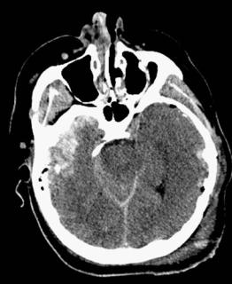
 B
B
■ Clinical Presentation
A 35-year-old who fell from a height of 14 feet.
Further Work-up
C D
84 |
RadCases.thieme.com |
RadCases Neuro Imaging |
|
■ Imaging Findings |
|
A B C D
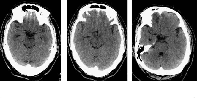
(A) Computed tomography (CT) of the brain shows contusions (arrows) and subarachnoid hemorrhage in both temporal lobes, which are more extensive on the left. Pneumocephalus (arrowheads) and soft-tissue swelling are present only on the left side. (B) CT of the brain shows contusion (black arrow) and subarachnoid hemorrhage (white arrow) in the right temporal lobe. Note the soft-tissue injuries on the left side. (C,D) Bone windows display fractures in the left temporal bone (arrows).
■ Di erential Diagnosis
•Coup and contrecoup cerebral injuries: These injuries show parenchymal contusions and extra-axial hemorrhages adjacent to the site of injury and in the opposite side of the brain. The contralateral parenchymal injuries tend to be more severe.
•Bilateral direct trauma: This is feasible in motor vehicle accidents. It presents with bilateral soft-tissue injuries.
•Aneurysmal rupture with subsequent fall: The traumatic lesions are disproportionate for this mechanism of injury. The aneurysmal subarachnoid hemorrhage is usually around the circle of Willis.
■Essential Facts
•Coup and contrecoup cerebral injuries are acceleration or deceleration injuries of the brain.
•The coup injury occurs at the site of the primary impact, which is identified by the associated scalp injuries or skull fractures.
•Epidural hematoma, contusion, or laceration of the brain surface often occurs at the site of a fracture, especially if it is depressed.
•The contrecoup injury arises on the side opposite the coup injury. Hemorrhagic cerebral contusions are more common and tend be larger on the side of the contrecoup injury, although both coup and contrecoup injuries can be hemorrhagic.
•A severe impact on a stationary head (e.g., a blow with a blunt object) results in skull fractures but generally does not cause contrecoup contusions. This is because, in such cases, the head does not accelerate or decelerate, and there is no brain lag.
•With frontal impact trauma, the brain moves over the floor of the anterior cranial fossa and slams into the sphenoid wings and petrous ridges, with resultant contusions in the inferior frontal, anterior temporal, and lateral temporal regions. Paramedian bony irregularities may cause superior frontal and parasagittal contusions.
¸Pearls & ˚ Pitfalls
¸Repeated imaging is indicated to monitor the size of the
hemorrhage and the development of delayed hemorrhage and vasogenic edema.
¸ Large hemorrhagic contusions commonly increase in size within the first 48 hours.
˚Contusions in the floor of the anterior cranial fossa and above the petrous ridge can be masked by volume averaging of the adjacent bone. In follow-up images, as edema develops, these contusions become evident.
85
Case 43
A B C
■ Clinical Presentation
A 35-year-old with headaches following an assault.
86 |
RadCases.thieme.com |
RadCases Neuro Imaging |
|
|
■ Imaging Findings |
|
|
A |
B |
|
C |
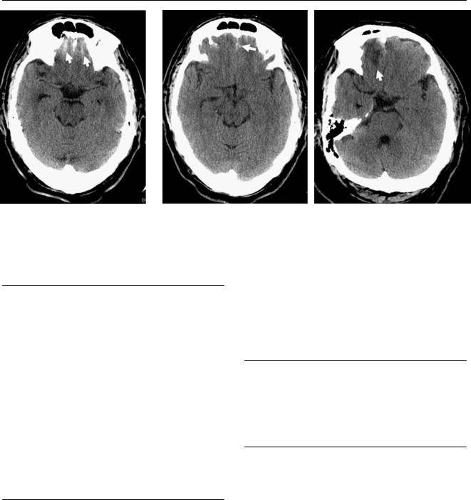
(A) Computed tomography (CT) demonstrates increased attenuation along the inferior frontal sulci (arrows). No fracture was identified in the images with bone algorithm. (B) CT demonstrates increased attenuation in the interhemispheric region (arrow). (C) Follow-up CT 2 days later shows low attenuation in the right frontal lobe inferiorly, consistent with edema (white arrow). The hemorrhage is still visible.
■ Di erential Diagnosis
•Traumatic subarachnoid hemorrhage (SAH): On CT, this shows high-attenuation blood within the basal cisterns and subarachnoid spaces. Traumatic hemorrhage is located in the convexities more frequently than in the basal cisterns. Nonvisualization of the interpeduncular cistern may be a clue that a small amount of isodense subarachnoid blood is present.
•Aneurysmal SAH: This presents as severe headache, described as the “worst headache of one’s life.” Hemorrhage tends to be located near the ruptured aneurysm, around the circle of Willis.
•Hemorrhagic contusion of the frontal lobe: This may be di cult to di erentiate from and may coexist with SAH. It can be masked by volume averaging with the bone in the floor of the anterior cranial fossa.
■ Essential Facts
•Small, bridging cortical vessels on the pia or arachnoidal leptomeninges crossing the subarachnoid space are injured.
•Blood from an intracerebral hematoma may decompress directly into the subarachnoid space or dissect into the ventricular system.
•When SAH occurs in conjunction with other forms of traumatic brain injury, it is often focal and located next to a contusion.
•The most frequent location is the convexity, followed by the fissures and basal cisterns.
•Hydrocephalus is the most common complication after SAH.
•SAH may induce cerebral vasospasm, which is more likely to occur when SAH is accompanied by subdural hematoma, intraventricular hemorrhage, cerebral contusion, or intracerebral hemorrhage.
■ Other Imaging Findings
•Fluid-attenuated inversion recovery imaging (FLAIR) images may detect small areas of acute or subacute SAH that are not detected by other magnetic resonance imaging sequences or CT scans.
¸Pearls & ˚ Pitfalls
¸The FLAIR sequence suppresses the intensity of cerebro-
spinal fluid (CSF) but does not di erentiate between abnormalities that appear as high signal intensity: hemorrhage, inflammatory exudate, neoplastic leptomeningeal infiltration, vascular slow flow, and hyperoxygenation during sedation or anesthesia.
˚In the setting of di use brain swelling, the elevated intracranial pressure causes an increase in the density of the subarachnoid space (pseudo-SAH) on CT by two mechanisms:
•The subarachnoid space contracts, and the amount of CSF in that space is decreased.
•The small pial vessels in the subarachnoid space become engorged and dilated, and a resultant increase in the blood pool contributes to the CT density.

87
Case 44
A  B
B 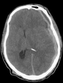
 C
C
■ Clinical Presentation
A 51-year-old man after a motor vehicle collision.