

Книги по МРТ КТ на английском языке / Neuro Imaging Redcases
.pdf138 RadCases.thieme.com |
RadCases Neuro Imaging |
|
|
■ Imaging Findings |
|
A B C 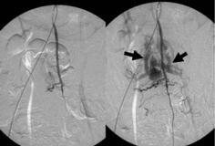 D
D
(A) Sagittal T2–weighted image (WI) of the thoracic spine shows multiple tiny flow voids in the posterior subarachnoid space (arrows). (B) Sagittal T2WI of the lumbar spine shows a tangle of vessels causing scalloping of the posterior margin of the S1 vertebral body (arrow). (C) Sagittal postcontrast T1WI in the lumbar region shows enhancement of the vascular structures within the thecal sac and epidural space (arrows). (D) Lateral projection of a spinal angiogram with injection in the artery of Adamkiewicz shows early opacification of large veins (arrows) in the epidural and subarachnoid spaces. No nidus is identified.
■ Di erential Diagnosis
•Spinal dural arteriovenous fistula: The fistula causes vessel dilatation and tortuosity, evidenced by signal voids around the cord. Gadolinium enhancement often facilitates recognition of the vessels. There may be T2 signal abnormality and patchy enhancement of the involved cord.
•Arachnoiditis: Arachnoiditis is inflammation of the meninges and subarachnoid space secondary to infectious, inflammatory, or neoplastic processes. T2WIs show loculation and obliteration of the subarachnoid space. The nerve roots show irregularity, clumping, and thickening. Peripheral adherence of the nerve roots to the walls of the thecal sac produces the so-called featureless, or empty, sac. There may be enhancement.
•Leptomeningeal carcinomatosis: Di use seeding of the leptomeninges by tumor metastases occurs most commonly in adults with breast carcinoma, lung carcinoma, or melanoma and in children with hematogenous malignancies or primitive neuroectodermal tumor. On magnetic resonance (MR) imaging, there is peripheral linear or nodular enhancement of the pia mater. In addition, enhancement of the nerve roots within the cauda equina is often seen, ranging from tiny nodules to large masses.
■Essential Facts
•Spinal vascular malformations comprise dural arteriovenous fistulas (DAVFs) and arteriovenous malformations (AVMs).
•DAVFs: acquired; fed by radiculomeningeal arteries; shunt located in the dura, near the spinal nerve root; venous congestion and intramedullary edema leading to chronic hypoxic myelopathy; treated with surgical ligation of the vein or glue embolization
•AVMs: inborn; fed by radiculomedullary (cord) arteries; intraor perimedullary shunt; may have a nidus or a fistulous transition from artery to vein; venous congestion, hemorrhage, steal phenomenon
•Selective spinal angiography is necessary to define the type of malformation and plan treatment.
¸Pearls & ˚ Pitfalls
¸A complete spinal angiogram includes selective injections
of the intercostal, lumbar, and internal iliac arteries. If no fistula is found, then the blood supply to the cervical spine, as well as the intracranial circulation, should be studied.
¸ Contrast-enhanced MR angiography may help to localize the enlarged feeders before conventional angiography.
˚Small vascular flow voids are usually present within the spinal subarachnoid space without mass e ect or cord signal abnormality.
139
Case 70
A 
 B
B
■ Clinical Presentation
A 17-year-old boy with progression of severe headache and lethargy that started 2 days prior.
Further Work-up
C D
140 RadCases.thieme.com |
RadCases Neuro Imaging |
|
|
■ Imaging Findings |
|
A B C D
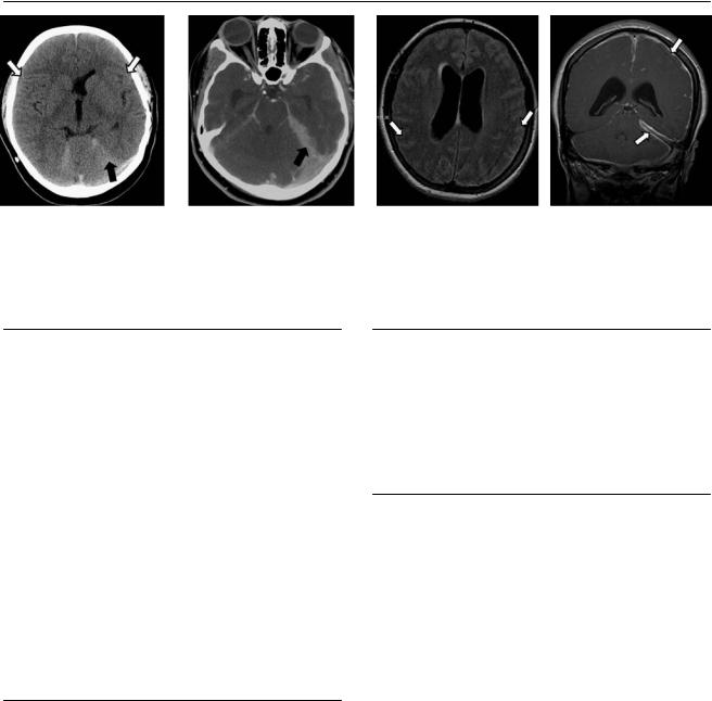
(A)Computed tomography (CT) without contrast shows e acement of the sulci (white arrows) and hyperdensity along the tentorium (black arrow).
(B)CT with contrast shows di use meningeal enhancement that is more prominent on the left tentorium (black arrow). (C) Axial fluid-attenuated inversion recovery (FLAIR) image shows dilated ventricles with increased signal in the leptomeninges (arrows). (D) Coronal T1-weighted image (WI) with contrast shows di use enhancement of the meninges, more prominent on the left side (arrows); the ventricles are dilated.
■ Di erential Diagnosis
•Bacterial meningitis: This is infectious leptomeningeal inflammation. Smooth, di use enhancement of the pia and arachnoid will be present. Enhancement is thicker in pyogenic meningitis than in viral meningitis. The sulci are e aced. Imaging findings can be normal in the initial phase.
•Tuberculous meningitis: Tuberculosis causes granulomatous meningitis. This is usually focal and occurs predominantly in the cisterns around the skull base. There is thickened (nodular) enhancement and possibly parenchymal extension (tuberculomas).
•Meningeal carcinomatosis: This is secondary extension of tumors to the meninges. It can a ect the dura mater, leptomeninges, or both. The primary lesion can be intraor extracranial. It can present as smooth or nodular enhancement. In the spine, drop metastases appear as masses or “sugar coating” after contrast administration. It is important to scan the entire neuraxis.
■ Essential Facts
•Infectious meningitis can be viral or bacterial.
•Bacterial meningitis secondary to meningococcal infection must be suspected in adolescents and young adults.
•Always use cerebrospinal fluid laboratories to the confirm diagnosis. Parenchymal extension is rare.
■ Other Imaging Findings
•CT findings can be normal or show slight leptomeningeal enhancement.
•FLAIR magnetic resonance imaging shows increased leptomeningeal signal; late enhancement makes this finding more conspicuous. T1WIs with contrast will show smooth, di use enhancement.
¸Pearls & ˚ Pitfalls
¸Delayed FLAIR images have been shown to be sensitive
for leptomeningeal disease if compared with the other magnetic resonance sequences.
˚Increased FLAIR leptomeningeal signal is nonspecific. It has also been described in subarachnoid hemorrhage and stroke, and after general anesthesia.
141
Case 71
A B C
D
■ Clinical Presentation
A 59-year-old man with a 1-month history of worsening back pain.
142 RadCases.thieme.com |
RadCases Neuro Imaging |
|
|
■ Imaging Findings |
|
A B C  D
D
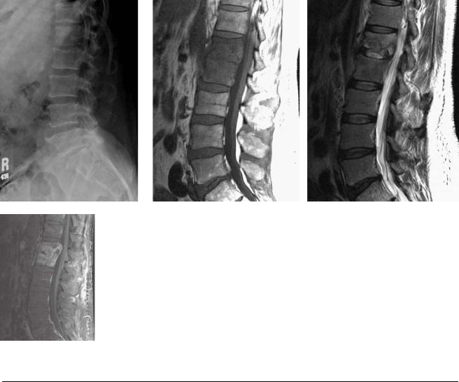
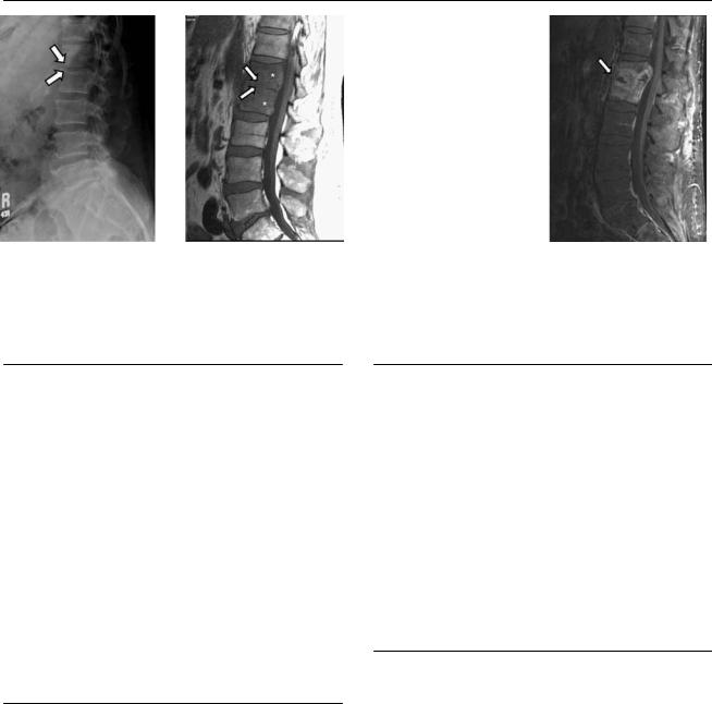
(A) Lateral view of the lumbar spine shows a decrease of the intervertebral space at L1-2 with poor definition of the adjacent end plates (arrows). (B) Sagittal T1-weighted image (WI) of the spine shows low signal in the L1 and L2 vertebral bodies (asterisks). The L1-2 disk shows low signal with poorly defined adjacent end plates (arrows). (C) Sagittal T2WI shows increased signal in the L1 and L2 vertebral bodies (asterisks) and in the L1-2 disk (arrows). (D) Sagittal fat-saturated T1WI of the lumbar spine with contrast shows di use enhancement of the L1 and L2 vertebral bodies and of the L1-2 disk (arrow).
■ Di erential Diagnosis
•Spondylodiskitis: This is pyogenic infection of the intervertebral disk and osteomyelitis of the adjacent vertebral bodies. The most frequent pathogen is Staphylococcus aureus. The disk and adjacent vertebral bodies enhance.
•Pott disease: Tuberculous osteomyelitis is also known as Pott disease. It is a granulomatous infection of the spine that causes large perivertebral abscesses with thin walls and end plate destruction. It involves more than two nonadjacent vertebral bodies, frequently with noncontiguous lesions that skip portions of the vertebral bodies. The midthoracic spine is more often a ected than the cervical and lumbar spine.
•Spinal metastasis: The spine is the most frequent location for skeletal metastasis. Involvement of the posterior components of the bone is common. Spinal metastasis enhances with contrast and spares the disks.
■Essential Facts
•Ill-defined perivertebral T2 signal abnormality and enhancement are frequent in spondylodiskitis.
•Erosion or destruction of the adjacent end plates occurs in 75% of cases.
•Paraspinal or epidural abscesses with thick walls may be present, although late in the disease.
•Spondylodiskitis usually involves the lumbar spine.
•Subligamentous spread of infection is rare, so there are no “skipped” portions of the vertebral bodies.
•Involvement of more than two vertebral bodies is unusual.
■ Other Imaging Findings
•Computed tomography shows normal or decreased density of the vertebral body and end plate destruction.
•On magnetic resonance imaging, the lesion will show low T1 signal of the vertebral body and ill-defined adjacent end plates, with increased T2 signal of the vertebral body and disk. T1 with contrast shows enhancement of the a ected segments with disk and di use soft-tissue enhancement. If abscess is present, there will be a thick enhancing wall.
•Fluorodeoxyglucose (FDG) positron emission tomography can be helpful to di erentiate degenerative from inflammatory end plate changes; degenerative changes do not show FDG uptake.
¸Pearls & ˚ Pitfalls
¸Noninfectious spondylodiskitis is called rheumatic spon-
dylodiskitis and is seen in 8% of patients with ankylosing spondylitis.
˚Tuberculous spondylitis starts in the anterior vertebral body and progresses to the posterior aspects.
˚The disk can be a ected late in Pott disease.
143
Case 72
A
■ Clinical Presentation
A 37-year-old man with immunosuppression and acute neurologic deficit.
Further Work-up
B C D
144 |
RadCases.thieme.com |
|
RadCases Neuro Imaging |
|
|
■ Imaging Findings |
|
|
|
A |
B |
C |
|
D |
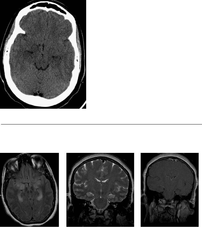
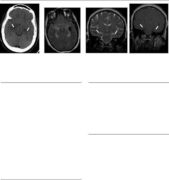
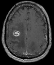
(A) Axial computed tomography (CT) without contrast shows decreased density of the medial aspect of both temporal lobes (arrows). (B) Axial fluid-at- tenuated inversion recovery (FLAIR) image shows high signal in the medial aspect of both temporal lobes with a lack of gray–white matter di erentiation (arrow). (C) Coronal T2-weighted image (WI) of the brain shows increased signal in the medial temporal lobes bilaterally (arrows). (D) Coronal T1WI of the brain with contrast shows patchy enhancement of the hippocampal region (arrows).
■ Di erential Diagnosis
•Herpes encephalitis: Herpes virus is the most frequent cause of infectious limbic encephalitis, which is characterized by cortical inflammation in the medial aspect of the temporal lobe, high FLAIR signal, loss of gray–white matter di erentiation, and restricted di usion. The onset is usually acute.
•Autoimmune limbic encephalitis: Causes may be paraneoplastic (associated with lung, thymus, breast, or testicular cancer) or nonparaneoplastic (rare, associated with volt- age-gated potassium channel antibody). Images appear like those of herpes encephalitis, but the acute clinical presentation is lacking. Autoimmune limbic encephalitis involves the limbic system, medial temporal lobe, cingulate gyrus, insula, and inferior frontal lobe. The involved areas show increased T2 and FLAIR signal, with patchy enhancement. There is no mass e ect. There is no restriction on di u- sion-weighted images (DWIs).
•Temporal lobe astrocytoma: This is a unilateral mass in the temporal lobe, with high T2 signal, no enhancement, and no restricted di usion. The symptoms are not acute.
■ Essential Facts
•The mortality rate is 50 to 70%.
•Herpes encephalitis can progress to necrotizing encephalitis.
•It is more frequent in patients with immunocompromise.
•It can also a ect other limbic structures, such as the insula, cingulate gyrus, and inferior frontal lobe.
•It is typically bilateral but can present on only one side.
•Mass e ect may be present in the acute phase.
■ Other Imaging Findings
•CT findings can be normal early in the disease; hypodensities in the medial temporal lobes are seen in late disease. CT can detect hemorrhage.
•Magnetic resonance imaging shows loss of gray–white matter di erentiation with increased T2/FLAIR signal in the medial temporal lobes involving the cortex.
Restricted di usion on DWIs; can show patchy enhancement with contrast. Gradient-echo imaging can detect hemorrhagic areas.
¸Pearls & ˚ Pitfalls
¸If a focal, well-defined area of enhancement is seen, sus-
pect a malignant tumor.
˚The presentation of congenital herpes simplex virus encephalitis is di erent from the presentation of herpes encephalitis in adults.

145
Case 73
A 
 B
B
C 
 D
D
■ Clinical Presentation
A 37-year-old man with severe headache and palsy of the left abducens nerve.
146 |
RadCases.thieme.com |
|
RadCases Neuro Imaging |
|
|
■ Imaging Findings |
|
|
|
A |
B |
C |
|
D |
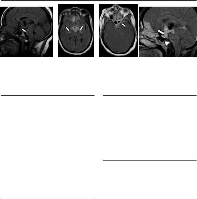
(A) Sagittal T1-weighted image (WI) shows a thickened pituitary stalk (white arrow) with e acement of the infundibulum (black arrow). (B) Axial fluidattenuated inversion recovery (FLAIR) image shows high signal in the perimesencephalic cistern (arrows) and infundibular region (asterisk). There is also increased signal in the frontal lobes. (C) Axial T1WI with contrast shows enhancement in the sellar region (asterisk) that extends to the cavernous sinus on the left (arrow). (D) Sagittal T1WI with contrast shows enhancement that involves the sellar region (white arrow), infundibulum (black arrow), and frontal region. Enhancement is also present in the prepontine cistern (arrowhead).
■ Di erential Diagnosis
•Tuberculous meningitis: This is a granulomatous infection of the basilar meninges. It is secondary to lung disease. Imaging shows thick nodular enhancement and associated parenchymal disease (tuberculomas). Tuberculous meningitis has a predilection for the suprasellar region.
•Meningeal sarcoidosis: Sarcoidosis causes a granulomatous meningitis with thick nodular enhancement. Less frequently, parenchymal extension through the VirchowRobin spaces is seen. It can involve the suprasellar region. Disease isolated to cranial nerves or the pituitary stalk is common.
•Nontuberculous infectious meningitis: This is leptomeningeal inflammation of viral or bacterial etiology. Meningococcal meningitis a ects adolescents and young adults.
Smooth, di use enhancement of the pia and arachnoid may be present. The sulci are e aced. Imaging findings can be normal in the initial phase. Parenchymal extension is rare.
■ Other Imaging Findings
•The results of computed tomography without contrast can be normal; in postcontrast studies, the basal cisterns and tuberculomas enhance.
•On magnetic resonance imaging (MRI), tuberculoma shows low T1 signal and increased T2 signal in the center of the area of necrosis.
•FLAIR image shows increased leptomeningeal signal. T1WI with contrast shows nodular enhancement of the meninges, solid or thick ring enhancement of tuberculoma.
¸Pearls & ˚ Pitfalls
¸Magnetization transfer MRI can help detect early involve-
ment of the meninges in patients with tuberculosis.
¸Consider the diagnosis of histiocytosis in children (4–6 years of age) with pituitary stalk involvement.
■ Essential Facts
•Tuberculomas can be solid or show thick ring enhancement.
•Parenchymal tuberculomas are secondary to hematogenous spread, typically through the Virchow-Robin spaces, as in cryptococcosis and sarcoidosis.
147
Case 74
A
■ Clinical Presentation
A 42-year-old patient infected with human immunodeficiency virus presenting with deteriorating mental status.
Further Work-up
B 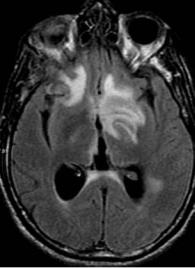 C
C 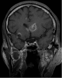
 D
D