

Книги по МРТ КТ на английском языке / Neuro Imaging Redcases
.pdf168 RadCases.thieme.com |
RadCases Neuro Imaging |
|
|
■ Imaging Findings |
|
A 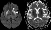 B
B 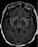 C
C 
 D
D
(A) Axial di usion-weighted image (WI) and apparent di usion coe cient map show areas of restriction in the caudate head and lenticular nuclei (arrowheads). (B) Axial fluid-attenuated inversion recovery magnetic resonance imaging (MRI) shows symmetric areas of increased T2 signal in the basal ganglia and thalami (asterisks). (C) Axial T2WI shows symmetric areas of increased T2 signal in the basal ganglia as well as in the thalami (asterisks). (D) Axial T1WI after gadolinium injection shows no enhancement (arrow).
■ Di erential Diagnosis
•Creutzfeldt-Jakob disease (CJD): Symmetric areas of restricted di usion and T2 hyperintensity involving the basal ganglia in a patient without acute alteration of mental status are highly suggestive of CJD. The lesions do not show mass e ect or enhancement.
•Hypoxic ischemic injury: This presents clinically as acute alteration of mental status after an episode of cerebral hypoperfusion. Restricted di usion and T2 signal changes in the basal ganglia and cerebral cortex are found on MRI. There is a predilection for the globus pallidus.
•Carbon monoxide poisoning: This is a combination of tissue hypoxia and direct carbon monoxide–mediated damage at the cellular level. Clinically, it may manifest acutely or produce a delayed neurologic syndrome. On MRI, necrosis in the globus pallidus and other basal ganglia is seen as low T1 and high T2 signal; hemorrhage will show high T1 and T2 signal. Carbon monoxide poisoning may also involve the white matter (predominantly periventricular) and cerebral cortex (with a predilection for the medial temporal lobes).
■Essential Facts
•CJD is a prion infectious disease characterized by progressive dementia, other neurologic abnormalities, and eventually death.
•Prions (proteinaceous infectious particles) are infective agents consisting of protein. They lack nucleic acid.
•CJD causes varying degrees of spongiform neuronal degeneration, neuronal loss, reactive astrocytic gliosis, and amyloid-like plaques that predominantly a ect gray matter structures.
•There are di erent subtypes: sporadic, familial, and acquired.
•The clinical presentation includes rapidly progressive dementia, motor dysfunction, myoclonus, and, in the later stages, akinetic mutism and death.
■ Other Imaging Findings
•MRI:
•Symmetric T2 hyperintensity involving the basal ganglia, in particular the corpus striatum
•Less commonly, asymmetric striatal involvement and signal abnormality in the thalamus and periaqueductal gray matter; cerebral cortical signal abnormality that is either symmetric or asymmetric
•No mass e ect or enhancement
•Progression to atrophy in the terminal stages
•At magnetic resonance spectroscopy, N-acetyl aspartate may be decreased in the early stage of disease.
•Single-photon emission computed tomography performed with technetium 99m–ethyl cysteinate dimer has been reported to show decreased cerebral blood flow in the regions of the brain that correspond to areas with abnormally high signal intensities on di usion-WIs.
¸Pearls & ˚ Pitfalls
¸MRI findings can precede the onset of characteristic clini-
cal disease, particularly with the use of di usion-WIs.
˚The pulvinar sign (symmetric high signal in the pulvinar compared with the signal in the remainder of the basal ganglia) is also seen occasionally with other diseases, such as limbic encephalitis.
169
Case 85
A B
C 
 D
D
■ Clinical Presentation
A 30-year-old with headache, weakness, and optic neuritis.

170 RadCases.thieme.com |
RadCases Neuro Imaging |
|
|
■ Imaging Findings |
|
A 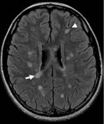 B
B 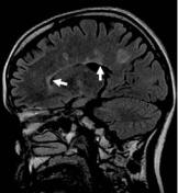 C
C 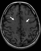
 D
D
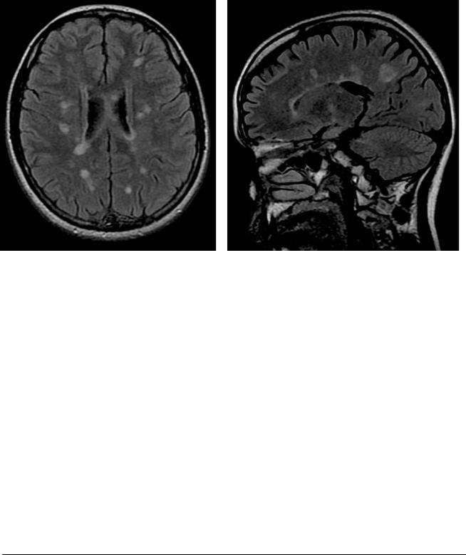
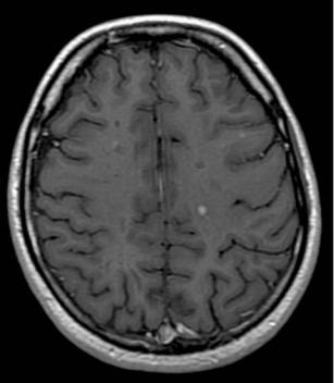
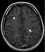
(A) Axial fluid-attenuated inversion recovery (FLAIR) image shows numerous round and oval areas of increased signal in the periventricular (arrow) and subcortical (arrowhead) white matter. (B) Sagittal FLAIR image demonstrates involvement of the transcallosal white matter tracts (arrows). (C) Axial T1weighted image (WI) shows low T1 signal (arrows) in some of the lesions. (D) Axial T1WI after gadolinium administration shows peripheral (arrowhead) and central (arrow) enhancement in some of the lesions.
■ Di erential Diagnosis
•Multiple sclerosis (MS): Characteristic MS lesions are periventricular in location and ovoid in shape, and they may or may not show enhancement, which is often ringlike. Lesions in the corpus callosum, the brainstem beyond the pons, and the spinal cord increase the specificity for MS.
•Chronic small-vessel ischemic disease: This is characterized by T2 hyperintensities in the white matter, which are frequently seen in patients of advanced age. The usual location is in the periventricular and subcortical white matter. Infratentorial lesions may present in the pons, but rarely elsewhere.
•Vasculitis: Vasculitis causes neurologic dysfunction and multiple focal ischemic lesions in persons of the same age range as those prone to MS. Abnormalities of the corpus callosum, optic nerves, and spinal cord may allow MS to be di erentiated from vascular disease.
■ Other Imaging Findings
•Conventional magnetic resonance imaging (MRI) findings:
•Lesions are bright on T2WIs and FLAIR images.
•On T1WIs, acute MS lesions are isointense to normal white matter and hypointense if chronic tissue injury or severe inflammatory edema has occurred; the accumulation of hypointense lesions (so-called black holes) may correlate with progression of disease and disability.
•The lesions enhance in the acute inflammatory phase and may vary in shape and size. They usually start as homogeneous enhancing nodules. Subsequent progression to ringlike enhancement is noted.
•Magnetization transfer imaging can detect areas of normal-appearing white matter with abnormal magnetization transfer ratios arising from myelin loss. It may also help to di erentiate MS plaques from small-vessel ischemic disease.
■ Essential Facts
•MS is an autoimmune inflammatory neurologic disease characterized by demyelination and axonal injury.
•MS presents as multiple lesions that are more periventricular than peripheral and have an ovoid shape; at the initial stage, the lesions are typically thin and appear to be linear (Dawson fingers). Other characteristics include dilated perivascular spaces; lesions in the subcortical region, brainstem, U-fibers, optic nerves, corpus callosum, and visual pathway; and generalized atrophy at a relatively young age.
•MS generally presents in young adults. A female predominance is noted. Clinical features include optic neuritis, transverse myelitis, internuclear ophthalmoplegia, and paresthesias or variable signs of neuropsychological dysfunction.
¸Pearls & ˚ Pitfalls
¸The ability to detect MS lesions with MRI increases with
the field strength.
¸The number and size of enhancing lesions detected on MRI can be increased with the use of a triple dose or three sequential single doses of gadolinium. Enhancement is suggestive of new lesion formation and inflammatory activity.
¸Numerous cortical lesions have been observed on histopathologic examination, but they are not commonly seen
on conventional MRI.
˚FLAIR images are usually optimized for contrast between lesions, white matter, and gray matter, and they may be less sensitive in detecting brainstem lesions. Proton-den- sity WIs and T2WIs are more reliable for the detection of lesions in this location.
171
Case 86
B
A
C  D
D
■ Clinical Presentation
A 42-year-old woman with paresthesias in both arms.

172 RadCases.thieme.com |
RadCases Neuro Imaging |
|
|
■ Imaging Findings |
|
A 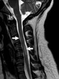 B
B 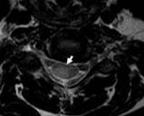 C
C 
 D
D
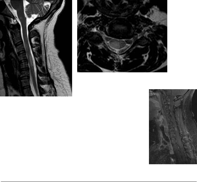
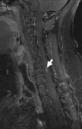
(A) Sagittal T2-weighted image (WI) of the cord shows multiple oval areas of increased signal in the ventral and dorsal cord spanning the length of one to two vertebral bodies (arrows). (B) Axial T2WI of the cervical spine shows a wedge-shaped peripheral cord lesion dorsally (arrow). (C) Axial T1WI before and after gadolinium injection. Note the enhancement of one of the cord lesions (arrow). (D) Sagittal fat-suppressed T1WI shows a cigar-shaped enhancing cord lesion that spans less than the height of one vertebral body (arrow).
■ Di erential Diagnosis
•Multiple sclerosis (MS) in the cord: This is characterized by peripherally located focal cord lesions that are less than two vertebral segments in length and occupy less than half of the cross-sectional area of the cord. In the axial images, the lesions may have a wedge shape, with the base at the cord surface, or a round shape if there is no contact with the cord surface.
•Devic neuromyelitis optica: This is a demyelinating disease characterized by bilateral visual disturbance and transverse myelopathy. It may have a monoor multiphasic course. Longitudinal, confluent lesions extending across five or more vertebral segments with hyperintensity on T2WIs are common. Cord swelling and enhancement may be present.
•Transverse myelitis (TM): TM is a clinical syndrome characterized by bilateral motor, sensory, and autonomic disturbances. There is perivascular monocytic and lymphocytic infiltration, demyelination, and axonal injury. TM may
be part of a multifocal central nervous system disease, a multisystemic disease, or an isolated, idiopathic entity. The lesions are centrally located and hyperintense, and they usually occupy more than two-thirds of the cross-sectional area of the cord and have a length of more than three to four vertebral segments. Cord expansion may or may not be present. Enhancement is infrequent. TM may be patchy or di use.
■Essential Facts
•MS is a chronic inflammatory demyelinating disease of the central nervous system that can lead to irreversible tissue loss or partial demyelination.
•The spinal cord is frequently involved in MS, either alone or with the brain.
•As many as 25% of cases have been found to involve only the spinal cord.
•The clinical presentation includes motor, sensory, or sphincter dysfunction.
•Most spinal cord lesions occur in the cervical region.
•Cord swelling is usually found only in the relapsingremitting form of MS.
•The incidence of enhancing lesions in the spine is significantly lower than the incidence in the brain.
•Cord atrophy can be due to inflammatory tissue injury, demyelination, and axonal loss.
■ Other Imaging Findings
•On magnetic resonance imaging (MRI), demyelinating plaques are hyperintense on T2WIs.
•In the spine, on sagittal sections, plaques have a cigar shape and may be located centrally, anteriorly, or
dorsally. Di use lesions are seen as mild intramedullary hyperintensities on T2WIs.
•In the brain, lesions are seen as multiple white matter lesions in the pericallosal region, brainstem, optic nerves, corpus callosum, and visual pathways.
•Chronic lesions are hypointense on T1WIs.
•Lesions, which may vary in shape and size, enhance in the acute inflammatory phase. The enhancement starts as homogeneous nodules and subsequently progresses to ringlike enhancing lesions.
¸Pearls & ˚ Pitfalls
¸Chronic foci of hypointensity on T1WIs of the brain,
known as black holes, are not present in the spinal cord.
˚Spinal cord imaging is prone to multiple artifacts:
•Motion: subject motion, blood flow, respiratory motion, swallowing, cerebrospinal fluid pulsation
•Susceptibility artifacts: interfaces (bone and soft tissue, fat and air), trabecular bone, metallic implants
•Chemical shift artifacts: epidural and vertebral fat
173
Case 87
A 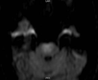
 B
B
■ Clinical Presentation
A 68-year-old with the recent onset of spastic quadriparesis.
Further Work-up
C D

174 RadCases.thieme.com |
RadCases Neuro Imaging |
|
|
■ Imaging Findings |
|
A  B
B 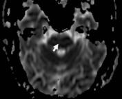 C
C 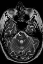
 D
D
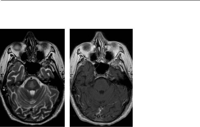
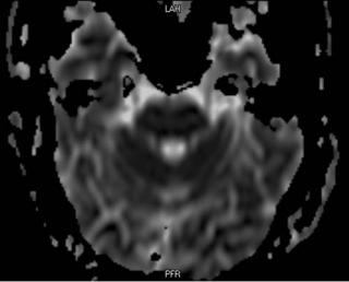
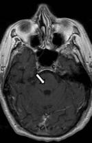
(A) Di usion-weighted image (WI) shows an area of increased signal without corresponding low signal on the apparent di usion coe cient (ADC) map, consistent with T2 shine-through (arrow). (B) ADC map does not show decreased signal in the area of di usion abnormality, indicating T2 shine-through (arrow). (C) Axial T2WI demonstrates an area of high signal in the central portion of the pons (arrow). (D) Axial T1WI after gadolinium contrast fails to demonstrate enhancement or T1 signal change.
■ Di erential Diagnosis
•Central pontine myelinolysis (CPM): CPM is an osmolar disturbance resulting in demyelination and is part of the osmotic demyelination syndrome. Acute demyelination in the pons results in symmetric areas of signal abnormality in the central pons on T2WIs. The appearance on di usionWIs is variable. No contrast enhancement is present.
•Posterior reversible encephalopathy syndrome: This is the result of neurotoxicity-induced vasogenic edema. It more frequently involves the cerebral hemispheres but may present with lesions in the basal ganglia, brainstem, and deep white matter (external/internal capsule).
•Acute pontine infarct: Patients present with the sudden onset of neurologic deficit. There is restricted di usion preceding a T2 signal increase. Vertebral or basilar occlusive changes may or may not be present.
■ Essential Facts
•Patients who are chronic alcoholics, malnourished, transplant recipients, or chronically debilitated are at risk.
•CPM is associated with electrolyte abnormalities, including hyponatremia, which is rapidly overcorrected.
•Extrapontine myelinolysis occurs in the white matter of the cerebellum, thalamus, globus pallidus, putamen, and lateral geniculate body.
•The clinical course is biphasic; generalized encephalopathy is caused by the hyponatremia, which usually improves transiently following an initial elevation of the sodium level.
•Spastic quadriparesis and pseudobulbar palsy develop 2 to 3 days later as a consequence of myelinolysis.
■ Other Imaging Findings
•Magnetic resonance imaging (MRI):
•Symmetric areas of signal intensity abnormality in the central pons are seen on T2WIs and fluid-attenuated inversion recovery images. The corticospinal tracts are spared.
•The condition may progress to classic hyperintense “trident-shaped” CPM, with sparing of the ventrolateral pons and corticospinal tracts.
•Restricted di usion may be seen before the T2 signal abnormalities.
¸Pearls & ˚ Pitfalls
¸The diagnosis of CPM is not ruled out in the setting of
normal imaging findings.
¸MRI changes may be delayed.
¸The severity of the MRI findings is not prognostic. ˚ In the absence of a clinical history of sodium shifts,
asymptomatic pontine lesions are unlikely to be CPM lesions.
Case courtesy of Alexander Simonetta, MD.
175
Case 88
A B
C D
■ Clinical Presentation
A 52-year-old with numbness in the left foot.
176 |
RadCases.thieme.com |
|
RadCases Neuro Imaging |
|
|
■ Imaging Findings |
|
|
|
A |
B |
C |
|
D |
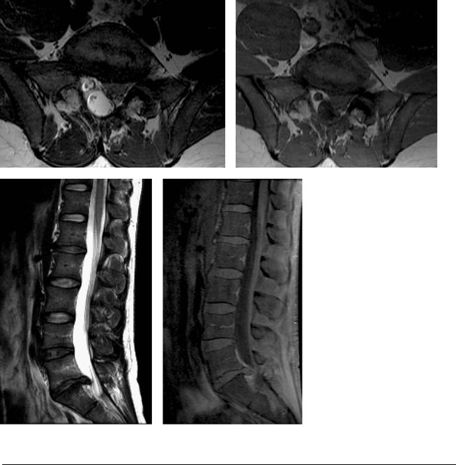
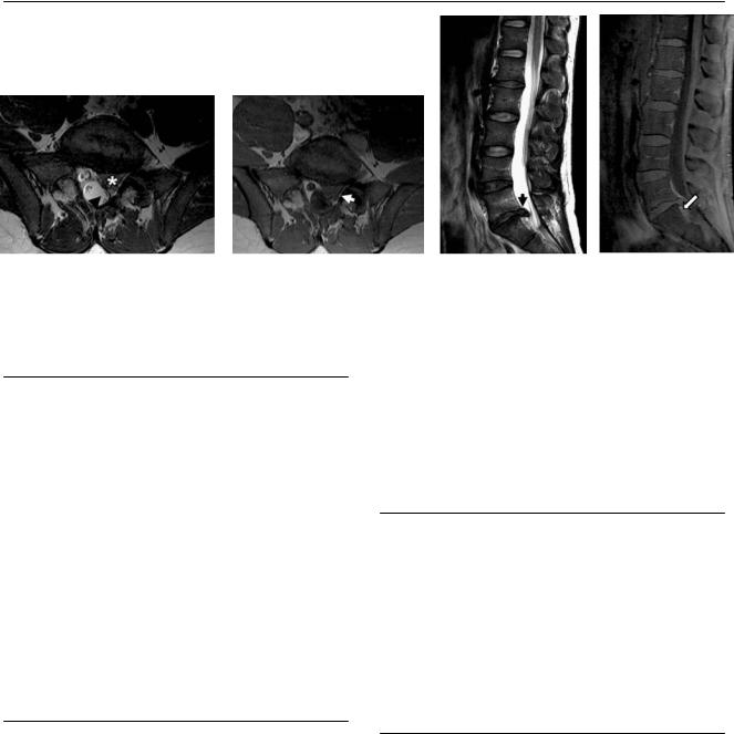
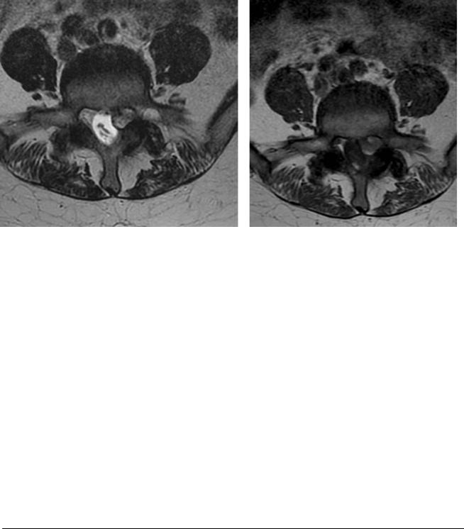
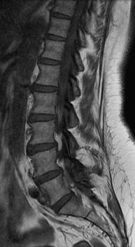
(A) Axial T2-weighted image (WI) at the level of the L5-S1 intervertebral disk shows an oval mass continuous with the disk occupying the left lateral recess (asterisk). Note the posterior displacement of the left S1 nerve root (arrowhead). (B) Axial T1WI at the level of the L5-S1 intervertebral disk. Note the thin plane of fat that separates the lesion from the facet joint (arrow). (C) Observe the continuity of the lesion with the L5-S1 disk on the sagittal T2WI (arrow).
(D) Sagittal fat-suppressed T1WI shows enhancement of the epidural plexus (arrow), but not of the disk herniation.
■ Di erential Diagnosis
•Disk herniation: This is a localized displacement of disk material beyond the limits of the intervertebral disk space. It encroaches on the epidural fat and sometimes the dural sac and nerve roots. The herniation is usually contiguous with the rest of the disk, but free fragments are possible. The herniation is usually isointense to the parent disk on T1and T2-weighted sequences.
•Facet synovial cyst: This frequently occupies the lateral recess and may extend to the central canal. Cysts can contain synovial serous fluid, more gelatinous material, air, or blood. They are continuous with the facet joint but may abut the disk as well.
•Epidural hematoma: This is an epidural collection with variable signal intensity depending on the blood products contained therein. Epidural hematoma may span more than one level.
•When there is no contiguity with the parent disk, the extruded material may be characterized as a sequestered or free fragment.
•Relative to the axial plane, the herniation may be central, right or left central, right or left subarticular, right or left foraminal, or right or left extraforaminal.
■ Other Imaging Findings
•Magnetic resonance imaging (MRI) is the technique most frequently used for the evaluation of disk herniation.
•Computed tomography is more accurate than MRI for the identification of calcified herniations and associated bony abnormalities, such as posterior accompanying osteophytes.
•In provocation lumbar diskography, direct disk stimulation is used to help identify the specific disk(s) responsible for lower back pain.
■ Essential Facts
•Degenerative disk disease is more common in the elderly population; however, acute herniations are frequent in the middle-aged population.
•Disk herniation is defined as the localized displacement of disk material beyond the normal margins of the intervertebral disk space.
•Protrusion: the greatest distance, in any plane, between the edges of the disk material beyond the disk space is less than the distance between the edges of the base in the same plane.
•Extrusion: any one distance between the edges of the disk material beyond the disk space is greater than the distance between the edges of the base in the same plane.
¸Pearls & ˚ Pitfalls
¸MRI can be used to follow nontreated disk herniations.
¸The main MRI findings useful to presume spontaneous
regression of disk herniations after 6 months are free fragments, T2-weighted hyperintense herniation, peripheral enhancement after gadolinium administration, and recent clinical onset.
˚After gadolinium injection, many herniations show a thin enhancing rim that is caused by the inflammatory reaction.
˚A contrast agent is more widely used in postoperative examinations because it is useful for di erentiating residual or recurrent herniations from scar tissue.
177
Case 89
A B
C 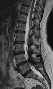
 D
D
■ Clinical Presentation
A 46-year-old with weakness and pain in the left leg.