

Книги по МРТ КТ на английском языке / Neuro Imaging Redcases
.pdf148 RadCases.thieme.com |
RadCases Neuro Imaging |
|
|
■ Imaging Findings |
|
A 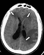 B
B 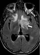 C
C 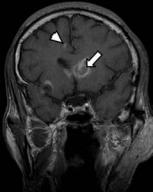
 D
D
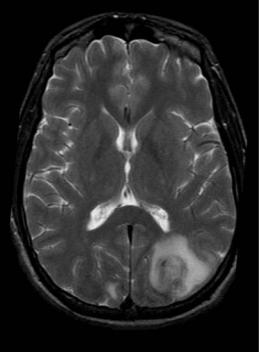
(A) Computed tomography (CT) of the brain without contrast shows areas of low attenuation in the white matter of both frontal lobes and in the left parietal region (arrows) related to vasogenic edema (the cortex is spared). There is e acement of the left frontal horn because of mass e ect. (B) Axial fluid-at- tenuated inversion recovery sequence shows high signal in the frontal lobes bilaterally and in the left basal ganglia (arrow). (C) Coronal T1-weighted image (WI) with contrast shows a lesion with ring enhancement in the head of the left caudate nucleus (arrow) and a small enhancing lesion in the gray–white matter junction in the medial right frontal lobe (arrowhead). (D) Axial magnetic resonance (MR) imaging with contrast shows an “eccentric target sign” in the right frontal lobe. There is a halo of ring enhancement with a nodular enhancing center (arrow).
■ Di erential Diagnosis
•Toxoplasmosis: This is the most common mass lesion in patients with acquired immunodeficiency syndrome
(AIDS). Multiple lesions with ring enhancement a ect the corticomedullary junction and basal ganglia. Toxoplasmosis does not extend to the ependymal surface. Eccentric target sign enhancement is highly suggestive of toxoplasmosis. Acute lesions can have vasogenic edema. Occasional hemorrhage (helps di erentiate toxoplasmosis from lymphoma) can be present.
•Primary central nervous system (CNS) lymphoma: CNS lymphoma causes basal ganglia and periventricular lesions that enhance. It frequently a ects the corpus callosum and can extend to the ependymal surface. Vasogenic edema is less frequent. No hemorrhage should be present.
•Metastases: These are usually white–gray matter junction masses. They are solid or necrotic with ring enhancement and vasogenic edema. They are rare in the basal ganglia (3%). Twenty to 50% are solitary lesions.
■Essential Facts
•In adults, most Toxoplasma gondii infections are subclinical, but severe infection can occur in patients with immunocompromise, such as those who have AIDS and malignancies.
•Toxoplasmosis is the most common opportunistic CNS infection in patients with AIDS (15–50%).
•Drug therapy does not eradicate T. gondii, and lifelong therapy to avoid relapse is often necessary in patients with immunocompromise.
■ Other Imaging Findings
•CT shows multiple areas of abnormal hypodensity in the basal ganglia and gray–white matter junction with ring enhancement.
•MR imaging: Lesions are hypoor isointense on T1WIs and hypoor isointense with surrounding vasogenic edema on T2WIs. Contrast will show an enhancing ring that can present with a central enhancing nodule (eccentric target sign).
¸Pearls & ˚ Pitfalls
¸It is extremely di cult to distinguish between toxoplas-
mosis and lymphoma on imaging. Empiric treatment is usually administered, and the lesions must be followed until they resolve completely.
¸If there is no improvement after treatment, thallium 201 single-photon emission computed tomography of the brain, positron emission tomography (results of both are positive for lymphoma), or MR spectroscopy can be used
to re-evaluate.
˚Di usion-weighted and apparent di usion coe cient lesion: white matter ratios greater than 1.6 favor toxoplasmosis over lymphoma.
149
Case 75
A
■ Clinical Presentation
A patient infected with human immunodeficiency virus presenting with headache, hemiparesis, and seizures.
Further Work-up
B 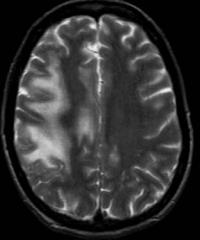 C
C 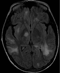
 D
D

150 RadCases.thieme.com |
RadCases Neuro Imaging |
|
|
■ Imaging Findings |
|
A 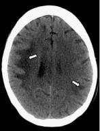 B
B 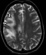 C
C 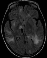
 D
D
(A) Computed tomography (CT) of the brain without contrast shows asymmetric patchy areas of low attenuation bilaterally (arrows) with involvement of the subcortical U-fibers. (B) Axial T2-weighted image (WI) shows patchy scattered areas of increased intensity bilaterally, more prominent on the right side, involving the subcortical U-fibers (arrows). (C) Axial fluid-attenuated inversion recovery (FLAIR) image shows multiple patchy areas of hyperintensity involving the subcortical white matter and the right thalamus (arrow). (D) Magnetic resonance (MR) imaging of the brain with contrast shows no enhancement (arrow).
■ Di erential Diagnosis
•Progressive multifocal leukoencephalopathy (PML): PML results in di use, patchy, asymmetric white matter lesions that are located bilaterally, mainly in the subcortical and periventricular white matter. PML has a predilection for the supratentorial region (parietal and frontal lobes) and thalamus (50% of patients). It spares the cortex and does not show mass e ect or contrast enhancement. Cortical atrophy is present in 70% of cases and ventricular dilatation in 50%. PML is hypodense on CT.
•Human immunodeficiency virus (HIV) encephalitis: This typically causes cortical atrophy and ventricular dilatation with di use, symmetric, periventricular white matter T2 hyperintensities. There is no mass e ect. The lesions are confluent and symmetric and do not enhance with contrast. The subcortical white matter is spared.
•Cytomegalovirus (CMV) infection: CMV infection causes encephalitis with periventricular white matter hyperintensities and ventriculitis with periventricular enhancement.
Imaging findings are nonspecific or absent.
■ Other Imaging Findings
•CT shows bilateral asymmetric areas of low attenuation.
•MR imaging: Lesions are hypointense on T1 and hyperintense on T2 and FLAIR. There is no di usion on di usionWIs. MR spectroscopy can detect high levels of choline and variable levels of myoinositol.
¸Pearls & ˚ Pitfalls
¸JC polyomavirus was formerly known as papovirus.
¸Rarely, PML can enhance.
¸The magnetization transfer ratio in PML is severely de-
creased in comparison with that in HIV encephalitis.
˚Look for involvement of the subcortical U-fibers to favor a diagnosis of PML.
■ Essential Facts
•PML is a fulminating opportunistic infection with JC polyomavirus (named for John Cunningham).
•It is a progressive demyelinating disease; the virus targets oligodendrocytes.
•Suspect PML in patients infected with HIV when patchy subcortical high signal appears on T2 or FLAIR.
•The corpus callosum can be involved.

151
Case 76
A 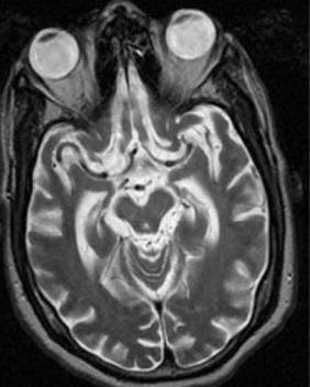
 B
B
C 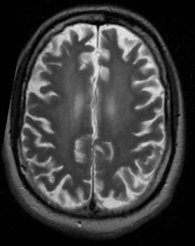
 D
D
■ Clinical Presentation
A 40-year-old man infected with human immunodeficiency virus presenting with progressive dementia and tremor.
152 |
RadCases.thieme.com |
|
RadCases Neuro Imaging |
|
|
■ Imaging Findings |
|
|
|
A |
B |
C |
|
D |
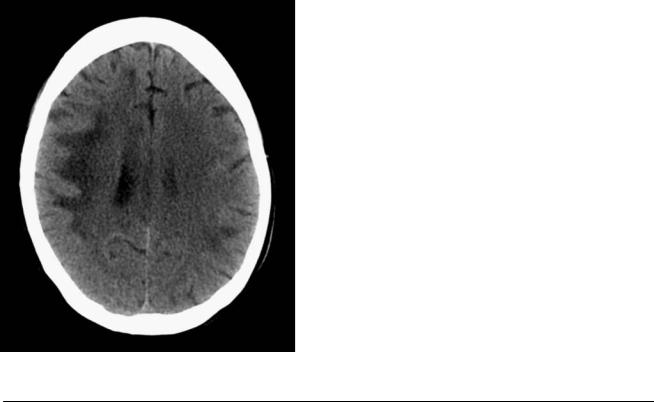
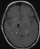
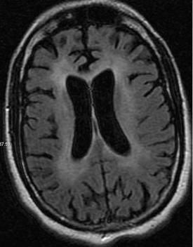
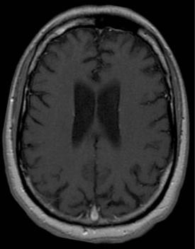
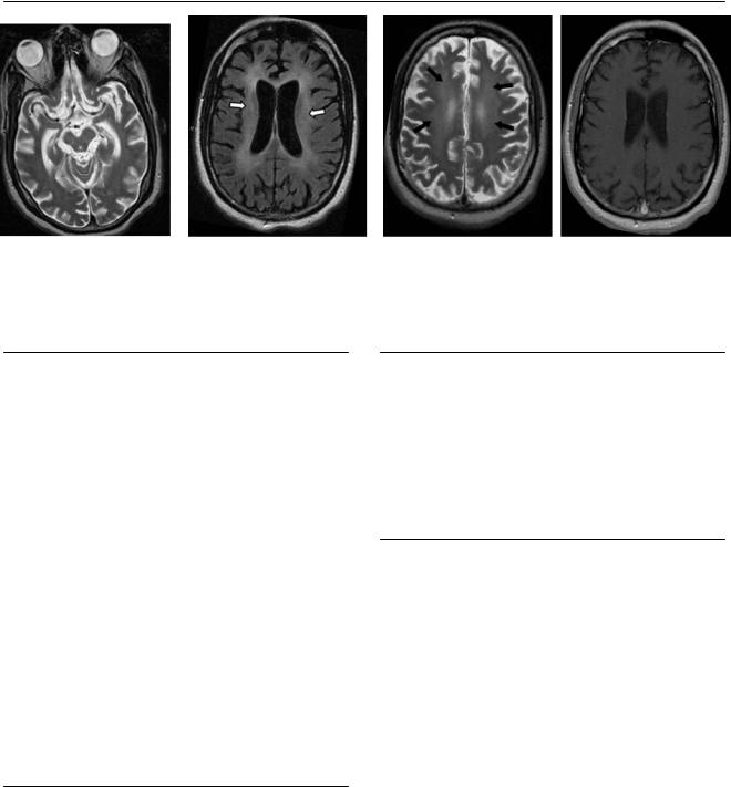
(A) Axial T2-weighted image (WI) shows di use cerebral atrophy. (B) Axial fluid-attenuated inversion recovery (FLAIR) image shows di use cerebral atrophy with symmetric, confluent periventricular white matter disease (arrows). The subcortical white matter is spared. (C) Axial T2WI shows the symmetric periventricular white matter hyperintensities (arrows), with sparing of the subcortical U-fibers. (D) Axial T1WI with contrast shows no enhancement of the periventricular lesions.
■ Di erential Diagnosis
•Human immunodeficiency virus (HIV) encephalopathy:
This is characterized by cortical atrophy, ventricular dilatation, and di use periventricular white matter T2 hyperintensities that are confluent and symmetric. The subcortical white matter is spared. It appears hypodense on computed tomography (CT). There is no enhancement or mass e ect.
•Progressive multifocal leukoencephalopathy (PML): Di use, patchy, asymmetric white matter lesions are located bilaterally, mainly in the subcortical and periventricular white matter. Involvement of the subcortical U-fibers is common. The lesions have a predilection for the supratentorial region (parietal and frontal lobes). Cortical atrophy and ventricular dilatation are less frequent. There is no mass e ect or enhancement.
•Cytomegalovirus (CMV) infection: Imaging is nonspecific, and findings can be normal. Encephalitis with periventricular white matter hyperintensities is common. Ventriculitis with periventricular enhancement is characteristic. CMV encephalitis is a rapidly advancing disease (progression is faster than in HIV leukoencephalitis) that leads to death, usually in 5 to 8 weeks.
■ Other Imaging Findings
•CT shows atrophy with confluent periventricular lowdensity lesions; there is no enhancement.
•Magnetic resonance (MR) imaging: Lesions are isoor hypointense on T1 and hyperintense on T2/FLAIR. They show no enhancement.
•MR spectroscopy shows high myoinositol and choline levels and reduced N-acetyl aspartate levels.
¸Pearls & ˚ Pitfalls
¸The magnetization transfer ratio in PML is severely de-
creased in comparison with that in HIV encephalitis.
˚The presence of ependymal enhancement favors CMV.
˚HIV leukoencephalitis does not present with mass e ect or enhancement.
˚In the late stages of HIV leukoencephalopathy, the lesions can involve all the white matter, including the subcortical fibers.
■ Essential Facts
•HIV encephalopathy is the most common neurologic disease of the central nervous system caused by the HIV itself.
•Others include vacuolar myelopathy, peripheral neuropathies, and polymyositis.
•HIV causes di use myelin loss and astroglial proliferation by monoand multinuclear macrophages.
•There is frontal dominance. The genu of the corpus callosum may be involved.

153
Case 77
A 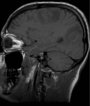
 B
B
C
D
■ Clinical Presentation
A 39-year-old drug addict with heart disease.
154 |
RadCases.thieme.com |
|
RadCases Neuro Imaging |
|
|
■ Imaging Findings |
|
|
|
A |
B |
C |
|
D |
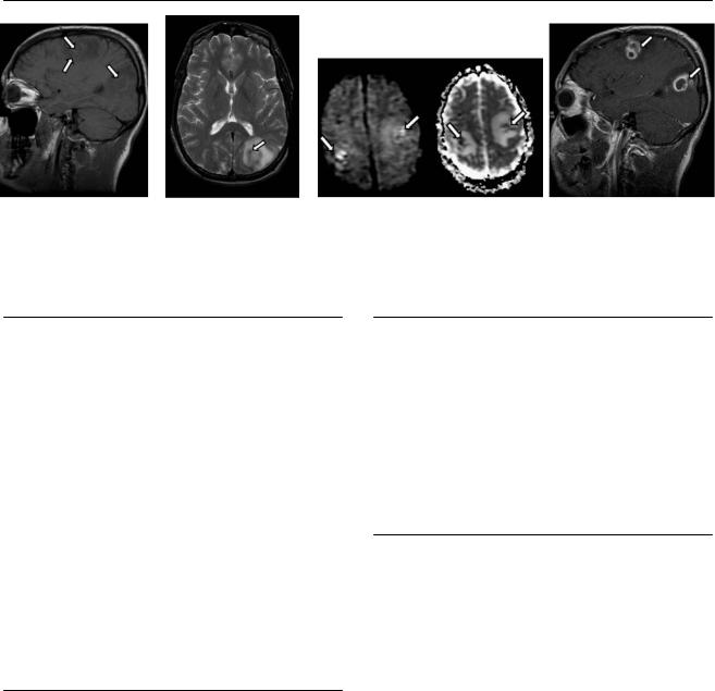
(A) Sagittal T1-weighted image (WI) shows heterogeneous subcortical nodules (arrows) surrounded by areas of low intensity related to vasogenic edema. The walls demonstrate areas of hyperintensity related to bleeding. (B) Axial T2WI shows vasogenic edema in the left occipital lobe with a central nodular area. There is an area of low signal in the lesion that is related to the bleed (arrow). (C) Di usion-WI and apparent di usion coe cient map show bilateral areas of restricted di usion (arrows). (D) Sagittal T1WI with contrast shows subcortical lesions with ring enhancement (arrows).
■ Di erential Diagnosis
•Septic emboli: Hematogenous dissemination of septic emboli may result from cardiac valvular disease. The lesions are usually in the territory of the middle cerebral artery (MCA) and are predominantly subcortical. Restricted diffusion, hemorrhage, and nodular or ring enhancement are common. Surrounding vasogenic edema may be present.
•Metastasis: Hematogenous metastasis occurs in the cortical or subcortical white matter, frequently in the territory of the MCA. Surrounding vasogenic edema is characteristic. There is no restricted di usion. Hemorrhage and nodular or ring enhancement may be present. A known primary tumor makes this diagnosis more likely.
•Aseptic emboli: Aseptic emboli are bilateral or unilateral small infarcts in the subcortical white matter without hemorrhage. They often occur in the territory of the MCA. There is no surrounding vasogenic edema, enhancement, or hemorrhage. Emboli may arise in the heart or large vessels, usually at the carotid bifurcation.
■Essential Facts
•Endocarditis must be suspected.
•Echocardiography and blood cultures are a part of the work-up.
•Staphylococcus aureus and Streptococcus viridans are the most common pathogens.
•In intravenous (IV) drug abusers, Staphylococcus infection and fungal septic emboli can occur.
■ Other Imaging Findings
•Computed tomography shows punctate areas of hypodensity that are hyperdense if hemorrhage is present.
•MRI:
•T1WIs: small areas of low signal
•T2WIs: vasogenic edema around a hyperintense lesion
•T1WIs with contrast: nodular or ring enhancement
•Di usion-WIs: restricted di usion
•Gradient-echo images: blooming in areas of hemorrhage
¸Pearls & ˚ Pitfalls
¸Hemorrhage is commonly associated with septic emboli.
¸In IV drug abusers with intracranial hemorrhage, a study
of the intracranial vasculature to exclude mycotic aneurysms is recommended.
˚Di usion-WIs and IV contrast can help to di erentiate between tumoral (no restriction, enhancement) and nontumoral (restriction, no enhancement) emboli to the brain.
155
Case 78
A 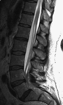 B
B 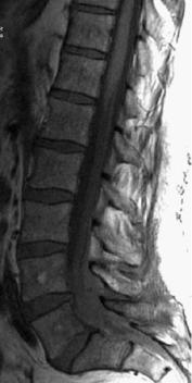
 C
C
D
■ Clinical Presentation
A patient with increased radicular pain and fever 1 month after laminectomy.
156 RadCases.thieme.com |
RadCases Neuro Imaging |
|
|
■ Imaging Findings |
|
A B C D
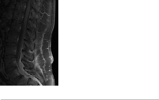
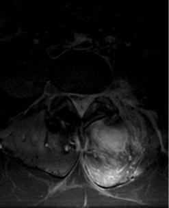
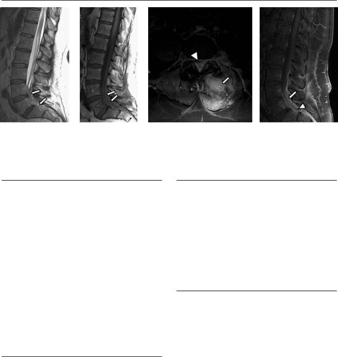
(A) Sagittal T2-weighted image (WI) shows heterogeneous signal in the lower lumbar spine (arrows). (B) Sagittal T1WI shows isointense signal in the lower lumbar spinal canal (arrows); the nerve roots are not seen. x(C) Fat-saturated axial T1WI with contrast shows enhancement of the left paraspinal muscles and the posterior aspect of the spinal canal (arrow) compressing the thecal sac (arrowhead). (D) Fat-saturated T1WI of the lumbar spine with contrast shows concentric anterior and posterior enhancement of the epidural space (arrow) and a central area without enhancement (arrowhead).
■ Di erential Diagnosis
•Epidural abscess: This is an infectious collection outside the dura mater that can be associated with osteomyelitis or paraspinal infection. Epidural abscess usually extends along two to nine vertebral segments. It demonstrates increased T2 signal in the epidural space compressing the thecal sac. There is avid enhancement and a thick capsule. Di usely increased T2 signal of the paraspinal soft tissues is noted.
•Epidural hemorrhage: This is characterized by a fluid collection outside the dura mater with variable signal intensity depending on the age of the hemorrhage. It usually extends beyond three consecutive vertebral segments. Epidural hemorrhage can have marginal enhancement.
•Epidural tumor: Epidural metastases are usually an extension of vertebral body lesions. Neurofibromas or schwannomas can occupy the epidural space, but they are commonly fusiform or dumbbell-shaped masses confined to one vertebral level and adjacent to the neural foramen.
■Essential Facts
•The primary source of infection is usually the vertebral body or disk, followed by the joint facet and perivertebral soft tissues.
•The center of the abscess shows fluid signal with thick peripheral enhancement.
■ Other Imaging Findings
•Computed tomography results can be normal. Look for findings of primary infection.
•Magnetic resonance imaging:
•T1WIs: The center is isointense to hypointense. If the protein level of the abscess contents is high, T1WIs can show increased signal.
•T2WIs: The signal is heterogeneous, mostly increased. There is di use high signal in the adjacent soft tissues.
•T1WIs with contrast show avid enhancement of the walls.
¸Pearls & ˚ Pitfalls
¸Di usion-WIs can help di erentiate abscess from hema-
toma.
˚The signal intensity of cerebrospinal fluid can be similar to that of abscess, making abscess di cult to detect on T2WIs.
157
Case 79
A
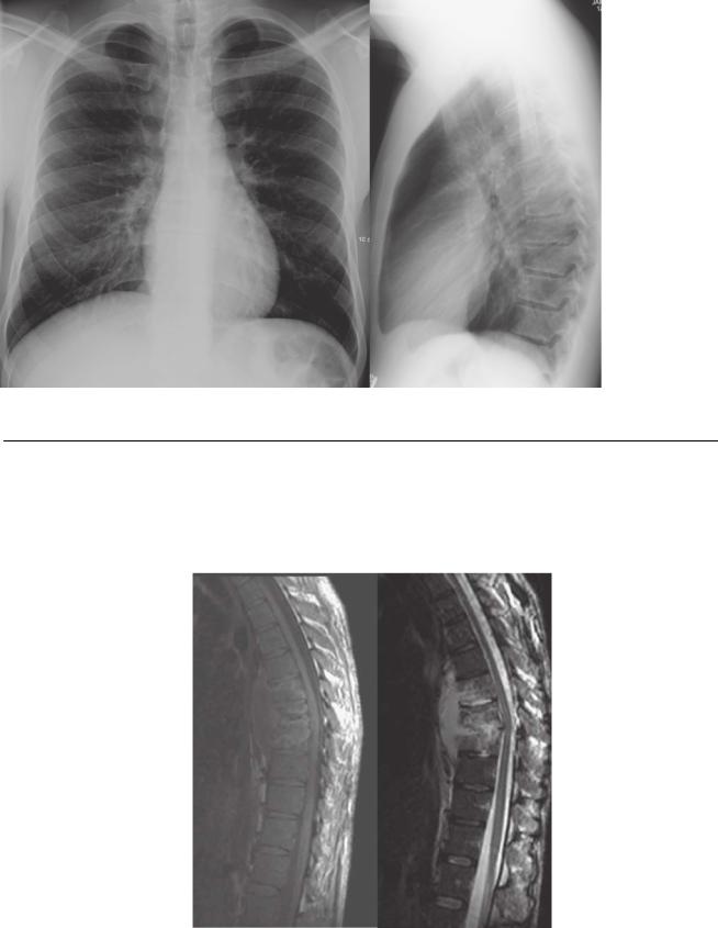
■ Clinical Presentation
A 45-year-old patient with chronic cough who is now experiencing persistent back pain.
Further Work-up
B 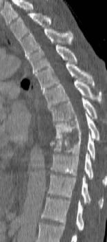 C
C  D
D