

Книги по МРТ КТ на английском языке / Neuro Imaging Redcases
.pdf88 |
RadCases.thieme.com |
RadCases Neuro Imaging |
|
■ Imaging Findings |
|
A B C
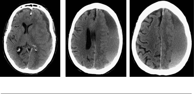
(A,B) Computed tomography (CT) scans with “brain windows” reveal a crescent-shaped extra-axial collection with high attenuation that is slightly heterogeneous (asterisk). It compresses the left lateral ventricle and causes rightward deviation of the midline structures (arrow). (C) Note the increased contrast between the hematoma (arrow) and the skull (arrowhead) in the “blood” window.
■ Di erential Diagnosis
•Acute subdural hematoma (SDH): Acute SDH is a crescentic, hyperdense collection between the dura and arachnoid membrane. Displacement of the cortex away from the inner table is common. It can cause midline shift with compression of the ipsilateral lateral ventricle and dilatation of the contralateral ventricle.
•Epidural hematoma: This is a collection of blood between the inner skull table and the dura. There is an adjacent skull fracture in 90% of cases. On CT, there is a hyperattenuating collection with a biconvex or lenticular shape and sharp margination. A low-attenuation swirl may be the result of blood from active bleeding mixing with serum from clotted blood.
•Subdural hygroma: This is a cerebrospinal fluid (CSF) collection that develops 6 to 30 days after a traumatic arachnoid tear. On CT, subdural hygromas have the same density as CSF. Blood vessels crossing through the extraaxial collection help di erentiate subdural hygromas from chronic SDHs.
■Essential Facts
•SDH is a collection of blood between the dura and arachnoid membrane.
•Bleeding may be from cerebral veins, venous sinuses, or cerebral contusions.
•Cerebral contusions are frequently associated with SDHs.
•The mass e ect of the collection is greater than expected because of swelling of the hemisphere.
•The most common locations are along the cerebral convexities, the falx cerebri, and the tentorium cerebelli.
•Bilateral SDHs may have a balancing e ect, so that there is no midline shift despite significant mass e ect.
•Small SDHs may be missed because of high convexity locations, beam-hardening artifacts, or narrow window settings.
•A wider “blood” window (width, 200; level, 70) helps to di erentiate pixels of similar brightness and so di erentiate acute blood from bone.
■ Other Imaging Findings
•Magnetic resonance imaging (MRI) is more sensitive than CT in the detection of small SDHs and tentorial or interhemispheric SDHs.
•The detection of SDH is critical in cases of suspected child abuse.
¸Pearls & ˚ Pitfalls
¸SDHs may cross suture lines but not dural reflections,
such as the falx and tentorium.
¸SDHs that are only a few hours old can have a mixed appearance of both hyperdense and hypodense regions because of the presence of uncoagulated blood before
clot formation.
˚In the setting of severe anemia, acute SDHs can be isodense to gray matter.
˚An ossified cerebral falx contains bone marrow with a high T1 signal, which may mimic a subdural hematoma on MRI.
89
Case 45
A B C
■ Clinical Presentation
A 65-year-old with headache and slow, progressive intellectual decline.
90 |
RadCases.thieme.com |
RadCases Neuro Imaging |
■ Imaging Findings
A B C
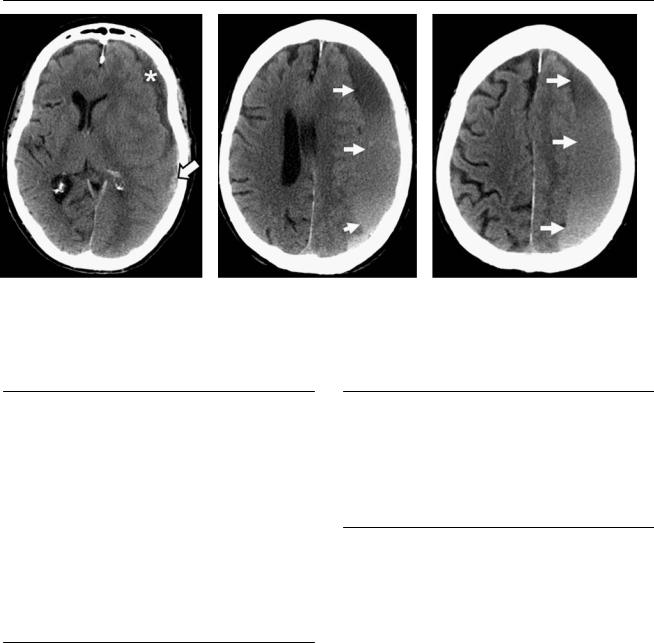
(A) Computed tomography (CT) at the level of the lateral ventricles shows a crescent-shaped extra-axial collection in the left convexity with high attenuation in the dependent portion (arrow) and fluid attenuation anteriorly (asterisk). (B,C) The slices in the superior aspect of the brain show a large collection (arrows) with layering of blood products causing hematocrit e ect. Note the relatively minimal mass e ect for the size of the hematoma.
■ Di erential Diagnosis
•Acute-on-chronic subdural hematoma (SDH): This is characterized by repeated hemorrhage into previous subdural collections. It may show layering of blood products.
•Acute SDH: The mass e ect from a collection that develops acutely is much more severe than that of a slowly accumulating hematoma.
•Epidural hematoma with active bleeding: This is a biconvex, hyperdense extra-axial fluid collection. There is adjacent skull fracture in 90% of cases. Epidural hematoma with active hemorrhage may contain alternating crescent-shaped regions of various densities that produce a “swirled” appearance.
■Essential Facts
•Chronic SDHs are more than 2 to 3 weeks old and are typically lower in attenuation than brain on CT.
•A history of trauma may be absent.
•Predisposing factors are older age, anticoagulant therapy, long-term hemodialysis, thrombocytopenia, and alcoholism.
•Ventricular decompression for hydrocephalus may cause SDHs.
•Acute-on-chronic SDHs develop from repeated hemorrhage into previous subdural collections.
•Fluid-blood levels are seen when sedimented fresh blood gravitates dependently and the proteinaceous fluid layers on top.
■ Other Imaging Findings
•Magnetic resonance imaging:
•This shows hematomas along the tentorium and falx that are more di cult to evaluate on axial CT images.
•Blood products of di erent ages can also be characterized to better advantage.
¸Pearls & ˚ Pitfalls
¸As the distance between the cerebral cortex and the dura
increases as a result of atrophy, small veins extending from the cortex to the dura (so-called bridging veins) become stretched and can be sheared by violent head motions, even if the head has not been struck by external objects.
¸Membranes can form within a chronic SDH, producing multiple compartments that have di erent attenuation
characteristics.
˚Subacute SDH becomes increasingly isodense to gray matter and di cult to detect on CT. When bilateral isodense SDHs are present, the mass e ects in the two hemispheres can be balanced, making detection even more di cult.
91
Case 46
A B
C
■ Clinical Presentation
A 7-month-old child after a fall.
92 |
RadCases.thieme.com |
RadCases Neuro Imaging |
|
|
■ Imaging Findings |
|
|
A |
B |
C |
|
|
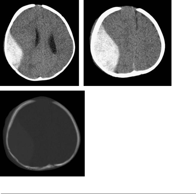
(A,B) Computed tomography (CT) scans of the brain demonstrate a lentiform collection of blood in the right frontoparietal convexity (asterisk). (C) Bone |
||
|
windows demonstrate a linear nondisplaced fracture in the right parietal bone (arrow). |
|
|
■ Di erential Diagnosis
•Epidural hematoma (EDH): This is a biconvex, hyperdense extra-axial fluid collection. There is adjacent skull fracture in 90% of cases. On occasion, subdural and epidural hematomas contain alternating crescent-shaped regions of various densities that produce a “swirled” appearance, considered an indication of active hemorrhage.
•Subdural hematoma: This is a crescentic, hyperdense collection between the arachnoid and dural membranes. There is displacement of the cortex away from the inner table. There is midline shift with compression of the ipsilateral lateral ventricle and dilatation of the contralateral ventricle.
•Meningioma: This dura-based tumor is isoattenuating to slightly hyperattenuating. Homogeneous and intense
enhancement after the injection of iodinated contrast material is characteristic. Perilesional edema may be extensive. Hyperostosis and intratumoral calcifications may be present.
■Essential Facts
•An EDH is a collection of blood between the inner skull table and the dura.
•There may be laceration of meningeal vessels, dural emissary veins, or sinuses.
•There is adjacent skull fracture in 90% of cases.
•Ninety-five percent are supratentorial.
•EDHs in the posterior fossa are rare and associated with higher morbidity and mortality than are supratentorial hematomas.
■ Other Imaging Findings
•Magnetic resonance imaging:
•Hyperacute and chronic EDHs exhibit signal intensity similar to that of cerebrospinal fluid on T1 and T2weighted images (WIs).
•The acute EDH is isointense on T1WIs and isoto hypointense on T2WIs.
•Subacute and early chronic EDHs are hyperintense on T1and T2WIs.
¸Pearls & ˚ Pitfalls
¸EDH does not cross cranial sutures, where the dura is
tightly adherent.
¸EDH may cross the falx or displace the dural venous sinuses from the inner table.
¸Emergent surgical treatment is nearly always needed.
˚EDH in the posterior fossa and along the floor of the anterior cranial fossa may be more di cult to visualize on CT.
93
Case 47
A 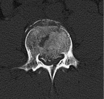 B
B
■ Clinical Presentation
A woman with severe back pain after being in an automobile-pedestrian accident.
Further Work-up
C 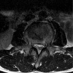 D
D

94 |
RadCases.thieme.com |
RadCases Neuro Imaging |
■ Imaging Findings
A 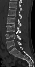 B
B 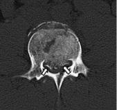 C
C 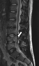
 D
D
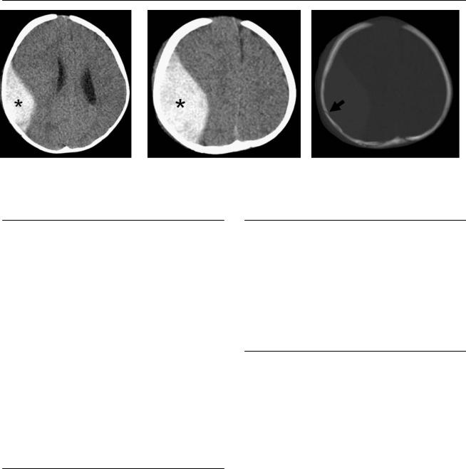
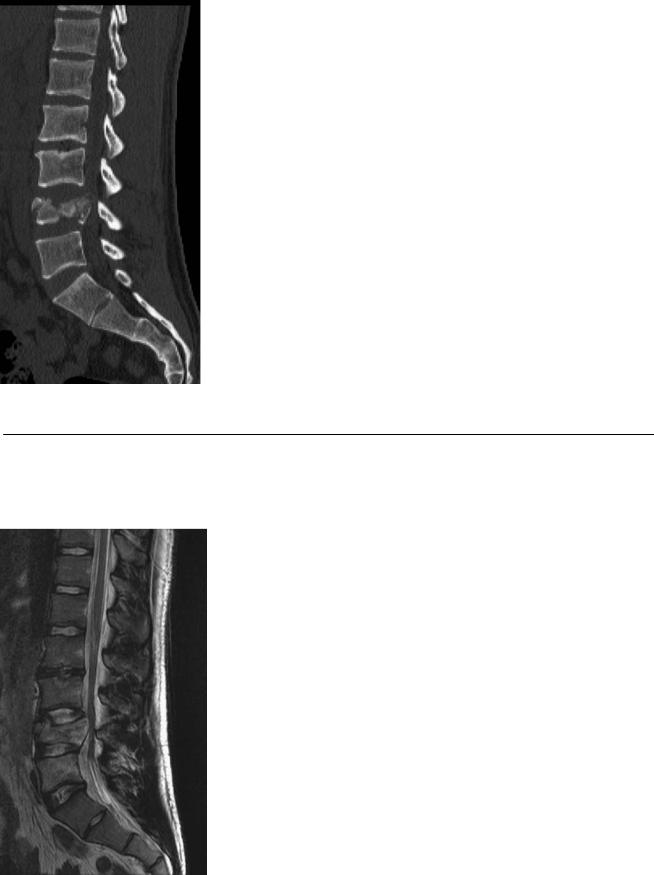
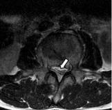
(A) Sagittal computed tomography (CT) scan of the lumbar spine shows a fracture of the L4 vertebral body involving all the walls, with retropulsion of fragments toward the spinal canal (arrow). (B) Axial CT scan of the lumbar spine shows a fracture of the L4 vertebral body involving all the walls, with retropulsion of fragments toward the spinal canal (arrows). There is also fracture of the posterior elements. (C) Sagittal T2-weighted image (WI) shows, in addition to the deformity of L4, bone marrow edema at L4 and L5. The narrowing of the canal and its e ect on the cauda equina (arrow) are better demonstrated. (D) Axial T2WI shows flattening of the thecal sac (arrow) secondary to stenosis caused by the posteriorly displaced fracture fragments.
■ Di erential Diagnosis
•Burst fracture: This is a compression fracture in which the entire vertebral body breaks. It results from axial loading that is most often secondary to motor vehicle accidents and falls. The axial load drives the intervertebral disk into the vertebral body below.
•Pathologic fracture: This occurs in a bone weakened as a result of neoplasm (multiple myeloma, metastatic bone disease). Fractures above the T7 level, fractures that present with a soft-tissue mass or osseous destruction, and fractures of the posterior part of the vertebral body are most likely to have a malignant etiology.
•Vertebral fracture due to osteomalacia: Osteomalacia causes bone softening and consequent biconcave deformity of the end plates (“codfish” vertebrae). In adulthood, these fractures resemble those found in osteoporosis. Usually, osseous deformities at other sites of the skeleton (e.g., protrusion acetabuli, pseudofractures [Looser zones] in the pelvis, and bone deformities) are also identified.
■Essential Facts
•Depending on the mechanism of injury, predictable patterns of fracture-dislocation are identified (hyperflexion, hyperflexion-rotation, hyperextension, hyperextensionrotation, vertical compression, or lateral flexion).
•In the lower cervical segment of C3-C7 and in the thoracolumbar spine, support is provided by three columns:
•Anterior column: the anterior vertebral bodies, anterior annulus fibrosus, and anterior longitudinal ligaments
•Middle column: posterior vertebral bodies, posterior longitudinal ligaments, and posterior annulus fibrosus
•Posterior column: posterior bony elements, ligamenta flava, and posterior ligaments
•The integrity of the middle column is the most crucial to spinal stability.
■ Other Imaging Findings
•Multidetector computed tomography (MDCT) is the modality of choice for the initial evaluation of bone injuries after blunt trauma to the cervical spine.
•When MDCT is not available, plain film remains the initial imaging modality. However, in patients who have unexplained focal pain, neurologic deficit with negative plain films, unexplained soft-tissue swelling, or abnormal plain films, additional evaluation with MDCT is indicated.
•Magnetic resonance imaging will show soft-tissue injury, spinal cord injury, intervertebral disk and ligament injury, and vascular injury.
¸Pearls & ˚ Pitfalls
¸Elderly patients usually have significant osteoporosis and
degenerative bone disease.
¸These patients may experience a significant fracture from a relatively minor trauma.
¸They also may have pathologic fractures.
˚Spinal cord injury in infants and young children often occurs without evidence of fracture or dislocation because of the inherent elasticity of their vertebral column, which makes the cord vulnerable to injury.
˚Patients who have spinal cord injury without radiographic abnormality often have a poor long-term prognosis.
95
Case 48
A B
C D
■ Clinical Presentation
A 1-year-old with the sudden onset of bilateral lower extremity weakness.
96 |
RadCases.thieme.com |
RadCases Neuro Imaging |
|
■ Imaging Findings |
|
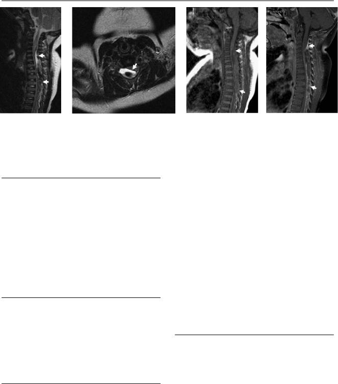
A B C D
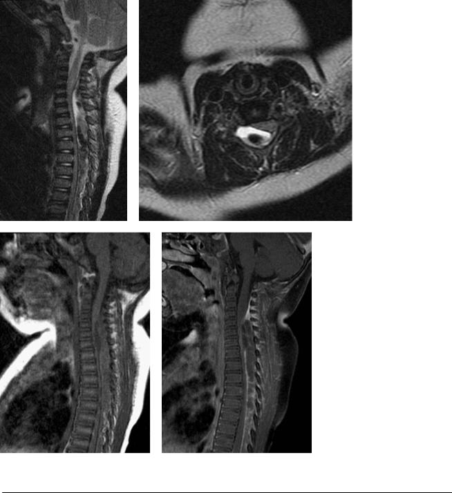
(A) Sagittal T2-weighted image (WI) shows a biconcave collection in the lower cervical and upper thoracic dorsal epidural space (arrows) with heterogeneous signal and areas of low signal. (B) In this axial image, the dura is visualized as a linear structure with low signal (arrow) between the anteriorly located cord and the hematoma. Note the compressive e ect on the cord, which does not show edema. (C,D) Sagittal T1WIs, preand postcontrast administration, show scattered linear areas of enhancement (arrows) within and at the periphery of the collection.
■ Di erential Diagnosis
•Epidural hematoma (EDH): EDH is an extra-axial collection of blood with heterogeneous signal intensity and a biconvex shape. Displacement of the dura toward the cord confirms the epidural location of the lesion.
•Epidural abscess: This is an expanding suppurative infection in the spinal epidural space. Epidural abscesses impinge on the spinal cord and may cause vascular compromise and infarction. The thoracic spine is more frequently involved.
•Epidural metastasis: This is usually associated with vertebral infiltration and pathologic fracture. It spares the disk.
■Essential Facts
•Spinal EDH:
•Etiology: trauma, anticoagulation therapy, vascular anomalies, spinal epidural procedures, spinal surgery, spontaneous
•Symptoms: severe localized back pain with delayed radicular radiation that may mimic disk herniation
•Treatment: emergency evacuation
■Other Imaging Findings
•Radiography and computed tomography (CT) are helpful in patients with a history of acute trauma to evaluate associated fractures or dislocation preoperatively and to assess the placement of instrumentation postoperatively.
•CT-myelography is indicated in patients who have spinal instrumentation, especially because the details of the spinal canal are often obscured by metal artifacts on magnetic resonance imaging (MRI). The hematoma is demonstrated as a biconvex filling defect in the spinal canal that usually extends over more than two vertebral levels.
•MRI:
•Sagittal MRI usually shows the biconvex hematoma in the dorsal epidural space, with well-defined contours and tapering of the superior and inferior margins.
•The hematomas show signal intensity that varies over time.
•Displaced dura on sagittal or axial MRI is an important finding that indicates the epidural location of the hematoma.
•The hematoma may demonstrate contrast enhancement.
¸Pearls & ˚ Pitfalls
¸Hyperintense cord lesions, which represent edema,
compression myelopathy, contusion injury, or infarction, decrease the possibility of neurologic recovery.
¸The cord involvement is better evaluated on axial than on sagittal images, particularly when the lesion is o the
midline.
˚Contrast enhancement of the cord parenchyma can be present and should not be misinterpreted as a neoplastic or an inflammatory cord lesion.

97
Case 49
A B
C
■ Clinical Presentation
A 37-year-old woman presenting with motor and sensory disturbances in the arms and legs after a motor vehicle accident.