

Книги по МРТ КТ на английском языке / Magnetic Resonance Imaging in Ischemic Stroke - K Sartor R 252 diger von Kummer Tobias Back
.pdf64 T. Back
Treatment e ects on DWI lesion growth
Cation channel blocker Glutamate antagonist (MK-801)
Normalized ADC lesion volume (%)
160untreated treated
140
120
100
80
60
40
0 |
1 |
2 |
3 |
4 |
5 |
6 |
MCA occlusion (h)
Volume of hemispheric DWI lesion
(Mean +/-SEM) mm3
180
150
120
90
60
30
0
Control
MK-801
|
|
|
|
** |
|
|
** |
** |
|
|
|
|
|
|
* |
** |
|
|
|
|
|
|
|
|
1 h |
2 h |
3 h |
4 h |
24 h |
MCA occlusion (h)
Fig. 4.16. Treatment effects by pharmacological intervention in models of permanent focal ischemia. Efficacy of cation channel blocker (left) or glutamate antagonist MK-801 (right) was monitored by using repetitive diffusion-weighted imaging (DWI). Lesions were defined by a change in the apparent diffusion coefficient (ADC, left) or by hyperintensity in DWI (right). Note that pharmacological intervention tends to slow down natural lesion enlargement rather than reversing ischemic lesions. [Reproduced with permission from Hoehn-Berlage et al. (1997), and Gill et al. (1996)]
lag from primary injury, also depending on the severity of the ischemic insult and the occurrence of reperfusion. Here, two aspects of delayed changes are to be addressed, namely the postischemic patterns of functional activation and the plasticity of the cortex as response to local injury. Of note, we stand at the very beginning of a better understanding of processes that reorganize injured brain.
4.4.1
Effects on Functional Activation
In Sect. 4.1.4 the detection of activated brain areas by BOLD or perfusion-weighted imaging was shown to depend on: (i) the ability of neurons to become activated and (ii) the neurovasculature to respond to neuronal activation by an (adequately matched) increase in local blood flow. During functional activation, a ≈ 6% increase in signal intensity is observed in BOLD images that is attributed to a decrease of the absolute concentration of dexoygenated hemoglobin
(Hbdeoxy) per voxel (Grüne et al. 1999). Hbdeoxy is paramagnetic and thereby decreases signal inten-
sity in T2*-weighted images. The signal increase is brought about by an excess increase in blood flow leading to excess concentration of oxygenated
hemoglobin (Hboxy) overriding the oxygen demand of the activated tissue. As a consequence, a perfect match of local blood flow and oxygen extraction
would result in an increase of absolute Hbdeoxy concentration per voxel (due to increased blood volume)
and, thus, to a negative BOLD signal. Under physiological conditions this match is imperfect so that a positive BOLD response to functional activation is the normal finding. Apart from this, functionally activated brain regions can be monitored by application of PI (i.e., bolus tracking MRI) that reveals a 70% increase in local blood flow or blood volume that matches very well CBF measurements by other methods documenting flow changes in the range of 70%-90% above baseline during functional activation (Schmitz et al. 1997).
In a recent study, rats underwent permanent MCA occlusion and functional activation was studied by bilateral forepaw stimulation and MRI 24 h after the insult (Reese et al. 2000). The CBF response was monitored in both hemispheres by using a T2*- sensitive imaging sequence and intravascular bolus of contrast agent. In the non-ischemic hemisphere, a proper activation due to forepaw stimulation was recorded, but on the affected side no response could be elicited. This failure of activation was first attributed to ischemic damage of the somato-
Insights from Experimental Studies |
65 |
sensory cortex. However, in animals that received neuroprotective treatment and in which the somatosensory cortex was spared from injury, still no response could be detected. The authors concluded that those cortical areas are functionally impaired although their afferent input remained to be unlesioned. More extended observations of functional recovery at 2 days and 2 weeks post occlusion were published by Dijkhuizen et al. (2001). At day 2 after stroke onset no functional activity was observed in the ischemic hemisphere. Interestingly, at this timepoint CO2-reactivity was preserved (or had recovered) and baseline flow was not increased, indicating the absence of severe vasodilation that would prohibit a further stimulation-induced vascular response. After 2 weeks, bilateral signs of activation were present in the infarct borderzone, both in and adjacent to the sensorimotor cortex. Neurobehavioral tests revealed a nearly complete recovery of forelimb function. Obviously, the cortical forelimb representation field has recruited periand contralesional functional fields in order to regain near-to- normal function.
In the pathophysiological state of resuscitation after global ischemia, the pattern of recovery is different. During postischemic hyperemia, autoregulation and CO2 reactivity are abolished and the former recovers earlier than the latter. The question arises whether the impaired functional coupling between metabolism and blood flow reflects disturbances of the functional integrity of the brain or just indicates impaired cerebrovascular reactivity. In studies by Schmitz et al. (1997, 1998), rats were exposed to 10-min cardiac arrest followed by reanimation and repeated MRI studies up to 7 days. ADC normalized within 45 min after resuscitation, but neurological scores and amplitudes of the sensory evoked potential (SEP) recovered at a much slower pace. While CO2 reactivity had returned to normal at 5 h after reanimation, the stimulus-induced CBF increase due to electrical forepaw stimulation recovered to only 40% of normal within 1 week (measured by laser-Doppler flowmetry) (Schmitz et al. 1997). Laser-Doppler flow measurements were precisely confirmed by PI (Schmitz et al. 1998). After 3 h of reperfusion, functional activity began to reappear, but the recovery of the BOLD signal progressed faster than that of the perfusion-weighted signal: the stim- ulus-induced signal intensity increase in T2* images at day 1 after resuscitation was already comparable to normal. The differences in the recovery of ADC, BOLD, and perfusion imaging were interpreted to relate to differences between metabolic and func-
tional recovery on one hand and between blood flow and oxygen extraction on the other. Thus, the much slower recovery of the CBF response to functional activation is not limited by an impaired cerebrovascular sensitivity. Since the BOLD signal is inversely proportional to the tissue oxygen extraction, it may be concluded that the decoupling between the T2*- weighted imaging behavior (rapidly restored) and PI responses to stimulation (sustainedly impaired) is due to reduced oxygen extraction upon activation.
4.4.2
Brain Plasticity and Stem Cell Implantation
Therapeutic strategies do not aim alone at preservation of lesioned tissue but also at functional restitution after completed stroke. There are two principle approaches to the latter: (i) mechanisms of brain plasticity and (ii) regeneration based on stem cell implantation.
The pattern and role of brain plasticity in stroke recovery has been incompletely characterized. Both ipsilesional and contralesional changes have been described in the previous section, but it remained unclear how these relate to functional recovery. In a recent investigation, brain activation patterns were correlated with tissue damage, hemodynamics, and neurologic status after temporary stroke, using functional MRI (Dijkhuizen et al. 2003). Functional activation and cerebrovascular reactivity maps were generated at days 1, 3 and 14 after 2-h MCA occlusion in rats. Significant activation responses in the contralesional hemisphere were detected at days 1 and 3. There was no correlation between activation parameters and perfusion status or cerebrovascular reactivity. The degree of shift of activation balance toward the contralesional hemisphere early after stroke increased with the extent of tissue injury. Functional recovery was associated mainly with preservation or restoration of activation in the ipsilesional hemisphere.
During neonatal development, the brain possesses the striking ability to transfer initially lost functions to new, unaffected cortical areas when irreversible lesions prohibit function of the original representation fields – an ability that is still to a lesser degree present in mature brain. This type of “plastic” response has been studied in rats with a well defined lesion of the somatosensory cortex that was induced 1 day after birth. Six months later functional MRI (fMRI) was performed with a forepaw stimulation paradigm when the animals showed no neurological
66 |
T. Back |
deficits. fMRI signal amplitude in activation areas was decreased on the ispilesional side with the occasional occurrence of contralesional activation in the secondary somatosensory cortex (Schwindt et al. 2004). Taking these and other results into account, neuromodulatory mechanisms enable the activation of previously inhibited but existing pathways that contribute to functional reorganization.
Stem cell implantation is a new intriguing way to promote regeneration. In vivo monitoring of stem cells after grafting is essential for a better understanding of their migrational dynamics and differentiation processes and of their regeneration potential. Using MRI at 78-Pm spatial resolution and cell labeling by a lipofection procedure with a MR contrast agent, focal cerebral ischemia was studied (Hoehn et al. 2002). Over a 3-week period, cell migration was observed along the corpus callosum to the lesioned hemisphere, and cells massively populated the borderzone of the damaged brain tissue on the hemisphere opposite to the implantation sites (Fig. 4.17). Obviously, embryonic stem cells have high migra-
tional dynamics, targeted to the cerebral lesion area. The translation of those results into clinical research needs to be shown in the future.
4.5
Outlook on Future Research
MRS is a promising tool to accompany imaging studies in cerebral ischemia. In vivo 31P MRS provides information on energy metabolism (ATP, PCr) and can be used to estimate intracellular pH from the chemical shift difference between Pi (inorganic phosphate) and PCr. Due to its relatively low sensitivity (7% of that of 1H) quite large voxels- of-interest have to be chosen which complicates studies in smaller animals. In vivo 1H spectroscopy offers insight into other metabolic arenas: during ischemia/hypoxia a prominent peak due to lactate can be detected that is not visible under normal physiological conditions. N-acetyl-aspar-
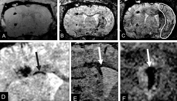
a |
|
b |
|
c |
|
|
|
|
|
d |
|
e |
|
f |
|
|
|
|
|
Fig. 4.17a–f. Coronal section through a rat brain at various times after implantation of embryonic stem cells contralateral to the induced focal ischemia. Data sets were recorded at the day of implantation (a) and at 6 (b) and 8 (c) days after implantation. The infarcted tissue area is outlined on (c). Note at 6 days (b) the discrete dark line (arrow in d, higher magnification) along the corpus callosum showing iron-oxide labeled cells migrating toward the lesioned hemisphere. At 8 days (c) a dark region becomes visible in the dorsal part of the lesioned territory reflecting first arrival of those cells. At higher magnification (d), the migration along the corpus callosum is better visible. The lining along the ventricular wall (e) and the accumulation of labeled stem cells on the choroid plexus (f) are presented in another example with high magnification. [With permission from Hoehn et al. (2002)]
Insights from Experimental Studies |
67 |
tate (NAA) is a substrate almost exclusively localized in neurons, the function of which is widely unknown. Its characteristic peak in proton spectra decreases in ischemic states, probably due to neuronal loss. Other peaks derived from proton spectra are those for glucose, (phospho)creatine, cholinecontaining compounds, glutamate/glutamine, and myo-inositol. They provide information of other aspects of ischemia-dependent changes in metabolism. Choline derivatives are involved in membrane function and fluidity, glutamate/glutamine point to the activity of the tricarboxylic acid cycle, and myo-inositol plays a role in the ion homeostasis that is known to be disturbed in acute ischemia. Perhaps the most interesting application of MRS is the multi-voxel approach of proton spectra called spectroscopic imaging (SI) (Brown et al. 1982). SI provides metabolic maps of the brain with increasing spatial resolution and has been successfully applied to animal and human stroke (Fig. 4.15). The high regional heterogeneity of cerebral infarcts is reflected by differential metabolic responses from the ischemic core and borderzone areas (Franke et al. 2000). Such metabolic profiles have already been used to characterize subtypes of human stroke (Liu et al. 2003) (see Chap. 11).
Technical progress and higher spatial and temporal resolution of MRI and MRS is going to trigger MR studies in small animals, especially in mice, although high-resolution MR methods have predominantly been applied to rat and cat models of ischemia. Gene-manipulated mice (knock-out, knock-in) offer a powerful way to gain insight into complex molecular interactions and intracellular signaling. Zaharchuk et al. (1997) showed for the first time that MRI can be used in mouse MCA occlusion to detect changes in T1, T2 and DWI. They also demonstrated that mice deficient of neuronal nitric oxide synthase had a smaller periinfarct zone (defined by ADC threshold) and attributed this finding to less severe metabolic changes after ischemia. Van Dorsten et al. (1999) investigated different wild-type mouse strains using PI and DWI and found significantly smaller lesion volumes in SV129 mice compared to C57Black/6 mice probably due to the smaller MCA territory of the former strain. Hence, differences in vascular anatomy have to be considered in the future when parent strains are selected for genetic engineering.
Finally, we have little doubt that MR methods will play a dominant and innovative role in future research of brain ischemia covering alterations of perfusion, metabolism and molecular signaling.
References
Alexis NE, Back T, Zhao W, Dietrich WD, Watson BD, Ginsberg MD (1996) Neurobehavioral consequences of induced spreading depression following photothrombotic middle cerebral artery occlusion. Brain Res 706:273-282
Antman EM, Giugliano RP, Gibson CM, McCabe CH, Coussement P, Kleiman NS, Vahanian A, Adgey AAJ, Menown I, Rupprecht H-J, van der Wieken R, Ducas J, Scherer J, Anderson K, van de Werf F, Braunwald E (1999) Abciximab facilitates the rate and extent of thrombolysis-Results of the thrombolysis in myocardial infarction (TIMI) 14 trial. Circulation 99:2720-2732
Arbelaez A, Castillo M, Mukherji SK (1999) Diffusion-weighted MR imaging of global cerebral anoxia. Am J Neuroradiol 20:999-1007
Astrup J, Symon L, Branston NM, Lassen NA (1977) Cortical evoked potential and extracellular K+ and H+ at critical levels of brain ischemia. Stroke 8:51-57
Astrup J, Siesjö BK, Symon L (1981) Thresholds in cerebral ischemia - the ischemic penumbra. Stroke 12:723-725
Back T, Hoehn-Berlage M, Kohno K, Hossmann K-A (1994a) Diffusion NMR imaging in experimental stroke: Correlation with cerebral metabolites. Stroke 25:494-500
Back T, Kohno K, Hossmann K-A (1994b) Cortical negative DC deflections following middle cerebral artery occlusion and KCl-induced spreading depression: effect on blood flow, tissue oxygenation and electroencephalogram. J Cereb Blood Flow Metab 14:12-19
Back T, Zhao W, Ginsberg MD (1995) Three-dimensional image analysis of brain glucose metabolism/blood flow uncoupling and its electrophysiological correlates in the acute ischemic penumbra following middle cerebral artery occlusion. J Cereb Blood Flow Metab 15:566-577
Back T, Ginsberg MD, Dietrich WD, Watson BD (1996) Induction of spreading depression in the ischemic hemisphere following experimental middle cerebral artery occlusion: Effect on infarct morphology. J Cereb Blood Flow Metab 16:202-213
Back T, Nedergaard M, Ginsberg MD (1998) The ischemic penumbra: Pathophysiology, and relevance of spreading depression-like phenomena. In: Ginsberg MD, Bogousslavsky J (eds) Cerebrovascular disease: pathophysiology, diagnosis, and management. Blackwell Scientific Publications, Malden, MA, USA, pp 276-286
Back T, Hirsch JG, Szabo K, Gass A (2000a) Failure to demonstrate peri-infarct depolarizations by repetitive MR diffusion imaging in acute human stroke. Stroke 31:2901-2906
Back T, Hoehn M, Mies G, Busch E, Schmitz B, Kohno K, Hossmann KA (2000b) Penumbral tissue alkalosis in focal cerebral ischemia: relationship to energy metabolism, blood flow, and steady potential. Ann Neurol 47:485-492
Back T, Schüler OG, Otto D, Culmsee C, Plesnila N, Krieglstein J, Oertel WH, Baethmann A (2002) Early versus delayed thrombolysis in embolic stroke: effects on blood flow, DC potential and infarct morphology. In: Krieglstein J, Klumpp S (eds) Pharmacology of cerebral ischemia. MedPharm Scientific Publishers, Stuttgart, pp 159-169
Back T, Otto D, Kittner D, Hemmen T, Oertel WH (2004) Failure to enhance thrombolytic therapy by neuroprotection with memantine in embolic stroke. Cerebrovasc Dis 17:59 Baird AE, Benfield A, Schlaug G, Siewert B, Lövblad K-O, Edelman RR, Warach S (1997) Enlargement of human cerebral
68
ischemic lesion volumes measured by diffusion-weighted magnetic resonance imaging. Ann Neurol 41:581-589
Barber PA, Darby DG, Desmond PM, Yang Q, Gerraty RP, Jolley D, Donnan GA, Tress BM, Davis SM (1998) Prediciton of stroke outcome with echoplanar perfusionand diffusionweighted MRI. Neurology 51:418-426
Bartus RT, Dean RL, Cavanaugh K, Eveleth D, Carriero DL, Lynch G (1995) Time-related neuronal changes following middle cerebral artery occlusion: Implications for therapeutic intervention and the role of calpain. J Cereb Blood Flow Metab 15:969-979
Belayev L, Zhao W, Pattany PM, Weaver RG, Huh PW, Lin B, Busto R, Ginsberg MD (1998) Diffusion-weighted magnetic resonance imaging confirms marked neuroprotective efficacy of albumin therapy in focal cerebral ischemia. Stroke 29:2587-2599
Belliveau JW, Kennedy DN Jr, McKinstry RC, Buchbinder BR, Weisskoff RM, Cohen MS, Vevea JM, Brady TJ, Rosen BR (1991) Functional mapping of the human visual cortex by magnetic resonance imaging. Science 254:716-719
Berek K, Lechleitner P, Luef G, Felber S, Saltuari L, Schinnerl A, Traweger C, Dienstl F, Aichner F (1995) Early determination of neurological outcome after prehospital cardiopulmonary resuscitation. Stroke 26:543-549
Böttiger BW, Schmitz B, Wiessner C, Vogel P, Hossmann KA (1998) Neuronal stress response and neuronal cell damage after cardiocirculatory arrest in rats. J CerebBlood Flow Metab 18:1077-1087
Branston NM, Hope DT, Symon L (1979) Barbiturates in focal ischemia of primate cortex: effects on blood flow distribution, evoked potential and extracellular potassium. Stroke 10:647-653
Branston NM, Ladds A, Symon L, Wang AD (1984) Comparison of the effects of ischaemia on early components of the somatosensory evoked potential in brainstem, thalamus, and cerebral cortex. J Cereb Blood Flow Metab 4:68-81
Branston NM, Strong AJ, Symon L (1977) Extracellular potassium activity, evoked potential and tissue blood flow. Relationships during progressive ischaemia of baboon cerebral cortex. J Neurol Sci 32:305-321
Brinker G, Franke C, Hoehn M, Uhlenkuken U, Hossmann KA (1999) Thrombolysis of cerebral clot embolism in rat: effect of treatment delay. NeuroReport 10:3269-3272
Brown TR, Kincaid BM, Ugurbil K (1982) NMR chemical shift imaging in three dimensions. Proc Natl Acad Sci USA 79:3523-3526
Busch E, Gyngell M, Eis M, Hoehn-Berlage M, Hossmann K- A (1996) Potassium-induced cortical spreading depression during focal cerebral ischemia in rats: contribution to lesion growth assessed by diffusion-weighted NMR and biochemical imaging. J Cereb Blood Flow Metab 16:1090-1099
Busch E, Kruger K, Hossmann K-A (1997) Improved model of thromboembolic stroke and rt-PA induced reperfusion in the rat. Brain Res 778:16-24
Busch E, Kruger K, Allegrini PR, Kerskens CM, Gyngell ML, Hoehn-Berlage M, Hossmann KA (1998) Reperfusion after thrombolytic therapy of embolic stroke in the rat: magnetic resonance and biochemical imaging. J Cereb Blood Flow Metab 18:407-418
Busch E, Beaulieu C, de Crespigny A, Kreischer S, Diener HC, Moseley ME (2002) Combined X-ray angiography and dif- fusion-perfusion MRI for studying stroke evolution after rt-PA treatment in rats. Brain Res 953:112-118
T. Back
Busza AL, Allen KL, King MD, van Bruggen N, Williams SR, Gadian DG (1992) Diffusion-weighted imaging studies of cerebral ischemia in gerbils: potential relevance to energy failure. Stroke 23:1602-1612
Butcher K, Parsons M, Baird T, Barber A, Donnan G, Desmond P, Tress B, Davis S (2003) Perfusion thresholds in acute stroke thrombolysis. Stroke 34:2159-2164
Chalela JA, Kang DW, Luby M, Ezzeddine M, Latour LL, Todd JW, Dunn B, Warach S (2004) Early magnetic resonance imaging findings in patients receiving tissue plasminogen activator predict outcome: insights into the pathophysiology of acute stroke in the thrombolysis era. Ann Neurol 55:105-112
Chen J, Jin K, Chen M, Pei W, Kawaguchi K, Greenberg DA, Simon RP (1997) Early detection of DNA strand breaks in the brain after transient focal ischemia: implications for the role of DNA damage in apoptosis and neuronal cell death. J Neurochem 69:232-245
Chen SD, Lee JM, Yang DI, Nassief A, Hsu CY (2002) Combination therapy for ischemic stroke: potential of neuroprotectants plus thrombolytics. Am J Cardiovasc Drugs 2:303-313
Choi DW (1992) Excitotoxic cell death. J Neurobiol 23:1261-1276
Chopp M, Zhang RL, Zhang ZG, Jiang Q (1999) The clot thick- ens-thrombolysis and combination therapies. Acta Neurochir Suppl (Wien) 73:67-71
Chu XP, Miesch J, Johnson M, Root L, Zhu XM, Chen D, Simon RP, Xiong ZG (2002) Proton-gated channels in PC12 cells. J Neurophysiol 87:2555-2561
De Crespigny AJ, Tsuura M, Moseley ME, Kucharczyk J (1993) Perfusion and diffusion MR imaging of thromboembolic stroke. J Magn Reson Imaging 3:746-754
Dereski MO, Chopp M, Knight RA, Rodolosi LC, Garcia JH (1993) The heterogeneous temporal evolution of focal ischemic neuronal damage in the rat. Acta Neuropathol (Berlin) 85:327-333
Derex L, Nighoghossian N, Hermier M, Adeleine P, Philippeau F, Honnorat J, Yilmaz H, Dardel P, Froment JC, Trouillas P (2004) Thrombolysis for ischemic stroke in patients with old microbleeds on pretreatment MRI. Cerebrovasc Dis 17:238-241
Dijkhuizen RM, de Graaf RA, Tulleken KA, Nicolay K (1999) Changes in the diffusion of water and intracellular metabolites after excitotoxic injury and global ischemia in neonatal rat brain. J Cereb Blood Flow Metab 19:341-349
Dijkhuizen RM, Ren J, Mandeville JB, Wu O, Ozdag FM, Moskowitz MA, Rosen BR, Finklestein SP (2001) Functional magnetic resonance imaging of reorganization in rat brain after stroke. Proc Natl Acad Sci USA 98:12766-12771
Dijkhuizen RM, Singhal AB, Mandeville JB, Wu O, Halpern EF, Finklestein SP, Rosen BR, Lo EH (2003) Correlation between brain reorganization, ischemic damage, and neurologic status after transient focal cerebral ischemia in rats: a functional magnetic resonance imaging study. J Neurosci 23:510-517
Dirnagl U, Pulsinelli W (1990) Autoregulation of cerebral blood flow in experimental focal brain ischemia. J Cereb Blood Flow Metab 10:327-336
Dirnagl U, Iadecola C, Moskowitz MA (1999) Pathobiology of ischaemic stroke: an integrated view. Trends Neurosci 22:391-397
Du C, Hu R, Csernansky CA, Hsu CY, Choi DW (1996) Very
Insights from Experimental Studies
delayed infarction after mild focal cerebral ischemia: a role for apoptosis? J Cereb Blood Flow Metab 16:195-201
Ebisu T, Katsuta K, Fujikawa A, Aoki I, Umeda M, Naruse S, Tanaka C (2001) Early and delayed neuroprotective effects of FK506 on experimental focal ischemia quantitatively assessed by diffusion-weighted MRI. Magn Reson Imaging 19:153-160
Endres M, Wang ZQ, Namura S, Waeber C, Moskowitz MA (1997) Ischemic brain injury is mediated by the activation of poly(ADP-ribose)polymerase. J Cereb Blood Flow Metab 17:1143-1151
Fiorelli M, Bastianello S, von Kummer R, del Zoppo GJ, Larrue V, Lesaffre E, Ringleb AP, Lorenzano S, Manelfe C, Bozzao L (1999) Hemorrhagic transformation within 36 hours of a cerebral infarct: relationships with early clinical deterioration and 3-month outcome in the European Cooperative Acute Stroke Study I (ECASS I) cohort. Stroke 30:2280-2284
Franke C, Brinker G, Pillekamp F, Hoehn M (2000) Probability of metabolic tissue recovery after thrombolytic treatment of experimental stroke: a magnetic resonance spectroscopic imaging study in rat brain. J Cereb Blood Flow Metab 20:583-591
Fujioka M, Taoka T, Matsuo Y, Mishima K, Ogoshi K, Kondo Y, Tsuda M, Fujiwara M, Asano T, Sakaki T, Miyasaki A, Park D, Siesjo BK (2003) Magnetic resonance imaging shows delayed ischemic striatal neurodegeneration. Ann Neurol 54:732-747
Furlan A, Higashida R, Wechsler L, Gent M, Rowley H, Kase C, Pessin M, Ahuja A, Callahan F, Clark WM, Silver F, Rivera F (1999) Intra-arterial prourokinase for acute ischemic stroke. The PROACT II study: a randomized controlled trial. Prolyse in acute cerebral thromboembolism. JAMA 282:2003-2011
Garcia JH, Kamijyo Y (1974) Cerebral infarction evolution of histopathological changes after occlusion of the middle cerebral artery in primates. Exp Neurol 33:408-421
Garcia JH, Yoshida Y, Chen H, Li Y, Zhang ZG, Lian J, Chen S, Chopp M (1993) Progression from ischemic injury to infarct following middle cerebral artery occlusion in the rat. Am J Pathol 142:623-635
Gill R, Andine P, Hillered L, Persson L, Hagberg H (1992) The effect of MK-801 on cortical spreading depression in the penumbral zone following focal ischemia in the rat. J Cereb Blood Flow Metab 12:371-379
Gill R, Sibson NR, Maskell L, Carpenter TA, Hall LD, Pickard JD (1996) The protective effect of MK-801 on infarct development over a period of 24 h as assessed by diffu- sion-weighted magnetic resonance imaging. NMR Biomed 9:241-248
Grotta J (1995) Why do all drugs work in animals but none in stroke patients? 2. Neuroprotective therapy. J Intern Med 237:89-94
Grubb NR, Fox KA, Smith K, Best J, Blane A, Ebmeier KP, Glabus MF, O’Carroll RE (2000) Memory impairment in out-of-hospital cardiac arrest survivors is associated with global reduction in brain volume, not focal hippocampal injury. Stroke 31:1509-1514
Grüne M, Pillekamp F, Schwindt W, Hoehn M (1999) Gradient echo time dependence and quantitative parameter maps for somatosensory activation in rats at 7 T. Magn Reson Med 42:118-126
Gyngell M, Busch E, Schmitz B, Kohno K, Back T, Hoehn-Ber- lage M, Hossmann K-A (1995) Evolution of acute focal
69
cerebral ischaemia in rats observed by localised 1H-MRS, diffusion-weighted MRI, and electrophysiological monitoring. NMR Biomed 8:206-214
Hacke W, Albers G, Al-Rawi Y, Bogousslavsky J, Davalos A, Eliasziw M,Fischer M,Furlan A,Kaste M,Lees KR,Soehngen M, Warach S; DIAS Study Group (2005) The Desmoteplase in Acutic Ischemic Stroke Trial (DIAS): a phase II MRIbased 9-hour window acute stroke thrombolysis trial with intravenous desmoteplase. Stroke 36:66–73
Hakim AM, Hogan MJ, Carpenter S (1992) Time course of cerebral blood flow and histological outcome after focal cerebral ischemia in rats. Stroke 23:1138-1143
Hallenbeck JM, Dutka AJ (1990) Background review and current concepts of reperfusion injury. Arch Neurol 47:1245-1254
Hata R, Maeda K, Hermann D, Mies G, Hossmann KA (2000) Evolution of brain infarction after transient focal cerebral ischemia in mice. J Cereb Blood Flow Metab 20:937-946
Heiss W-D (1983) Flow thresholds to functional and morphological damage of brain tissue. Stroke 14:329-331
Heiss W-D, Rosner G (1983) Functional recovery of cortical neurons as related to degree and duration of ischemia. Ann Neurol 14:294-301
Heiss W-D, Huber M, Fink GR, Herholz K, Pietrzyk U, Wagner R, Wienhard K (1992) Progressive derangement of periinfarct viable tissue in ischemic stroke. J Cereb Blood Flow Metab 12:193-203
Heiss W-D, Graf R, Wienhard K, Lottgen J, Saito R, Fujita T, Rosner G, Wagner R (1994) Dynamic penumbra demonstrated by sequential multitracer PET after middle cerebral artery occlusion in cats. J Cereb Blood Flow Metab 14:892-902
Heiss W-D, Thiel A, Grond M, Graf R (1999) Contribution of immediate and delayed ischaemic damage to the volume of final infarcts. Lancet 353:1677-1678
Helpern JA, Dereski MO, Knight RA, Ordidge RJ, Chopp M, Qing ZX (1993) Histopathological correlations of nuclear magnetic resonance imaging parameters in experimental cerebral ischemia. Magn Reson Imaging 11:241-246
Hilger T, Niessen F, Diedenhofen M, Hossmann KA, Hoehn M (2002) Magnetic resonance angiography of thromboembolic stroke in rats: indicator of recanalization probability and tissue survival after recombinant tissue plasminogen activator treatment. J Cereb Blood Flow Metab 22:652-662
Hoehn M (2003) Functional magnetic resonance imaging. In: Van Bruggen N, Roberts T (eds) Biomedical imaging in experimental neuroscience. CRC Press, Boca Raton, FL, USA, pp 93-135
Hoehn M, Nicolay K, Franke C, van der Sanden B (2001) Application of magnetic resonance to animal models of cerebral ischemia. J Magn Reson Imaging 14:491-509
Hoehn M, Kustermann E, Blunk J, Wiedermann D, Trapp T, Wecker S, Focking M, Arnold H, Hescheler J, Fleischmann BK, Schwindt W, Buhrle C (2002) Monitoring of implanted stem cell migration in vivo: a highly resolved in vivo magnetic resonance imaging investigation of experimental stroke in rat. Proc Natl Acad Sci USA 99:16267-16272
Hoehn-Berlage M (1995) Diffusion-weighted NMR imaging: application to experimental focal cerebral ischemia. NMR Biomed 8:345-358
Hoehn-Berlage M, Eis M, Back T, Kohno K,Yamashita K (1995a) Changes of relaxation times T1, T2 and apparent diffusion coefficient ADC after permanent MCA occlusion in the rat:
70
Temporal evolution, regional extent, and comparison with histology. Magn Reson Med 34:824-834
Hoehn-Berlage M, Norris DG, Kohno K, Mies G, Leibfritz D, Hossmann KA (1995b) Evolution of regional changes in apparent diffusion coefficient during focal ischemia of rat brain: the relationship of quantitative diffusion NMR imaging to reduction in cerebral blood flow and metabolic disturbances. J Cereb Blood Flow Metab 15:1002-1011
Hoehn-Berlage M, Hossmann K-A, Busch E, Eis M, Schmitz B, Gyngell M (1997) Inhibition of nonselective cation channels reduces focal ischemic injury of rat brain. J Cereb Blood Flow Metab 17:534-542
Hossmann KA (1997) Reperfusion of the brain after global ischemia: hemodynamic disturbances. Shock 8:95-101 Hossmann K-A (1987) Pathophysiology of cerebral infarction.
In: Vinken PJ, Bruyn GW, Klawans HL (eds) Handbook of clinical neurology. Elsevier, Amsterdam, pp 107-153
Hossmann K-A (1991) Animal models of cerebral ischemia. 1. Review of literature. Cerebrovasc Dis 1:2-15
Hossmann K-A (1994) Viability thresholds and the penumbra of focal ischemia. Ann Neurol 36:557-565
Hossmann KA, Hoehn-Berlage M (1995) Diffusion and perfusion MR imaging of cerebral ischemia. Cerebrovasc Brain Metab Rev 7:187-217
Hossmann KA, Schuier FJ (1980) Experimental brain infarcts in cats. I. Pathophysiological observations. Stroke 11:583-592
Hossmann KA, Zimmermann V (1974) Resuscitation of the monkey brain after 1 h complete ischemia. Brain Res 81:59-74
Hossmann KA, Schmidt-Kastner R, Ophoff BG (1987) Recovery of integrative central nervous function after one hour global cerebro-circulatory arrest in normothermic cat. J Neurol Sci 77:305-320
Hossmann KA, Fischer M, Bockhorst K, Hoehn-Berlage M (1994) NMR imaging of the apparent diffusion coefficient (ADC) for the evaluation of metabolic suppression and recovery after prolonged cerebral ischemia. J Cereb Blood Flow Metab 14:723-731
Hyder F, Behar KL, Martin MA, Blamire AM, Shulman RG (1994) Dynamic magnetic resonance imaging of the rat brain during forepaw stimulation. J Cereb Blood Flow Metab 14:649-655
Iijima T, Mies G, Hossmann K-A (1992) Repeaed negative DC deflections in rat cortex following middle cerebral artery occlusion are abolished by MK-801: effect on volume of ischemic injury. J Cereb Blood Flow Metab 12:727-733
Jacewicz M, Tanabe J, Pulsinelli WA (1992) The CBF threshold and dynamics for focal cerebral infarction in spontaneously hypertensive rats. J Cereb Blood Flow Metab 12:359-370
Jiang Q, Zhang ZG, Chopp M, Helpern JA, Ordidge RJ, Garcia JH, Marchese BA, Qing ZX, Knight RA (1993) Temporal evolution and spatial distribution of the diffusion constant of water in rat brain after transient middle cerebral artery occlusion. J Neurol Sci 120:123-130
Jiang Q, Zhang RL, Zhang ZG, Ewing JR, Jiang P, Divine GW, Knight RA, Chopp M (2000) Magnetic resonance imaging indexes of therapeutic efficacy of recombinant tissue plasminogen activator treatment of rat at 1 and 4 hours after embolic stroke. J Cereb Blood Flow Metab 20:21-27
Jones TH, Morawetz RB, Crowell RM, Marcoux FW, Fitzgibbon SJ, DeGirolami U, Ojemann RG (1981) Thresholds of
T. Back
focal cerebral ischemia in awake monkeys. J Neurosurg 54:773-782
Jorgensen L, Torvik A (1969) Ischaemic cerebrovascular diseases in an autopsy series. 2. Prevalence, location, pathogenesis, and clinical course of cerebral infarcts. J Neurol Sci 9:285-320
Junge CE, Sugawara T, Mannaioni G, Alagarsamy S, Conn PJ, Brat DJ, Chan PH, Traynelis SF (2003) The contribution of protease-activated receptor 1 to neuronal damage caused by transient focal cerebral ischemia. Proc Natl Acad Sci USA 100:13019-13024
Karonen JO, Vanninen RL, Liu Y, Ostergaard L, Kuikka JT, Nuutinen J, Vanninen EJ, Partanen PLK, Vainio PA, Korhonen K, Perki J, Roivainen R, Sivenius J, Aronen HJ (1999) Combined diffusion and perfusion MRI with correlation to single-photon emission CT in acute ischemic stroke. Ischemic penumbra predicts infarct growth. Stroke 30:1583-1590
Kidwell CS, Alger JR, Di Salle F, Starkman S, Villablanca P, Bentson J, Saver JL (1999) Diffusion MRI in patients with transient ischemic attacks. Stroke 30:1174-1180
Kidwell CS, Saver JL, Mattiello J, Starkman S, Vinuela F, Duckwiler G, Gobin YP, Jahan R, Vespa P, Kalafut M, Alger JR (2000) Thrombolytic reversal of acute human cerebral ischemic injury shown by diffusion/perfusion magnetic resonance imaging. Ann Neurol 47:462-469
Kidwell CS, Saver JL, Villablanca JP, Duckwiler G, Fredieu A, Gough K, Leary MC, Starkman S, Gobin YP, Jahan R, Vespa P, Liebeskind DS, Alger JR, Vinuela F (2002) Magnetic resonance imaging detection of microbleeds before thrombolysis: an emerging application. Stroke 33:95-98
Kidwell CS, Alger JR, Saver JL (2003) Beyond mismatch: evolving paradigms in imaging the ischemic penumbra with multimodal magnetic resonance imaging. Stroke 34:2729-2735
Kirino T, Sano K (1984) Selective vulnerability in the gerbil hippocampus following transient ischemia. Acta Neuropathol (Berl) 62:201-208
Knight RA, Ordidge RJ, Helpern JA, Chopp M, Rodolosi LC, Peck D (1991) Temporal evolution of ischemic damage in rat brain measured by proton nuclear magnetic resonance imaging. Stroke 22:802-808
Knight RA, Dereski MO, Helpern JA, Ordidge RJ, Chopp M (1994) Magnetic resonance imaging assessment of evolving focal cerebral ischemia. Comparison with histopathology in rats. Stroke 25:1252-1261; discussion 1261-1252
Knight RA, Barker PB, Fagan SC, Li Y, Jacobs MA, Welch KM (1998) Prediction of impending hemorrhagic transformation in ischemic stroke using magnetic resonance imaging in rats. Stroke 29:144-151
Kobatake K, Sako K, Izawa M, Yamamoto YL, Hakim AM (1984) Autoradiographic determination of brain pH following middle cerebral artery occlusion in the rat. Stroke 15:540-547
Kohno K, Back T, Hoehn-Berlage M, Hossmann K-A (1995a) A modified rat model of middle cerebral artery thread occlusion under electrophysiological control for magnetic resonance investigations. Magn Reson Imag 13:65-71
Kohno K, Hoehn-Berlage M, Mies G, Back T, Hossmann K-A (1995b) Relationship between diffusion-weighted magnetic resonance images, cerebral blood flow and energy state in experimental brain infarction. Magn Reson Imag 13:73-80
Insights from Experimental Studies
Kucharczyk J, Mintorovitch J, Moseley ME, Asgari HS, Sevick RJ, Derugin N, Norman D (1991) Ischemic brain damage: reduction by sodium-calcium ion channel modulator RS87476. Radiology 179:221-227
Kuroiwa T, Nagaoka T, Ueki M,Yamada I, Miyasaka N, Akimoto H (1998) Different apparent diffusion coefficient: water content correlations of gray and white matter during early ischemia. Stroke 29:859-865
Lassen NA, Vorstrup S (1984) Ischemic penumbra results in incomplete infarction: is the sleeping beauty dead? Stroke 15:755
Le Bihan D, Breton E, Lallemand D, Grenier P, Cabanis E, LavalJeantet M (1986) MR imaging of intravoxel incoherent motions: application to diffusion and perfusion in neurologic disorders. Radiology 161:401-407
Leao AAP (1944) Spreading depression of activity in the cerebral cortex. J Neurophysiol 7:359-390
Lin W, Lee JM, Lee YZ, Vo KD, Pilgram T, Hsu CY (2003) Temporal relationship between apparent diffusion coefficient and absolute measurements of cerebral blood flow in acute stroke patients. Stroke 34:64-70
Liu Y, Karonen JO, Vanninen RL, Ostergaard L, Roivainen R, Nuutinen J, Perkio J, Kononen M, Hamalainen A, Vanninen EJ, Soimakallio S, Kuikka JT, Aronen HJ (2000) Cerebral hemodynamics in human acute ischemic stroke: a study with diffusionand perfusion-weighted magnetic resonance imaging and SPECT. J Cereb Blood Flow Metab 20:910-920
Liu YJ, Chen CY, Chung HW, Huang IJ, Lee CS, Chin SC, Liou M (2003) Neuronal damage after ischemic injury in the middle cerebral arterial territory: deep watershed versus territorial infarction at MR perfusion and spectroscopic imaging. Radiology 229:366-374
Lo EH, Matsumoto K, Pierce AR, Garrido L, Luttinger D (1994) Pharmacologic reversal of acute changes in diffusionweighted magnetic resonance imaging in focal cerebral ischemia. J Cereb Blood Flow Metab 14:597-603
Marrannes R, Willems R, DePrins E, Wauquier A (1988) Evidence for a role of the N-methyl-D-aspartate (NMDA) receptor in cortical spreading depression in the rat. Brain Res 457:226-240
Mies G, Iijima T, Hossmann K-A (1993) Correlation between periinfarct DC shifts and ischemic neuronal damage in rat. NeuroReport 4:709-711
Minematsu K, Li L, Sotak CH, Davis MA, Fisher M (1992) Reversible focal ischemic injury demonstrated by diffu- sion-weighted magnetic resonance imaging in rats. Stroke 23:1304-1311
Minematsu K, Fisher M, Li L, Davis MA, Knapp AG, Cotter RE, McBurney RN, Sotak CH (1993a) Effects of a novel NMDA antagonist on experimental stroke rapidly and quantitatively assessed by diffusion-weighted MRI. Neurology 43:397-403
Minematsu K, Fisher M, Li L, Sotak CH (1993b) Diffusion and perfusion magnetic resonance imaging studies to evaluate a noncompetitive N-methyl-D-aspartate antagonist and reperfusion in experimental stroke in rats. Stroke 24:2074-2081
Mintorovitch J, Moseley ME, Chileuitt L, Shimizu H, Cohen Y, Weinstein MD (1991) Comparison of diffusionand T2weighted MRI for early detection of cerebral ischemia and reperfusion in rats. Magn Reson Med 18:39-50
Molina CA, Alvarez-Sabin J, Montaner J, Abilleira S, Arenil-
71
las JF, Coscojuela P, Romero F, Codina A (2002) Throm- bolysis-related hemorrhagic infarction: a marker of early reperfusion, reduced infarct size, and improved outcome in patients with proximal middle cerebral artery occlusion. Stroke 33:1551-1556
Morawetz RB, DeGirolami U, Ojemann RG, Marcoux FW, Crowell RM (1978) Cerebral blood flow determined by hydrogen clearance during middle cerebral artery occlusion in unanesthetized monkeys. Stroke 9:143-149
Moseley ME, Cohen Y, Mintorovitch J, Chileuitt L, Shimizu H, Kucharczyk JF, Wendland MF, Weinstein PR (1990) Early detection of regional cerebral ischemia in cats: comparison of diffusionand T2-weighted MRI and spectroscopy. Magn Reson Med 14:330-346
Moseley ME, Butts K, Yenari MA, de Crespigny A (1995) Clinical aspects of DWI. NMR Biomed 8:387-396
Mukherjee P, Bahn MM, McKinstry RC, Shimony JS, Cull TS, Akbudak E, Snyder AZ, Conturo TE (2000) Differences between gray matter and white matter water diffusion in stroke: diffusion-tensor MR imaging in 12 patients. Radiology 215:211-220
Muller TB, Haraldseth O, Jones RA, Sebastiani G, Lindboe CF, Unsgard G, Oksendal AN (1995) Perfusion and dif- fusion-weighted MR imaging for in vivo evaluation of treatment with U74389G in a rat stroke model. Stroke 26:1453-1458
Nedergaard M, Astrup J (1986) Infarct rim: effect of hyperglycemia on direct current potential and [14C]2-deoxyglucose phosphorylation. J Cereb Blood Flow Metab 6:607-615
Nedergaard M, Gjedde A, Diemer NH (1986) Focal ischemia of the rat brain: autoradiographic determination of cerebral glucose utilization, glucose content, and blood flow. J Cereb Blood Flow Metab 6:414-424
Neumann-Haefelin T, Wittsack HJ, Wenserski F, Siebler M, Seitz R, Modder U, Freund H-J (1999) Diffusionand per- fusion-weighted MRI. The DWI/PI mismatch region in acute stroke. Stroke 30:1591-1597
Neumann-Haefelin T, Kastrup A, de Crespigny A, Ringer TM, Sun GH, Yenari MA, Moseley ME (2001) MRI of subacute hemorrhagic transformation in the rat suture occlusion model. Neuro Report 12:309-311
Neumann-Haefelin C, Brinker G, Uhlenkuken U, Pillekamp F, Hossmann KA, Hoehn M (2002) Prediction of hemorrhagic transformation after thrombolytic therapy of clot embolism: an MRI investigation in rat brain. Stroke 33:1392-1398
Nicotera P (2003) Molecular switches deciding the death of injured neurons. Toxicol Sci 74:4-9
Niessen F, Hilger T, Hoehn M, Hossmann KA (2003) Differences in clot preparation determine outcome of recombinant tissue plasminogen activator treatment in experimental thromboembolic stroke. Stroke 34:2019-2024
NINDS rt-PA Stroke Study Group (1995) Tissue plasminogen activator for acute ischemic stroke. N Engl J Med 333:1581-1587
Olah L, Wecker S, Hoehn M (2000) Secondary deterioration of apparent diffusion coefficient after 1-hour transient focal cerebral ischemia in rats. J Cereb Blood Flow Metab 20:1474-1482
Olah L, Wecker S, Hoehn M (2001) Relation of apparent diffusion coefficient changes and metabolic disturbances after 1 hour of focal cerebral ischemia and at different reperfusion phases in rats. J Cereb Blood Flow Metab 21:430-439
72
Opitz E, Schneider M (1950) Über die Sauerstoffversorgung des Gehirns und den Mechanismus der Mangelwirkungen. Ergeb Physiol 46:126-260
Ouyang YB, Tan Y, Comb M, Liu CL, Martone ME, Siesjo BK, Hu BR (1999) Survivaland death-promoting events after transient cerebral ischemia: phosphorylation of Akt, release of cytochrome C and activation of caspase-like proteases. J Cereb Blood Flow Metab 19:1126-1135
Parsons MW, Barber PA, Chalk J, Darby DG, Rose S, Desmond PM, Gerraty RP, Tress BM, Wright PM, Donnan GA, Davis SM (2002) Diffusionand perfusion-weighted MRI response to thrombolysis in stroke. Ann Neurol 51:28-37
Paschen W, Hossmann K-A, van den Kerckhoff W (1983) Regional assessment of energy-producing metabolism following prolonged complete ischemia of cat brain. J Cereb Blood Flow Metab 3:321-329
Peters O, Back T, Lindauer U, Busch C, Megow D, Dreier J, Dirnagl U (1998) Increased formation of reactive oxygen species following permanent and reversible middle cerebral artery occlusion in the rat. J Cereb Blood Flow Metab 18:196-205
Pillekamp F, Grune M, Brinker G, Franke C, Uhlenkuken U, Hoehn M, Hossmann K (2001) Magnetic resonance prediction of outcome after thrombolytic treatment. Magn Reson Imaging 19:143-152
Reese T, Porszasz R, Baumann D, Bochelen D, Boumezbeur F, McAllister KH, Sauter A, Bjelke B, Rudin M (2000) Cytoprotection does not preserve brain functionality in rats during the acute post-stroke phase despite evidence of non-infarc- tion provided by MRI. NMR Biomed 13:361-370
Roussel SA, van Bruggen N, King MD, Houseman J, Williams SR, Gadian DG (1994) Monitoring the initial expansion of focal ischaemic changes by diffusion-weighted MRI using a remote controlled method of occlusion. NMR Biomed 7:21-28
Russell RW, Simcock JP, Wilkinson IM, Frears CC (1970) The effect of blood pressure changes on the leptomeningeal circulation of the rabbit. Brain 93:491-504
Schaafsma A, de Jong BM, Bams JL, Haaxma-Reiche H, Pruim J, Zijlstra JG (2003) Cerebral perfusion and metabolism in resuscitated patients with severe post-hypoxic encephalopathy. J Neurol Sci 210:23-30
Schmidt-Kastner R, Paschen W, Ophoff BG, Hossmann KA (1989) A modified four-vessel occlusion model for inducing incomplete forebrain ischemia in rats. Stroke 20:938-946
Schmitz B, Bottiger BW, Hossmann KA (1997) Functional activation of cerebral blood flow after cardiac arrest in rat. J Cereb Blood Flow Metab 17:1202-1209
Schmitz B, Bock C, Hoehn-Berlage M, Kerskens CM, Bottiger BW, Hossmann KA (1998) Recovery of the rodent brain after cardiac arrest: a functional MRI study. Magn Reson Med 39:783-788
Schüler OG, Eriskat J, Baethmann AJ, Back T (2001a) Thrombolysis induces a reperfusion-dependent inhibition of periinfarct depolarizations in experimental thromboembolic stroke. J Cereb Blood Flow Metab 21:S396
Schüler OG, Plesnila N, Otto D, Baethmann AJ, Back T (2001b) Early thrombolysis inhibits periinfarct depolarizations in embolic MCA occlusion. NeuroReport 12:3943-3946
Schwindt W, Burke M, Pillekamp F, Luhmann HJ, Hoehn M (2004) Functional magnetic resonance imaging and somatosensory evoked potentials in rats with a neonatally induced freeze lesion of the somatosensory cortex. J Cereb Blood Flow Metab 24:1409-1418
T. Back
Seega J, Elger B (1993) Diffusionand T2-weighted imaging: evaluation of oedema reduction in focal cerebral ischaemia by the calcium and serotonin antagonist levemopamil. Magn Reson Imaging 11:401-409
Sevick RJ, Kucharczyk JF, Mintorovitch J, Moseley ME, Derugin N, Norman D (1990) Diffusion-weighted MR imaging in acute cerebral ischemia: Comparison and correlation with histopathology. Acta Neurochir [Suppl] 51:210-212
Shima T, Hossmann KA, Date H (1983) Pial arterial pressure in cats following middle cerebral artery occlusion. 1. Relationship to blood flow, regulation of blood flow and electrophysiological function. Stroke 14:713-719
Shuaib A, Yang Y, Nakada MT, Li Q, Yang T (2002) Glycoprotein IIb/IIIa antagonist, murine 7E3 F(ab’) 2, and tissue plasminogen activator in focal ischemia: evaluation of efficacy and risk of hemorrhage with combination therapy. J Cereb Blood Flow Metab 22:215-222
Siesjö BK (1978) Brain energy metabolism. Wiley, New York Siesjö BK (1988) Mechanisms of ischemic brain damage. Crit
Care Med 16:954-963
Silva MD, Omae T, Helmer KG, Li F, Fisher M, Sotak CH (2002) Separating changes in the intraand extracellular water apparent diffusion coefficient following focal cerebral ischemia in the rat brain. Magn Reson Med 48:826-837
Singhal AB, Topcuoglu MA, Koroshetz WJ (2002) Diffusion MRI in three types of anoxic encephalopathy. J Neurol Sci 196:37-40
Smith ML, Auer RN, Siesjö BK (1984) The density and distribution of ischemic brain injury in the rat following 2-10 min of forebrain ischemia. Acta Neuropathol (Berl) 64:319-332
Snider BJ, Du C, Wei L, Choi DW (2001) Cycloheximide reduces infarct volume when administered up to 6 h after mild focal ischemia in rats. Brain Res 917:147-157
Straub S, Junghans U, Jovanovic V, Wittsack HJ, Seitz RJ, Siebler M (2004) Systemic thrombolysis with recombinant tissue plasminogen activator and tirofiban in acute middle cerebral artery occlusion. Stroke 35:705-709
Strong AJ, Venables GS, Gibson G (1983) The cortical ischaemic penumbra associated with occlusion of the middle cerebral artery in the cat: 1. Topography of changes in blood flow, potassium ion activity, and EEG. J Cereb Blood Flow Metab 3:86-96
Strong AJ, Fabricius M, Boutelle MG, Hibbins SJ, Hopwood SE, Jones R, Parkin MC, Lauritzen M (2002) Spreading and synchronous depressions of cortical activity in acutely injured human brain. Stroke 33:2738-2743
Szafer A, Zhong J, Anderson AW, Gore JC (1995) Diffusionweighted imaging in tissues: theoretical models. NMR Biomed 8:289-296
Takano K, Latour LL, Formato JE, Carano RAD, Helmer KG, Hasegawa Y, Sotak CH, Fisher M (1996) The role of spreading depression in focal ischemia evaluated by diffusion mapping. Ann Neurol 39:308-318
Tamura A, Graham DI, McCulloch J, Teasdale GM (1981) Focal cerebral ischemia in the rat: 2. Regional blood flow determined by [14C]iodoantipyrine autoradiography following middle cerebral artery occlusion. J Cereb Blood Flow Metab 1:61-69
Thornton JS, Ordidge RJ, Penrice J, Cady EB, Amess PN, Punwani S, Clemence M, Wyatt JS (1998) Temporal and anatomical variations of brain water apparent diffusion coefficient in perinatal cerebral hypoxic-ischemic injury: relationships to cerebral energy metabolism. Magn Reson Med 39:920-927
Insights from Experimental Studies
Tyson GW, Teasdale GM, Graham DI, McCulloch J (1984) Focal cerebral ischemia in the rat: Topography of hemodynamic and histopathological changes. Ann Neurol 15:559-567
Van der Toorn A, Sykova E, Dijkhuizen RM, Vorisek I, Vargova L, Skobisova E, van Lookeren Campagne M, Reese T, Nicolay K (1996) Dynamic changes in water ADC, energy metabolism, extracellular space volume, and tortuosity in neonatal rat brain during global ischemia. Magn Reson Med 36:52-60
Van Dorsten FA, Hata R, Maeda K, Franke C, Eis M, Hossmann KA, Hoehn M (1999) Diffusionand perfusion-weighted MR imaging of transient focal cerebral ischaemia in mice. NMR Biomed 12:525-534
Van Dorsten FA, Olah L, Schwindt W, Grune M, Uhlenkuken U, Pillekamp F, Hossmann KA, Hoehn M (2002) Dynamic changes of ADC, perfusion, and NMR relaxation parameters in transient focal ischemia of rat brain. Magn Reson Med 47:97-104
Van Lookeren Campagne M, Thomas GR, Thibodeaux H, Palmer JT, Williams SP, Lowe DG, van Bruggen N (1999) Secondary reduction in the apparent diffusion coefficient of water, increase in cerebral blood volume, and delayed neuronal death after middle cerebral artery occlusion and early reperfusion in the rat. J Cereb Blood Flow Metab 19:1354-1364
Verheul HB, Balazs R, Berkelbach van der Sprenkel JW, Tulleken CA, Nicolay K, Tamminga KS, van Lookeren Campagne M (1994) Comparison of diffusion-weighted MRI with changes in cell volume in a rat model of brain injury. NMR Biomed 7:96-100
Von Kummer R, Holle R, Rosin L, Forsting M, Hacke W (1995) Does arterial recanalization improve outcome in carotid territory stroke? Stroke 26:581-487
Waltz AG (1970) Effect of Pa CO2 on blood flow and microvasculature of ischemic and nonischemic cerebral cortex. Stroke 1:27-37
Waltz AG, Sundt TM Jr (1968) Influence of systemic blood pressure on blood flow and microcirculation of ischemic cerebral cortex: a failure of autoregulation. Prog Brain Res 30:107-112
73
Warach S (2001) New imaging strategies for patient selection for thrombolytic and neuroprotective therapies. Neurology 57:S48-S52
Warach S, Chien D, Li W, Ronthal M, Edelman RR (1992) Fast magnetic resonance diffusion-weighted imaging of acute human stroke. Neurology 42:1717-1723
Warach S, Gaa J, Siewert B, Wielopowski P, Edelman RR (1995) Acute human stroke studied by whole brain echo planar diffusion-weighted magnetic resonance imaging. Ann Neurol 37:231-241
Weir CJ, Kaste M, Lees KR (2004) Targeting neuroprotection clinical trials to ischemic stroke patients with potential to benefit from therapy. Stroke 35:2111-2116
Welsh FA, Yuen G, Placantonakis DG, Vu TQ, Haiss F, O’Hearn E, Molliver ME, Aicher SA (2002) Why do Purkinje cells die so easily after global brain ischemia? Aldolase C, EAAT4, and the cerebellar contribution to posthypoxic myoclonus. Adv Neurol 89:331-359
Wijdicks EF, Campeau NG, Miller GM (2001) MR imaging in comatose survivors of cardiac resuscitation. Am J Neuroradiol 22:1561-1565
Yenari MA, de Crespigny A, Palmer JT, Roberts S, Schrier SL, Albers GW, Moseley ME, Steinberg GK (1997) Improved perfusion with rt-PA and hirulog in a rabbit model of embolic stroke. J Cereb Blood Flow Metab 17:401-411
Yoneda Y, Tokui K, Hanihara T, Kitagaki H, Tabuchi M, Mori E (1999) Diffusion-weighted magnetic resonance imaging: detection of ischemic injury 39 minutes after onset in a stroke patient. Ann Neurol 45:794-797
Zaharchuk G, Hara H, Huang PL, Fishman MC, Moskowitz MA, Jenkins BG, Rosen BR (1997) Neuronal nitric oxide synthase mutant mice show smaller infarcts and attenuated apparent diffusion coefficient changes in the peri-inf- arct zone during focal cerebral ischemia. Magn Reson Med 37:170-175
Zhang RL, Chopp M, Zhang ZG, Jiang Q, Ewing JR (1997) A rat model of focal embolic cerebral ischemia. Brain Res 766:83-92
Zivin JA (1997) Factors determining the therapeutic window for stroke. Neurology 50:599-603