

Книги по МРТ КТ на английском языке / Magnetic Resonance Imaging in Ischemic Stroke - K Sartor R 252 diger von Kummer Tobias Back
.pdf200 |
A. Gass and H. Ay |
Fig. 13.3. FLAIR (left column) and diffu- sion-weighted transverse images (right column) in three patients with typical acute new ischemic subcortical lesions. Acute small subcortical lesions occurred in the pons (top row), internal capsule (center row), and lateral to the body of the right lateral ventricle
13.4
White Matter Lesions in the Elderly
In normal elderly controls, non-specific periventricular and subcortical T2-hyperintense lesions of the brain are a common finding and are reported in 27%-92% of the elderly population (Breteler et al. 1994; de Leeuw et al. 2001). In asymptomatic individuals over 50 years of age, changes in the white matter on T2-weighted, proton density and FLAIR images are frequently found, varying from a few scattered lesions in the centrum semiovale or peri-
ventricular region to a rim of high signal intensity around the ventricles, and to extensive hyperintense changes of most of the periventricular and lobar white matter. These changes increase with age, cerebrovascular disease and hypertension (Breteler et al. 1994; DeCarli et al. 1995).
The exact nature of these findings remains controversial. A number of investigators have postulated various explanations for this observation including atrophic perivascular demyelination (Kirkpatrick and Hayman 1987), ischemia (Meguro et al. 1990), and infarction with gliosis
Microangiographic Disease and Lacunar Stroke |
201 |
Fig. 13.4. Transverse FLAIR images of a CADASIL patient. Note the location of white matter lesions in the external capsule and close to the temporal pole anterior to the frontal horns of the lateral ventricles
(Marshall et al. 1988). Clinico-pathological studies indicate that diffuse hyperintensities on T2- weighted scans represent ischemic damage of the subcortical fiber system (Awad et al. 1986; Fazekas et al. 1998; Scheltens et al. 1995). The Fazekas group related the histopathologic changes associated with incidental white matter signal hyperintensities on MRIs from 11 elderly patients (age range, 52–82 years) to a descriptive classification for such abnormalities (Fazekas et al. 1993). They found that punctuate, early confluent, and confluent white matter hyperintensities corresponded to increasing severity of ischemic tissue damage, ranging from mild perivascular alterations to large areas with variable loss of fibers, multiple small cavitations, and marked arteriolosclerosis. Microcystic infarcts and patchy rarefaction of myelin were also characteristic for irregular periventricu-
lar high signal intensity, while hyperintense periventricular caps and a smooth halo, however, were of nonischemic origin and constituted areas of demyelination associated with subependymal gliosis and discontinuity of the ependymal lining. In a recent study, similar regional distribution of white matter lesions in vascular dementia, Alzheimer’s disease and healthy aging was found suggesting a common (vascular) pathogenic factor in all three groups (Gootjes et al. 2004). In a cohort of elderly normal controls with incidental white matter lesions, no hyperintense signal abnormality was identified on DWI which may be viewed as an indication that acute focal ischemia is a rather rare event in this group of patients and that white matter lesions can also be the result of more slowly evolving tissue degeneration not presenting with reductions of the ADC (Szabo et al. 2004).
202 |
A. Gass and H. Ay |
White matter lesions in the healthy elderly population progress as shown in a study by Schmidt and coworkers (2003) who studied 296 volunteers aged 50-75 years at baseline, 3 years and 6 years. They found that the lesion grade at baseline was the only significant predictor of lesion progression. Punctuate white matter lesions showed no progression and may be viewed as comparably benign, whereas early confluent white matter abnormalities predicted new lesion accumulation and appear to be morphological and clinical predictors of worsening (Pantoni 2004). Several studies have reported a significant correlation between white matter lesions and reduced performance in specific cognitive domains in non-demented elderly subjects, particularly with a history of hypertension (Skoog et al. 1996; Ylikoski et al. 1993), while the Austrian Stroke Prevention Study did not find an association between the evolution of white matter lesions and cognitive functioning (Schmidt et al. 2002). A recent magnetization transfer MRI study also indicated that brain tissue abnormalities in otherwise normal elderly subjects with nonspecific white matter hyperintensities extend beyond the macroscopic white matter lesions visualized on conventional magnetic resonance images (Mezzapesa et al. 2003).
13.5
Subcortical Lesions and Dementia
In order of prevalence, vascular dementia is the second most common type of dementia after dementia in Alzheimer’s disease (Dubois and Hebert 2001). Although concepts of vascular dementia have historically been based on stroke and the multi-infarct model (Hachinski et al. 1974) which requires multiple large cortical infarcts for dementia to develop, it is increasingly recognized that vascular dementia can result from several other – predominantly subcortical – vascular pathologies such as ischemic, hypoperfusive, or hemorrhagic brain lesions and can manifest with a spectrum of clinical dementia syndromes and psychiatric symptoms including depression, apathy, disinhibitive behavior, paranoia, and emotional lability (Esiri et al. 1997; Pohjasvaara et al. 2003; Rockwood et al. 2003). Subcortical vascular dementia may be caused by small vessel disease with lacunar infarct and ischemic white matter lesions as the primary types of brain lesions. The
onset of dementia in these patients is insidious, and correlations between symptoms and MRI features are unclear. The clinical features include gait disorder/gait apraxia, urinary incontinence, personality changes, nighttime confusion, and difficulty with activities of daily living. Focal signs were also seen in the group of patients with pathologically confirmed subcortical arteriosclerotic encephalopathy and stroke-like episodes (Tarvonen-Schroder et al. 1996).
The relationship between white matter abnormalities detected by MRI and intellectual decline is less clear, probably because MRI detects preclinical stages, and decompensation due to loss of resources is a multifactorial process. Studies in patient groups with small vessel disease (Sabri et al. 1999; Mungas et al. 2001) found only relatively weak correlations between lesion load on T2-weighted imaging and dementia, while in studies that only included moderate or severe periventricular white matter lesions, a clearer association between diffuse white matter abnormalities detected by MRI and dementia emerged (BrantZawadzki et al. 1985; Junque et al. 1990; Kertesz et al. 1990). This was also underlined in a comparative study of T2and T1hypointense lesion loads (indicative of severe tissue damage) (Gass et al. 1998). In patients with MRI-detected white matter lesions, the severity of intellectual decline correlated both with the size of the lateral ventricles (Tanaka et al. 1989) and with the degree of callosal atrophy (Yamauchi et al. 2000), presumably reflecting the severity of associated white matter tissue loss. In conclusion, the majority of small subcortical lesions seen in the white matter on MRI are not associated with clinical findings, but they may be early indicators of vascular disease that may progress to more extensive tissue changes and dementia. Recent evidence suggests, that morphological changes are important and that the degree and distribution of white matter damage separates the individuals with normal intellect from the patients with dementia.
New MR techniques may also have a role in this regard. In a recent study, diffusion tensor MRI (DTI) indices correlated more strongly with cognitive function than T2 lesion volume in patients with white matter lesions (O’Sullivan et al. 2004). The correlation was strongest for diffusivity of normal appearing white matter and remained significant after controlling for conventional MRI parameters, including brain parenchymal volume and T1 and T2 lesion load.

Microangiographic Disease and Lacunar Stroke |
203 |
13.6
Hereditary Disorders Associated with White Matter Lesions
The genetic contribution to stroke and vascular dementia is important. The underlying genetic defects for several monogenic disorders have already been identified (Joutel et al. 1996).
Cerebral autosomal dominant arteriopathy with subcortical infarcts and leukoencephalopathy (CADASIL) is a monogenic cause of ischemic small vessel disease and stroke in middle-aged individuals. Clinical manifestations include TIAs and strokes (80%), cognitive deficits (50%), migraine with aura (40%), psychiatric disorders (30%), and epilepsy (10%) (Dichgans et al. 1998). Mean age at onset is 46 years and MRI reveals a combination of small lacunar lesions and diffuse white matter abnormalities (Auer et al. 2001). T2 hyperintensity of the white matter of the temporal poles and involvement of the external capsule is a MRI finding in CADASIL infrequent in other forms of microangiopathy that may help to raise the suspicion of the disease (O’Sullivan et al. 2001). Recently, a peculiar sign that appears to be of high specificity has
been described in CADASIL patients with extensive white matter abnormality. It was best appreciated on FLAIR images that lacunar lesions can be present at the level of the junction of gray and white matter (van den Boom et al. 2002). The underlying vascular lesion is a unique non-amyloid angiopathy involving small arteries (100-400 µm) and capillaries primarily in the brain. Characteristically, ultrastructurally osmiophilic inclusion bodies are found in brain vessels, and these changes have also been seen on skin biopsy that is a standard diagnostic test for CADASIL. Mutations in the NOTCH3 gene which codes for a large transmembrane receptor have been identified (Joutel et al. 1996). Expression of NOTCH3 is restricted to vascular smooth muscle cells. The mutations result in a selective accumulation of the extracellular domain of the receptor within blood vessels (Joutel et al. 2000).
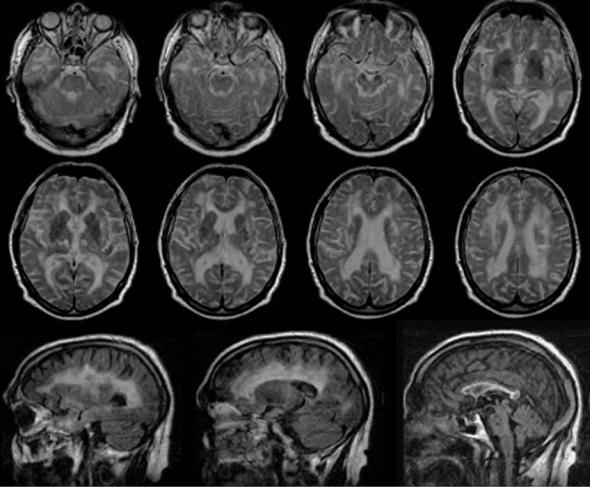
Another hereditary entity that has generated increasing interest in the past years is cerebral amyloid angiopathies (CAA). CAA are a heterogeneous group of disorders characterized by deposition of amyloid in the walls of leptomeningeal and cerebral cortical blood vessels. Clinical features include recurrent or multiple lobar hemorrhages, cogni-
Fig. 13.5. T2*-weighted gradient echo images in a patient with biopsy proven cerebral amyloid angiopathy (CAA). Extensive low signal abnormality is noted on the brain surfaces and in the parenchyma indicating the previous hemorrhages. Periventricular high intensity lesions, a common finding in CAA is also noted
204 |
A. Gass and H. Ay |
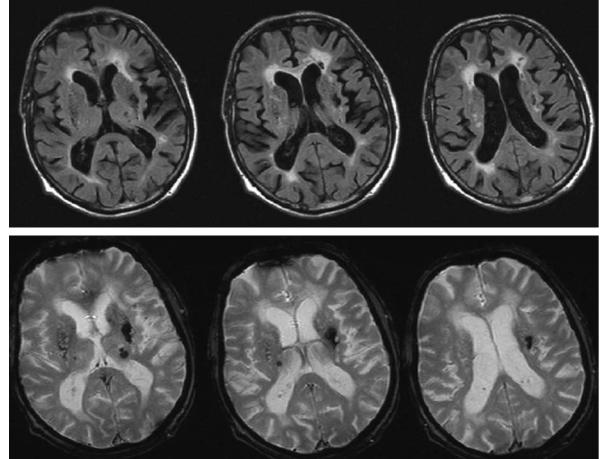
Fig. 13.6. FLAIR (top row) and T2*-weighted gradient echo images (bottom row) in a patient with hypertension showing a typical lesion pattern. Note subcortical lesions including lacunar lesions with severe tissue destruction and CSF like, low signal intensity (e.g. lateral to the anterior horn of the left lateral ventricle) on FLAIR images. On T2*-weighted images residuals from hemorrhagic lesions and small bleeds are appreciated mainly in the basal ganglia
tive deterioration, and ischemic stroke. MRI displays diffuse white matter abnormalities and focal lesions that can be ischemic or hemorrhagic. Vessels show amyloid deposition, cracking of single layers, microaneurysm formations and fibroid necrosis. The rupture of the structurally weakened arteries results in cerebral hemorrhage, characteristically in the cortices rather than the subcortical regions. CAA has usually been diagnosed postmortem. Although neuropathologic examination remains the definitive diagnostic approach to CAA, recent data suggest that a reliable diagnosis can be reached from clinical and MRI information alone using the Boston criteria (Greenberg et al. 1996; Knudsen et al. 2001), which include clinical data, MRI or CT demonstrating multiple hemorrhages restricted to lobar, cortical, or cortico-subcortical regions, and pathologic tissue (evacuated hematoma or cortical
biopsy). Several autosomal dominantly inherited forms of cerebral amyloid angiopathy can be differentiated by genetic, biochemical and pathological findings (Greenberg et al. 1999).
13.7
Microangiopathic Disease and Hemorrhage
The presence of microhemorrhages has been reported in the last 5 years in patients with white matter disease and cognitive decline, with and lately also without a history of intracranial hemorrhage or cerebral infarction. The potential of MRI to reveal residues of intracerebral bleeding throughout life rests on its high sensitivity to iron-containing compounds. At the site of intracerebral hemorrhage
Microangiographic Disease and Lacunar Stroke |
205 |
(ICH), hemosiderin remains stored in macrophages and leads to focal dephasing of the MRI signal. This causes areas of past bleeding to appear dark on T2*- weighted images. Techniques with high sensitivity to differences in magnetic susceptibility, such as the gradient-echo sequence, enhance these effects and allow detection of even minor hemosiderin deposition (Atlas et al. 1988). MRI is invaluable for obtaining this information because most small bleedings remain clinically undetected.
Scharf et al. (1994) were the first to present MRI evidence of previous, clinically silent intracerebral bleeds that they termed hemorrhagic lacunes. This type of lesion was significantly more frequent in patients with an intracerebral hematoma. In a series of 120 patients with primary ICH, Offenbacher and coworkers (1996) observed multiple foci of MRI signal loss compatible with old microbleeds in 28 individuals. In parallel, other investigators noticed similar MRI lesions in 9 of 15 patients with lobar hemorrhage (Greenberg et al. 1996). These abnormalities were considered to be evidence of previous petechial bleeds. Cortical and lobar ICH, predominantly of elderly patients, has been frequently related to amyloid angiopathy (Vinters 1987; Itoh et al. 1993). Also in this setting, MRI evidence of past bleedings has been reported (Greenberg et al. 1996). Furthermore, a cross-sectional study has also documented the accumulation of silent microbleeds over a short period of time in a series of patients with lobar hemorrhage and the diagnosis of probable or possible CAA (Greenberg et al. 1999).
Roob et al. (1999) noted focal areas of signal loss on gradient-echo T2*-weighted MRI in 18 of 280 (6.4%) participants in a cross-sectional study of neurologically asymptomatic elderly volunteers. These lesions were seen in cortico-subcortical regions of the brain, in the basal ganglia, and in infratentorial locations. Individuals with MRI evidence of microbleeds were significantly older, had a significantly higher frequency of hypertension, and had a higher rate of early confluent white matter lesions and lacunes. Foci of signal loss in cortico-subcortical regions were significantly less often associated with hypertension than microbleeds in the basal ganglia or infratentorial regions. These associations support the role of microangiopathy in the pathogenesis of focal T2* hypointensities.
Hypertension is regarded as the main clinically observable risk factor to cause microbleeds (Roob et al. 1999). Recent findings suggest that microbleeds on T2*-weighted MRI are an indicator of advanced small artery disease of the brain with an increased
risk for bleeding (Kato et al. 2002). With more clinical significance of such findings, MRI demonstration of microbleeds could gain use to identify patients who have an increased risk for intracerebral bleeding complications resulting from therapy that affects blood clotting, This result should be taken into consideration when treating patients with stroke (Kidwell et al. 2002).
References
Alexander GE, DeLong MR, Strick PL (1986) Parallel organization of functionally segregated circuits linking basal ganglia and cortex. Annu Rev Neurosci 9:357-381
Arboix A, Marti-Vilalta JL, Pujol J, Sanz M (1990) Lacunar cerebral infarct and nuclear magnetic resonance. A review of sixty cases. Eur Neurol 30:47-51
Atlas SW, Mark AS, Grossman RI, Gomori JM (1988) Intracranial hemorrhage: gradient-echo MR imaging at 1.5 T. Comparison with spin-echo imaging and clinical applications. Radiology 168:803-807
Auer DP, Putz B, Gossl C, Elbel G, Gasser T, Dichgans M (2001) Differential lesion patterns in CADASIL and sporadic subcortical arteriosclerotic encephalopathy: MR imaging study with statistical parametric group comparison. Radiology 218:443-451
Awad IA, Spetzler RF, Hodak JA, Awad CA, Carey R (1986) Incidental subcortical lesions identified on magnetic resonance imaging in the elderly. I. Correlations with age and cerebrovascular risk factors. Stroke 17:1084-1089
Ay H, Buonanno FS, Rordorf G, Schaefer PW, Schwamm LH, Wu O, Gonzalez RG, Yamada K, Sorensen GA, Koroshetz WJ (1999a) Normal diffusion-weighted MRI during stroke-like deficits. Neurology 52:1784-1792
Ay H, Oliveira-Filho J, Buonanno FS, Ezzeddine M, Schaefer PW, Rordorf G, Schwamm LH, Gonzalez RG, Koroshetz WJ (1999b) Diffusion-weighted imaging identifies a subset of lacunar infarction associated with embolic source. Stroke 30:2644-2650
Bamford J (2001) Classical lacunar syndromes. In: Bogousslavsky J, Caplan L (eds) Sreoke syndromes. Cambridge University Press, Cambridge, pp 583-589
Bamford J, Bogousslavsky J (eds) (2002) Subcortical stroke, 2nd edn. Oxford Medical Publications, Oxford, pp 27-34 Bamford J, Sandercock P, Jones L, Warlow C (1987) The natural
history of lacunar infarction: the Oxfordshire Community Stroke Project. Stroke 18:545-551
Benito-Leon J, Alvarez-Linera J, Porta-Etessam J (2001;Detection of acute pontine infarction by diffusion-weighted MRI in capsular warning syndrome. Cerebrovasc Dis 11:350-351
Bogousslavsky J, Van Melle G, Regli F (1988) The Lausanne Stroke Registry: analysis of 1,000 consecutive patients with first stroke. Stroke 19:1083-1092
Brant-Zawadzki M, Fein G, Van Dyke C, Kiernan R, Davenport L, de Groot J (1985) MR imaging of the aging brain: patchy white-matter lesions and dementia. Am J Neuroradiol 6:675-682
Breteler MM, van Swieten JC, Bots ML, Grobbee DE, Claus JJ,
206
van den Hout JH, van Harskamp F, Tanghe HL, de Jong PT, van Gijn J (1994) Cerebral white matter lesions, vascular risk factors, and cognitive function in a population-based study: the Rotterdam Study. Neurology 44:1246–1252
DeCarli C, Murphy DGM, Tranh M, Grady CL, Haxby JV, Gillette JA et al (1995) The effect of white matter hyperintensity volume on brain structure, cognitive performance, and cerebral metabolism of glucose in 51 healthy adults. Neurology 45:2077-2084
DeCarli C, Miller BL, Swan GE, Reed T, Wolf PA, Garner J, Jack L, Carmelli D (1999) Predictors of brain morphology for the men of the NHLBI Twin Study. Stroke 30:529–536
De Leeuw FE, de Groot JC, Oudkerk M, Witteman JC, Hofman A, van Gijn J, Breteler MM (1999) A follow-up study of blood pressure and cerebral white matter lesions. Ann Neurol 46:827–833
De Leeuw FE, de Groot JC, Achten E, Oudkerk M, Ramos LM, Heijboer R et al (2001) Prevalence of cerebral white matter lesions in elderly people: a population based magnetic resonance imaging study. The Rotterdam Scan Study. J Neurol Neurosurg Psychiatry 70:9-14
Dichgans M, Joutel A (2002) Cerebrovascular disorders. In: Rimoin D, Connor JM, Pyeritz RE, Korf B (eds) Emery and Rimoin’s principles and practice of medical genetics. Churchill Livingstone, London, pp 3209-3230
Dichgans M, Mayer M, Uttner I, Bruning R, Muller-Hocker J, Rungger G, Ebke M, Klockgether T, Gasser T (1998) The phenotypic spectrum of CADASIL: clinical findings in 102 cases. Ann Neurol 44:731-739
Donnan GA, Tress BM, Bladin PF (1982) A prospective study of lacunar infarction using computerized tomography. Neurology 32:49-56
Dubois MF, Hebert R (2001) The incidence of vascular dementia in Canada: a comparison with Europe and East Asia. Neuroepidemiology 20:179-187
Englund E (2002) Neuropathology of white matter lesions in vascular cognitive impairment. Cerebrovasc Dis 13 [Suppl 2]:11-15
Erkinjuntti T, Benavente O, Eliasziw M, Munoz DG, Sulkava R, Haltia M, Hachinski V (1996) Diffuse vacuolization (spongiosis) and arteriolosclerosis in the frontal white matter occurs in vascular dementia. Arch Neurol 53:325-332
Esiri MM, Wilcock GK, Morris JH (1997) Neuropathological assessment of the lesions of significance in vascular dementia. J Neurol Neurosurg Psychiatry 63:749-753
Fazekas F, Kleinert R, Offenbacher H, Schmidt R, Kleinert G, Payer F, Radner H, Lechner H (1993) Pathologic correlates of incidental MRI white matter signal hyperintensities. Neurology 43:1683-1689
Fazekas F, Schmidt R, Kleinert R, Kapeller P, Roob G, Flooh E (1998) The spectrum of age-associated brain abnormalities: their measurement and histopathological correlates. J Neural Transm [Suppl] 53:31-39
Fisher CM (1965) Lacunes: small, deep cerebral infarcts. Neurology 15:774-784
Fisher CM (1979) Capsular infarcts: the underlying vascular lesions. Arch Neurol 36:65-73
Fisher CM (1982) Lacunar strokes and infarcts: a review. Neurology 32:871-876
Fisher CM (1991) Lacunar infarcts: a review. Cerebrovasc Dis 1:311-320
Furuta A, Ishii N, Nishihara Y, Horie A (1991) Medullary arteries in aging and dementia. Stroke 22:442-446
A. Gass and H. Ay
Gass A, Oster M, Cohen S, Daffertshofer M, Schwartz A, Hennerici MG (1998) Assessment of T2and T1-weighted MRI brain lesion load in patients with subcortical vascular encephalopathy. Neuroradiology 40:503-506
Gass A,Ay H, Szabo K, Koroshetz WJ (2004) Diffusion-weighted MRI for the “small stuff ”: the details of acute cerebral ischaemia. Lancet Neurol 3:39-45
Gerraty RP, Parsons MW, Barber PA, Darby DG, Desmond PM, Tress BM, Davis SM (2002) Examining the lacunar hypothesis with diffusion and perfusion magnetic resonance imaging. Stroke 33:2019-2024
Greenberg SM, Finklestein SP, Schaefer PW (1996a) Petechial hemorrhages accompanying lobar hemorrhage: detection by gradient-echo MRI. Neurology 46:1751-1754
Greenberg SM, Briggs ME, Hyman BT et al (1996b) Apolipoprotein E e4 is associated with the presence and earlier onset of hemorrhage in cerebral amyloid angiopathy. Stroke 27:1333–1337
Greenberg SM, O’Donnell HC, Schaefer PW, Kraft E (1999) MRI detection of new hemorrhages: potential marker of progression in cerebral amyloid angiopathy. Neurology 53:1135-1138
Gootjes L, Teipel SJ, Zebuhr Y, Schwarz R, Leinsinger G, Scheltens P, Moller HJ, Hampel H (2004) Regional distribution of white matter hyperintensities in vascular dementia, Alzheimer’s disease and healthy aging. Dement Geriatr Cogn Disord 18:180-188
Hachinski VC, Lassen NA, Marshall J (1974) Multi-infarct dementia. A cause of mental deterioration in the elderly. Lancet 2:207-210
Hachinski VC, Potter P, Merskey H (1987) Leukoaraiosis. Arch Neurol 44:21-23
Hommel M, Besson G, Le Bas JF, Gaio JM, Pollak P, Borgel F, Perret J (1990) Prospective study of lacunar infarction using magnetic resonance imaging. Stroke 21:546-554
Inzitari D, Diaz F, Fox A, Hachinski VC, Steingart A, Lau C, Donald A, Wade J, Mulic H, Merskey H (1987) Vascular risk factors and leuko-araiosis. Arch Neurol 44:42-47
Ishii N, Nishihara Y, Imamura T (1986) Why do frontal lobe symptoms predominate in vascular dementia with lacunes? Neurology 36:340-345
Itoh Y, Yamada M, Hayakawa M, Otomo E, Miyatake T (1993) Cerebral amyloid angiopathy: a significant cause of cerebellar as well as lobar cerebral hemorrhage in the elderly. J Neurol Sci 116:135-141
Joutel A, Corpechot C, Ducros A,Vahedi K, Chabriat H, Mouton P, Alamowitch S, Domenga V, Cecillion M, Marechal E, Maciazek J, Vayssiere C, Cruaud C, Cabanis EA, Ruchoux MM, Weissenbach J, Bach JF, Bousser MG, Tournier-Lasse- rve E (1996) Notch3 mutations in CADASIL, a hereditary adult-onset condition causing stroke and dementia. Nature 383:707-710
Joutel A, Andreux F, Gaulis S, Domenga V, Cecillon M, Battail N, Piga N, Chapon F, Godfrain C, Tournier-Lasserve E (2000) The ectodomain of the Notch3 receptor accumulates within the cerebrovasculature of CADASIL patients. J Clin Invest 105:597-605
Junque C, Pujol J, Vendrell P, Bruna O, Jodar M, Ribas JC, Vinas J, Capdevila A, Marti-Vilalta JL (1990) Leuko-araiosis on magnetic resonance imaging and speed of mental processing. Arch Neurol 47:151-156
Kang DW, Chalela JA, Ezzeddine MA, Warach S (2003) Association of ischemic lesion patterns on early diffusion-
Microangiographic Disease and Lacunar Stroke
weighted imaging with TOAST stroke subtypes. Arch Neurol 60:1730-1734
Kato H, Izumiyama M, Izumiyama K, Takahashi A, Itoyama Y (2002) Silent cerebral microbleeds on T2*-weighted MRI: correlation with stroke subtype, stroke recurrence, and leukoaraiosis. Stroke 33:1536-1540
Kertesz A, Black SE, Tokar G, Benke T, Carr T, Nicholson L (1988) Periventricular and subcortical hyperintensities on magnetic resonance imaging. ‘Rims, caps, and unidentified bright objects’. Arch Neurol 45:404-408
Kertesz A, Polk M, Carr T (1990) Cognition and white matter changes on magnetic resonance imaging in dementia. Arch Neurol 47:387-391
Kidwell CS, Saver JL, Villablanca JP, Duckwiler G, Fredieu A, Gough K, Leary MC, Starkman S, Gobin YP, Jahan R, Vespa P, Liebeskind DS, Alger JR, Vinuela F (2002) Magnetic resonance imaging detection of microbleeds before thrombolysis: an emerging application. Stroke 33:95-98
Kirkpatrick JB, Hayman LA (1987) White-matter lesions in MR imaging of clinically healthy brains of elderly subjects: possible pathologic basis. Radiology 162:509-511
Knudsen KA, Rosand J, Karluk D, Greenberg SM (2001) Clinical diagnosis of cerebral amyloid angiopathy: validation of the Boston criteria. Neurology 56:537-539
Kuker W, Weise J, Krapf H, Schmidt F, Friese S, Bahr M (2002) MRI characteristics of acute and subacute brainstem and thalamic infarctions: value of T2and diffusion-weighted sequences. J Neurol 249:33-42
Liao D, Cooper L, Cai J, Toole JF, Bryan NR, Hutchinson RG, Tyroler HA (1996) Presence and severity of cerebral white matter lesions and hypertension, its treatment, and its control. The ARIC study. Atherosclerosis risk in communities study. Stroke 27:2262–2270
Liao D, Cooper L, Cai J, Toole J, Bryan N, Burke G, Shahar E, Nieto J, Mosley T, Heiss G (1997) The prevalence and severity of white matter lesions, their relationship with age, ethnicity, gender, and cardiovascular disease risk factors: the ARIC Study. Neuroepidemiology 16:149–162
Lie C, Hirsch JG, Rossmanith C, Hennerici MG, Gass A (2004) Clinicotopographical correlation of corticospinal tract stroke: a color-coded diffusion tensor imaging study. Stroke. 35:86-92
Lindgren A, Norrving B, Rudling O, Johansson BB (1994) Comparison of clinical and neuroradiological findings in first-ever stroke. A population-based study. Stroke 25:1371-1377
Lindgren A, Staaf G, Geijer B, Brockstedt S, Stahlberg F, Holtas S, Norrving B (2000) Clinical lacunar syndromes as predictors of lacunar infarcts. A comparison of acute clinical lacunar syndromes and findings on diffusion-weighted MRI. Acta Neurol Scand 101:128-134
Lodder J, Boiten J (1993) Incidence, natural history, and risk factors in lacunar infarction. Adv Neurol 62:213-227
Longstreth WT Jr, Manolio TA, Arnold A, Burke GL, Bryan N, Jungreis CA, Enright PL, O’Leary D, Fried L (1996) Clinical correlates of white matter findings on cranial magnetic resonance imaging of 3301 elderly people. The cardiovascular health study. Stroke 27:1274-1282
Lovblad KO, Laubach HJ, Baird AE, Curtin F, Schlaug G, Edelman RR, Warach S (1998) Clinical experience with diffu- sion-weighted MR in patients with acute stroke. AJNR Am J Neuroradiol 19:1061-1066
Marshall VG, Bradley WG Jr, Marshall CE, Bhoopat T, Rhodes
207
RH (1988) Deep white matter infarction: correlation of MR imaging and histopathologic findings. Radiology 167:517-522
McQuinn BA, O’Leary DH (1987) White matter lucencies on computed tomography, subacute arteriosclerotic encephalopathy (Binswanger’s disease), and blood pressure. Stroke 18:900-905
Mega MS, Cummings JL (1994) Frontal-subcortical circuits and neuropsychiatric disorders. J Neuropsychiatry Clin Neurosci 6:358-70
Meguro K, Hatazawa J, Yamaguchi T, Itoh M, Matsuzawa T, Ono S, Miyazawa H, Hishinuma T, Yanai K, Sekita Y et al (1990) Cerebral circulation and oxygen metabolism associated with subclinical periventricular hyperintensity as shown by magnetic resonance imaging. Ann Neurol 28:378-383
Mezzapesa DM, Rocca MA, Pagani E, Comi G, Filippi M (2003) Evidence of subtle gray-matter pathologic changes in healthy elderly individuals with nonspecific white-matter hyperintensities. Arch Neurol 60:1109-1112
Mungas D, Jagust WJ, Reed BR et al (2001) MRI predictors of cognition in subcortical ischemic vascular disease and Alzheimer’s disease. Neurology 57:2229–2235
O’Donnell HC, Rosand J, Knudsen KA, Furie KL, Segal AZ, Chiu RI, Ikeda D, Greenberg SM (2000) Apolipoprotein E genotype and the risk of recurrent lobar intracerebral hemorrhage. N Engl J Med 342:240-245
Offenbacher H, Fazekas F, Schmidt R, Koch M, Fazekas G, Kapeller P (1996) MR of cerebral abnormalities concomitant with primary intracerebral hematomas. Am J Neuroradiol 17:573-578
Oliveira-Filho J, Ay H, Schaefer PW, Buonanno FS, Chang Y, Gonzalez RG, Koroshetz WJ (2000) Diffusion-weighted magnetic resonance imaging identifies the “clinically relevant” small-penetrator infarcts. Arch Neurol 57:1009-1014 Oliveira-Filho J, Ay H, Koroshetz WJ, Buonanno FS (2001) Localization of clinical syndromes using DWI: two examples of the “capsular” warning syndrome. J Neuroimaging
11:44-47
O’Sullivan M, Jarosz JM, Martin RJ, Deasy N, Powell JF, Markus HS (2001) MRI hyperintensities of the temporal lobe and external capsule in patients with CADASIL. Neurology 56:628-634
O’Sullivan M, Morris RG, Huckstep B, Jones DK, Williams SCR, Markus HS (2004) Diffusion tensor MRI correlates with executive dysfunction in patients with ischaemic leukoaraiosis. J Neurol Neurosurg Psychiatry 75:441-447
Pantoni L, Garcia JH (1997) Pathogenesis of leukoaraiosis: a review. Stroke 28:652–659
Pantoni L, Basile AM, Pracucci G, Asplund K, Bogousslavsky J, Chabriat H, Erkinjuntti T, Fazekas F, Ferro JM, Hennerici M, O’brien J, Scheltens P,Visser MC, Wahlund LO, Waldemar G, Wallin A, Inzitari D (2005) Impact of age-related cerebral white matter changes on the transition to disability - the LADIS study: rationale, design and methodology. Neuroepidemiology 24:51-62
Petty GW, Brown RD Jr, Whisnant JP, Sicks JD, O’Fallon WM, Wiebers DO (2000) Ischemic stroke subtypes: a popula- tion-based study of functional outcome, survival, and recurrence. Stroke 31:1062-1068
Pohjasvaara T, Mantyla R, Ylikoski R, Kaste M, Erkinjuntti T (2003) Clinical features of MRI-defined subcortical vascular disease. Alzheimer Dis Assoc Disord 17:236-242
208
Ravens JR (1978) Vascular changes in the human senile brain. Adv Neurol 20:487-501
Révész T, Hawkins CP, du Boulay EP, Barnard RO, McDonald WI (1989) Pathological findings correlated with magnetic resonance imaging in subcortical arteriosclerotic encephalopathy (Binswanger’s disease). J Neurol Neurosurg Psychiatry 52:1337-1344
Rockwood K, Burns A, Gauthier S, DeKosky ST (2003) Vascular cognitive impairment. Lancet Neurol 2:89-98
Roman GC (1987) Senile dementia of the Binswanger type. A vascular form of dementia in the elderly. JAMA 258:1782-1788
Roman GC, Royall DR (1999) Executive control function: a rational basis for the diagnosis of vascular dementia. Alzheimer Dis Assoc Disord 13 [Suppl 3]:S69-S80
Roman GC, Erkinjuntti T, Wallin A, Pantoni L, Chui HC (2002) Subcortical ischaemic vascular dementia. Lancet Neurol 1:426-436
Roob G, Schmidt R, Kapeller P, Lechner A, Hartung HP, Fazekas F (1999) MRI evidence of past cerebral microbleeds in a healthy elderly population. Neurology 52:991-994
Sabri O, Ringelstein EB, Hellwig D et al (1999) Neuropsychological impairment correlates with hypoperfusion and hypometabolism but not with severity of white matter lesions on MRI in patients with cerebral microangiopathy. Stroke 30:556–566
Scharf J, Brauherr E, Forsting M, Sartor K (1994) Significance of haemorrhagic lacunes on MRI in patients with hypertensive cerebrovascular disease and intracerebral haemorrhage. Neuroradiology 36:504-508
Scheltens P, Barkhof F, Leys D, Wolters EC, Ravid R, Kamphorst W (1995) Histopathologic correlates of white matter changes on MRI in Alzheimer’s disease and normal aging. Neurology 45:883-888
Scheltens P, Erkinjunti T, Leys D, Wahlund LO, Inzitari D, del Ser T, Pasquier F, Barkhof F, Mantyla R, Bowler J, Wallin A, Ghika J, Fazekas F, Pantoni L (1998) White matter changes on CT and MRI: an overview of visual rating scales. European Task Force on Age-Related White Matter Changes. Eur Neurol 39:80-89
Schmidt R, Schmidt H, Kapeller P, Enzinger C, Ropele S, Saurugg R, Fazekas F (2002) The natural course of MRI white matter hyperintensities. J Neurol Sci 203-204:253-257
Schmidt R, Enzinger C, Ropele S, Schmidt H, Fazekas F (2003) Austrian stroke prevention study. Progression of cerebral white matter lesions: 6-year results of the Austrian stroke prevention study. Lancet 361:2046-2048
Singer MB, Chong J, Lu D, Schonewille WJ, Tuhrim S, Atlas SW (1998) Diffusion-weighted MRI in acute subcortical infarction. Stroke 29:133-136
Skoog I, Berg S, Johansson B, Palmertz B, Andreasson LA (1996) The influence of white matter lesions on neuropsychological functioning in demented and non-demented 85-year-olds. Acta Neurol Scand 93:142-148
Summergrad P, Peterson B (1989) Binswanger’s disease, part
A. Gass and H. Ay
I. The clinical recognition of subcortical arteriosclerotic encephalopathy in elderly neuropsychiatric patients. J Geriatr Psychiatry Neurol 2:123-133
Szabo K, Bäzner H, Kern R, Blahak C, Hennerici MG, Gass A (2004) Lack of incidental DWI hyperintensity in healthy elderly individuals. Cerebrovasc Dis [Suppl] 5:75
Tanaka Y, Tanaka O, Mizuno Y, Yoshida M (1989) A radiologic study of dynamic processes in lacunar dementia. Stroke 20:1488-1493
Tarvonen-Schroder S, Roytta M, Raiha I, Kurki T, Rajala T, Sourander L (1996) Clinical features of leuko-araiosis. J Neurol Neurosurg Psychiatry 60:431-436
Tekin S, Cummings JL (2002) Frontal-subcortical neuronal circuits and clinical neuropsychiatry: an update. J Psychosom Res 53:647-654
Tuszynski MH, Petito CK, Levy DE (1989) Risk factors and clinical manifestations of pathologically verified lacunar infarctions. Stroke 20:990-999
Van den Boom R, Lesnik Oberstein SA, van Duinen SG, Bornebroek M, Ferrari MD, Haan J, van Buchem MA (2002) Subcortical lacunar lesions: an MR imaging finding in patients with cerebral autosomal dominant arteriopathy with subcortical infarcts and leukoencephalopathy. Radiology 224:791-796
Van Dijk EJ, Breteler MM, Schmidt R, Berger K, Nilsson LG, Oudkerk M, Pajak A, Sans S, de Ridder M, Dufouil C, Fuhrer R, Giampaoli S, Launer LJ, Hofman A; CASCADE Consortium (2004) The association between blood pressure, hypertension, and cerebral white matter lesions: cardiovascular determinants of dementia study. Hypertension 44:625-630
Vermeer SE, Hollander M, van Dijk EJ, Hofman A, Koudstaal PJ, Breteler MM (2003) Silent brain infarcts and white matter lesions increase stroke risk in the general population: the Rotterdam scan study. Stroke 34:1126-1129
Wiszniewska M, Devuyst G, Bogousslavsky J, Ghika J, van Melle G (2000) What is the significance of leukoaraiosis in patients with acute ischemic stroke? Arch Neurol 57:967– 973
Wolfe N, Linn R, Babikian VL, Knoefel JE, Albert ML (1990) Frontal systems impairment following multiple lacunar infarcts. Arch Neurol 47:129-132
Yamauchi H, Fukuyama H, Shio H (2000) Corpus callosum atrophy in patients with leukoaraiosis may indicate global cognitive impairment. Stroke 31:1515-1520
Ylikoski R, Ylikoski A, Erkinjuntti T, Sulkava R, Raininko R, Tilvis R (1993) White matter changes in healthy elderly persons correlate with attention and speed of mental processing. Arch Neurol 50:818-824
Yonemura K, Kimura K, Minematsu K, Uchino M,Yamaguchi T (2002) Small centrum ovale infarcts on diffusion-weighted magnetic resonance imaging. Stroke 33:1541-1544
You R, McNeil JJ, O’Malley HM, Davis SM, Donnan GA (1995) Risk factors for lacunar infarction syndromes. Neurology 45:1483-1487

Territorial and Embolic Infarcts |
209 |
14 Territorial and Embolic Infarcts
José M. Ferro
CONTENTS
14.1Introduction 209
14.2Clinical MR Patterns of
Acute Territorial Infarcts 210
14.2.1 Middle Cerebral Artery (MCA) Infarcts 210
14.2.1.1Superficial Middle Cerebral Artery (MCA) Infarcts 210
14.2.1.2 |
Large Middle Cerebral Artery (MCA) Infarcts |
211 |
|||
14.2.1.3 |
MCA Anterior or Superior Division Infarcts |
211 |
|||
14.2.1.4 |
MCA Posterior or Inferior Division Infarcts |
211 |
|||
14.2.1.5 |
Cortical Branch Syndromes 211 |
|
|
|
|
14.2.2 |
Subcortical Hemispheric Infarcts |
212 |
|
|
|
14.2.2.1 |
Anterior Choroidal Artery Infarcts |
212 |
|
|
|
14.2.2.2 |
Thalamic Infarcts |
212 |
|
|
|
14.2.3 |
Posterior Cerebral Artery (PCA) Infarcts |
214 |
|||
14.2.4 |
Anterior Cerebral Artery (ACA) Infarcts |
214 |
|
||
14.2.5 |
Combined Hemispheric Infarcts |
215 |
|
|
|
14.2.6 |
Brain Stem Infarcts |
215 |
|
|
|
14.2.6.1Mesencephalic Infarcts 215
14.2.6.2Pontine Infarcts 216
14.2.6.3Medullary Infarcts 217
14.2.6.4 |
Stroke Patterns in Basilar Artery Occlusion |
217 |
|
14.2.7 |
Cerebellar Infarcts 218 |
|
|
14.2.7.1 |
Superior Cerebellar Artery (SCA) |
218 |
|
14.2.7.2 |
Anterior Inferior Cerebellar Artery (AICA) |
218 |
|
14.2.7.3 |
Posterior Inferior Cerebellar Artery (PICA) |
218 |
|
14.2.7.4 |
Pseudotumoral Cerebellar Infarcts |
219 |
|
14.2.8 |
Acute Multiple Brain Infarcts (AMBI) 219 |
|
|
14.3Cardioembolic Infarcts 220
14.3.1 Infarcts with Hemorrhagic Transformation 221 References 221
14.1 Introduction
Despite the advances in neuroimaging, the early identification of ischemic stroke subtypes and patterns has several heuristic values, besides academic and research interest. It helps the physician to answer the patient’s and their relatives’ anxieties concerning the risk of early death, disability, stroke
J. M. Ferro, MD, PhD
Neurology Service, Department of Neurosciences and Mental Health, Hospital de Santa Maria, University of Lisbon, Av. Prof. Egas Moniz, 1649–035 Lisbon, Portugal
recurrence and length of hospital stay. It guides the neurologist to choose the most cost-effective ancillary procedures and the therapy with best efficacy. It also helps the hospital manager to calculate the average cost of care for stroke subtypes.
Prior to the use of magnetic resonance (MR) and in particular diffusion-weighted (DWI) MR, our clinico-anatomic concepts of acute stroke patterns were based on post-mortem correlations and on computerized tomography (CT). Post-mortem correlations are an accurate method with two obvious biases: (1) delay between acute clinical stroke pattern and death and (2) mortality bias. The introduction and dissemination of MR provided the neurological community with an opportunity to verify the validity of the classical description of acute stroke patterns. The yield of DWI for the detection of potentially relevant findings in acute stroke patients includes: (1) detecting an acute ischemic lesion in a territory different from that suspected clinically; (2) revealing multiple lesions in different vascular territories, when clinical examination and CT indicated a unifocal lesion; (3) clarifying whether a lesion seen on CT or MR is indeed acute or long-standing (Albers et al. 2000; Lee et al. 2000; Lindgren et al. 2000; Oliveira-Filho et al. 2000).
The early identification of the clinical and MR patterns of acute territorial infarcts can help the managing physician concerning prediction of outcome, risk of early death and dependency, risk of recurrence, stroke mechanism and etiology, selection of ancillary procedures, selection of best (effective and safe) anti-thrombotic treatment, risk of complications, length of stay and cost of hospital care.
Using only a few neurological findings the Oxfordshire Community Stroke Project (OCSP) classification allocates strokes to four subgroups, locating them either in the territory of the anterior (total anterior circulation infarct, TACI; partial anterior circulation infarct, PACI; lacunar infarct, LACI) and the posterior circulation, (posterior circulation infarct, POCI) (Bamford et al. 1991). The OCSP is a clinical syndromic classification, which