

Книги по МРТ КТ на английском языке / Magnetic Resonance Imaging in Ischemic Stroke - K Sartor R 252 diger von Kummer Tobias Back
.pdf220 |
J. M. Ferro |
internal carotid artery produces mainly multiple infarcts with three distinctive patterns: (1) territorial, either multiple cortical, cortical and deep or single, (2) borderzone, alone or in combination with a territorial infarct (Fig. 14.7), (3) bilateral lesions (Kang et al. 2003).
14.3
Cardioembolic Infarcts
Decreased consciousness at onset, sudden onset to maximal defect, rapid regression of symptoms, aphasia, neglect and visual field defects are more common in cardioembolic than other types of stroke, Headache, seizure at onset or onset during activity are not specific for cardioembolic stroke (Arboix et al. 1999; Kittner et al. 1992; Minematsu et al. 1992; Ramirez-Lassepas et al. 1987; Timsit et al. 1992). Stroke syndromes such as isolated Wernicke’s aphasia, global aphasia without hemiparesis, Wallenberg’s syndrome and top-of-the-basilar syndrome are suggestive of cardioembolism.
Simultaneous or sequential strokes in different arterial territories, multi-level posterior circulation infarcts, simultaneous infarcts in the three subterritories (superficial anterior, superficial posterior and deep) of the MCA, and hemorrhagic transformation of an ischemic infarct also point to a cardiac origin of the stroke (Arquizan et al. 1997; Ay et al. 1999; Ferro 2003a,b). Occlusion of the carotid artery by a mobile thrombus, early recanalization of an occluded vessel and the identification of microembolism in both MCAs are all highly indicative of a cardiac source of emboli.
DWI can demonstrate scattered lesions in acute stroke patients with initial negative CT (Koennecke et al. 2001) associated with an embolic etiology. In patients presenting with classical lacunar syndromes, DWI can demonstrate lesions not apparent in CT or conventional MR in addition to the lesion in the territory of a single perforator. The most common associated lesions are punctuate subsidiary lesions in the territory of the leptomeningeal arteries (Ay et al. 1990).
Multiple scattered cerebral infarcts can be demonstrated by DWI in highly emboligenic conditions
Fig. 14.7. Combined junctional and cortical infarcts in a case of carotid occlusion
Territorial and Embolic Infarcts
such as endocarditis (Singhal et al. 2002) and cardiac surgery, and cause both focal syndromes and diffuse encephalopathy which are often found in such conditions (Wityk et al. 2001).
14.3.1
Infarcts with Hemorrhagic Transformation
About 40% of ischemic infarcts show hemorrhagic transformation on CT; this proportion is even higher on MRI (Egelhof et al. 1998). Cardioembolism accounts for the vast majority of infarcts with hemorrhagic transformation. TACIs, patients who are not fully alert, severe strokes (NIHSS > 14 points), proximal occlusion and delayed recanalization (> 6 h) predict hemorrhagic transformation. Hypodensity on CT > 1/3 of MCA territory and diabetes are associated with an increased risk of hemorrhagic transformation after thrombolysis (Alexandrov et al. 1997; de Freitas et al. 2001; Ferro 2003a,b; Fisher and
Adams 1951; Jörgensen and Torvic 1969; Molina et al. 2001; Moulin et al. 2000; Nighoghossian et al. 2002). In MR imaging, ischemic brain regions destined to hemorrhagic transformation have lower apparent diffusion coefficient values and persistently delayed perfusion compared to regions which will not suffer hemorrhagic transformation (Tong et al. 2000, 2001; Selim et al. 2002). Old microbleeds detected by T2*-weighted sequences, indicating severe microangiopathy or amyloid angiopathy (Fig. 13.5, Chap. 13), are also a marker for hemorrhagic transformation in particular after thrombolytic therapy. Gadolinium enhancement patterns (vascular, meningeal and parenchymal) are also predictors of hemorrhagic transformation. Vascular enhancement defined as tubular enhancing vessels located in the sulci supplying the ischemic territory, is the most common pattern (Vo et al. 2003).
References
Albers GW, Lansberg MG, Norbash AM et al (2000) Yield of diffusion-weighted MRI for detection of potentially relevant findings in stroke patients. Neurology 54:1562– 1567
Alexandrov AV, Black SE, Ehrlich LE et al (1997) Predictors of hemorrhagic transformation occurring spontaneously and on anticoagulants in patients with acute ischemic stroke. Stroke 28:1198–1202
Altieri M, Metz RJ, Muller C et al (1999) Multiple brain infarcts: clinical and neuroimaging patterns using diffu- sion-weighted magnetic resonance. Eur Neurol 42:76–82
221
Amarenco P (1991) The spectrum of cerebellar infarctions. Neurology 41:973–979
Amarenco P (1993) Les infarctus du cervelet et leurs mécanismes. Rev Neurol (Paris) 149:728–748
Amarenco P, Rosengart A, de Witt LD et al (1993) Anterior inferior cerebellar artery territory infarcts: mechanisms and clinical features. Arch Neurol 50:154–161
Amarenco P, Lévy C, Cohen A et al (1994) Causes and mechanisms of territorial and nonterritorial cerebellar infarcts in 115 consecutive cases. Stroke 25:105–112
Arboix A, Oliveres M, Massons J et al (1999) Early differentiation of cardioembolic from atherothrombotic cerebral infarction: a multivariate analysis. Eur J Neurol 6:677–683 Arquizan C, Lamy C, Mas J-L (1997) Infarctus cérébraux multiples simultanés sus-tentoriels. Rev Neurol (Paris)
153:748–753
Ay H, Oliveira-Filho J, Buonanno FS et al (1999) Diffusionweighted imaging identifies a subset of lacunar infarction associated with embolic source. Stroke 30:2644–2650
Ay H, Uluc K, Arsava EM et al (2002) Unusually prolonged progressing stroke: an expanding anterior cerebral artery infarction. Cerebrovasc Dis 13:64–66
Bamford J, Sandercock P, Dennis M et al (1991) Classification and natural history of clinically identifiable subtypes of cerebral infarction. Lancet 337:1521–1526
Bang OY, Heo JH, Kim JY et al (2002) Middle cerebral artery stenosis is a major clinical determinant in striatocapsular small, deep infarction. Arch Neurol 59:259–263
Boccardi E, Della Sala S, Motto C et al (2002) Utilisation behaviour consequent to bilateral SMA softening. Cortex 38:289–308
Bogousslavsky J (1993) Subcortical infarcts. In: Fisher M, Bogousslavsky J (eds) Current review of cerebrovascular diasease. Current Medicine, Philadelphia, pp 31–40
Bogousslavsky J, Caplan LR (1993) Vertebrobasilar occlusive disease: review of selected aspects. 3 thalamic infarcts. Cerebrovasc Dis 3:193–205
Bogousslavsky J, Caplan LR (2001) Stroke syndromes, 2nd edn. Cambridge University Press, Cambridge
Bogousslavsky J, Regli F, Uske A (1988) Thalamic infarcts: clinical syndromes, etiology and prognosis. Neurology 38:837–848
Bogousslavsky J, Maeder P, Regli F et al (1994) Pure midbrain infarction: clinical syndromes, MRI, and etiologic patterns. Neurology 44:2032–2040
Brandt T, Steinke W, Thie A et al (2000) Posterior cerebral artery territory infarcts: clinical features, infarct topography, causes and outcome. Multicenter results and a review of the literature. Cerebrovasc Dis 10:170–182
Cals N, Devuyst G, Afsar N et al (2002) Pure superficial posterior cerebral artery territory infarction in The Lausanne Stroke Registry. J Neurol 249:855–861
De Freitas GR, Carruzzo A, Tsiskaridze A et al (2001) Massive haemorrhagic transformation in cardioembolic stroke: the role of arterial wall trauma and dissection. J Neurol Neurosurgery Psychiatry 70:672–674
Della Sala S, Francescani A, Spinnler H (2002) Gait apraxia after bilateral supplementary motor area lesion. J Neurol Neurosurg Psychiatry 72:77–85
Devuyst G, Bogousslavsky J, Meuli R et al (2002) Stroke or transient ischemic attacks with basilar artery stenosis or occlusion: clinical patterns and outcome. Arch Neurol 59:567–573
222
Donnan GA,Yasaka M (1998) Lacunes and lacunar syndromes. In: Ginsberg MD, Bogousslavsky J (eds) Cerebrovascular diseases. Pathophysiology, diagnosis and management, vol II. Blackwell Science, Massachusetts, pp 1090–1102
Ebrahim S, Harwood R (1999a) Mortality. In: Ebrahim S, Harwood R (eds) Stroke. Epidemiology, evidence and clinical practice. Oxford University Press, Oxford, pp 217–228
Ebrahim S, Harwood R (1999b) Recovery. In: Ebrahim S, Harwood R (eds) Stroke. Epidemiology, evidence and clinical practice. Oxford University Press, Oxford, pp 264–281
Egelhof T, Essig M, von Kummer R, Dörfler A, Winter R, Sartor K (1998) Der acute ischämische Hirninfarkt: eine prospective, serielle Untersuchung mit der Magnetresonanztomographie. Fortschr Röntgenstr 168:222–227
Ferro JM (2003a) Brain embolism - answers to practical questions. J Neurol 250:139–147
Ferro JM (2003b) Cardioembolic stroke: an update. Lancet Neurol 2:177–188
Fisher CM, Adams RD (1951) Observations on brain embolism with special reference to the mechanism of hemorrhagic infarction. J Neuropathol Exp Neurol 10:92–93
Georgiadis AL, Yamamoto Y, Kwan ED et al (1999) Anatomy of sensory findings in patients with posterior cerebral artery territory infarction. Arch Neurol 56:835–838
Gerraty RP, Parsons MW, Barber A et al (2002) Examining the lacunar hypothesis with diffusion and perfusion magnetic resonance imaging. Stroke 33:2019–2024
Glass TA, Nennessey PM, Pazdera L et al (2002) Outcome at 30 days in the New England Medical Center Posterior Circulation Registry. Arch Neurol 59:369–376
Gorelick PB (1996) Atlas of cerebrovascular disease. Current Medicine, Philadelphia
Han SW, Sohn YH, Lee PH et al (2000) Pure homonymous hemianopia due to anterior choroidal artery territory infarction. Eur Neurol 43:35–38
Heinsius T, Bogousslavsky J, van Melle G (1998) Large infarcts in the middle cerebral artery territory. Etiology and outcome patterns. Neurology 50:341–350
Hornig CR, Rust DS, Busse O et al (1994) Space-occupying cerebellar infarction: clinical course and prognosis. Stroke 25:372–374
Jongen JCF, Franke CL, Ramos LMP et al (2004) Direction of flow in posterior communicating artery on magnetic resonance angiography in patients with occipital lobe infarcts. Stroke 35:104–108
Jörgensen L, Torvik A (1969) Ischaemic cerebrovascular disease in an autopsy series: part prevalence, location, pathogenesis and clinical course of cerebral infarcts. J Neurol Sci 9:285–320
Kang DW, Chalela JA, Ezzeddine MA et al (2003a) Association of ischemic lesion patterns on early diffusion-weighted imaging with TOAST stroke subtypes. Arch Neurol 60:1730–1734
Kang DW, Chu K, Ko SB et al (2003b) Lesion patterns and mechanism of ischemia in internal carotid artery disease: a diffusion-weighted imaging study. Arch Neurol 59:1577– 1582
Kasner SE, Demchuk AM, Berrouschot J et al (2001) Predictors of fatal brain edema in massive hemispheric ischemic stroke. Stroke 32:2117–2123
Kittner SJ, Sharkness CM, Sloan MA et al (1992) Infarcts with a cardiac source of embolism in the NINDS Stroke Data Bank: neurologic examination. Neurology 42:299–302
J. M. Ferro
Koennecke H-C, Bernarding J, Braun J et al (2001) Scattered brain infarct pattern on diffusion-wheighted magnetic resonance imaging in patients with acute ischemic stroke. Cerebrovasc Dis 11:157–163
Koh MG, Phan TG, Atkinson JLD et al (2000) Neuroimaging in deteriorating patients with cerebellar infarcts and mass effect. Stroke 31:2062–2067
Kumral E (2001) Multiple, multilevel and bihemispheric infarcts. In: Bogousslavsky J, Caplan L (eds) Stroke syndromes. Cambridge University Press, Cambridge, pp 499– 511
Kumral E, Evyapan D, Balkir K et al (2001) Bilateral thalamic infarction. Clinical, etiological and MRI correlates. Acta Neurol Scand 103:35–42
Kumral E, Afsar N, Kirbas D et al (2002a) Spectrum of medial medullary infarction: clinical and magnetic resonance imaging findings. J Neurol 249:85–93
Kumral E, Bayulkem G, Akyol A et al (2002b) Mesencephalic and associated posterior circulation infarcts. Stroke 33:2224–2231
Kumral E, Bayulkem G, Evyapan D (2002c) Clinical spectrum of pontine infarction clinical-MRI correlations. J Neurol 249:1659–1670
Kumral E, Bayulkem G, Evyapan D et al (2002d) Spectrum of anterior cerebral artery territory infarction: clinical and MRI findings. Eur J Neurol 9:615–624
Lee LJ, Kidwell CS, Alger J et al (2000) Impact on stroke subtype diagnosis of early diffusion-weighted magnetic resonance imaging and magnetic resonance angiography. Stroke 31:1081–1089
Lee MS, Oh SH, Lee KR (2002) Transient repetitive movements of the limbs in patients with acute basilar artery infarction. Neurology 59:1116–1117
Lee PH, Bang OY, Oh SH et al (2003) Subcortical white matter infarcts. Comparison of superficial perforating artery and internal borderzone infarcts using diffusion-weighted magnetic resonance imaging. Stroke 34:2630–2635
Leys D, Mounier-Vehier F, Lavenu I et al (1994) Anterior choroidal artery territory infarcts. Study of presumed mechanisms. Stroke 25:837-842
Lie C, Hirsch JG, Roßmanith C et al (2004) Clinicotopographical correlation of corticospinal tract stroke. A color-coded diffusion tensor imaging study. Stroke 35:86–93
Lindgren A, Staaf G, Geijer B et al (2000) Clinical lacunar syndromes as predictors of lacunar infarcts. A comparison of acute clinical lacunar syndromes and findings on diffu- sion-weighted MRI. Acta Neurol Scand 101:128–134
Lindley RI, Warlow CP, Wardlaw JM et al (1993) Interobserver reliability of a clinical classification of acute cerebral infarction. Stroke 24:1801–1804
Martin PJ, Chang HM, Wityk R et al (1998) Midbrain infarction: associations and aetiologies in the New England Medical Center Posterior Circulation Registry. J Neurol Neurosurg Psychiatry 64:392–395
Mead GE, Shingler H, Farrell A et al (1998) Carotid disease in acute stroke. Age Ageing 27:677–682
Mead GE, Lewis SC, Wardlaw JM et al (2000) How well does Oxfordshire Community Stroke Project classification predict the site and size of the infarct on brain imaging? J Neurol Neurosurg Psychiatry 68:558–562
Melo TP, Bogousslavsky J, Moulin T et al (1992) Thalamic ataxia. J Neurol 239:331–337
Melo TP, Bogousslvasky J (1992) Hemiataxia-hypesthesia:
Territorial and Embolic Infarcts
a thalamic stroke syndrome. J Neurol Neurosurg Psych 55:581–584
Min WK, Park KK, Kim YS et al (2000) Atherothrombotic middle cerebral artery territory infarction. Topographic diversity with common occurrence of concomitant small cortical and subcortical infarcts. Stroke 31:2055–2061
Minematsu K, Yamaguchi T, Omae T (1992) Spectacular shrinking deficit: rapid recovery from a major hemispheric syndrome by migration of an embolus. Neurology 42:157–162
Molina CA, Montaner J, Abilleira S et al (2001) Timing of spontaneous recanalization and risk of hemorrhagic transformation in acute cardioembolic stroke. Stroke 32:1079–1084
Montavont A, Nighoghossian N, Hermier M et al (2003) Hemiplegia in posterior cerebral artery occlusion: acute MRI assessment. Cerebrovasc Dis 16:452–453
Moulin T, Crepin-Leblond T, Chopard JL et al (1993) Hemorrhagic infarcts. Eur Neurol 34:64–77
Moulin T, Tatu L, Vuillier F et al (2000) Role of a stroke data bank in evaluating cerebral infarction subtypes: patterns and outcome of 1.776 consecutive patients from the Besancon stroke registry. Cerebrovasc Dis 10:261–271
Nighoghossian N, Hermier M, Adeleine P et al (2002) Old microbleeds are a potential risk factor for cerebral bleeding after ischemic stroke. A gradient-echo T2* - weighted brain MRI study. Stroke 33:735–742
Oliveira-Filho J, Ay H, Schaefer PW et al (2000) Diffusionweighted magnetic resonance imaging identifies the “clinically relevant” small-penetrator infarcts. Arch Neurol 57:1009–1014
Pinto AN, Melo TP, Lourenço ME et al (1998) Can a clinical classification of stroke predict complications and treatments during hospitalization? Cerebrovasc Dis 8:204–209
Ramirez-Lassepas M, Cipolle RJ, Bjork RJ et al (1987) Can embolic stroke be diagnosed on the basis of neurologic clinical criteria? Arch Neurol 44:87–89
Rieke K, Kreiger D, Adams HP et al (1993) Therapeutic strategies in space-occupying cerebellar infarction based on clinical, neuroradiological and neurophysiological data. Cerebrovasc Dis 3:45–55
Roh JK, Kang DW, Lee SH et al (2000) Significance of acute multiple brain infarction on diffusion-weighted imaging. Stroke 31:688–694
Saeki N, Shimazaki K, Yamaura A (1999) Isolated infarction in the territory of lateral posterior choroidal arteries. J Neurol Neurosurg Psychiatry 67:413–415
Saposnik G, Caplan LR (2001) Convulsive-like movements in brainstem stroke. Arch Neurol 58:654–657
Schmahmann JD (2003) Vascular syndromes of the thalamus. Stroke 34:2264–2278
Schwarz S, Egelhof T, Schwab S et al (1997) Basilar artery embolism. Clinical syndrome and neuroradiologic patterns in patients without permanent occlusion of the basilar artery. Neurology 49:1346–1352
Schwarz S, Schwab S, Hacke W (2001) Large and panhemispheric infarcts. In: Bogousslavsky J, Caplan L (eds) Stroke syndromes. Cambridge University Press, Cambridge, pp 490–498
Selim M, Fink JN, Kumar S et al (2002) Predictors of hem-
223
orrhagic transformation after intravenous recombinant tissue plasminogen activator: prognostic value of the initial apparent diffusion coefficient and diffusion-weighted lesion volume. Stroke 33:2047–2052
Singer MB, Chong J, Lu D et al (1998) Diffusion-weighted MRI in acute subcortical infarction. Stroke 29:133–136
Singhal AB, Topcuoglu MA, Buonanno FS (2002) Acute ischemic stroke patterns in infective and nonbacterial thrombotic endocarditis: a diffusion-weighted magnetic resonance imaging study. Stroke 33:1267–1273
Steinke W, Mangold J, Schwartz A et al (1997) Mechanisms of infarction in the superficial posterior cerebral artery territory. J Neurol 244:571–578
Studer A, Georgiadis D, Baumgartner RW (2003) Ischemic infarct involving all arterial territories of the thalamus. Acta Neurol Scand 107:423–425
Timsit SG, Sacco RL, Mohr JP et al (1992) Early clinical differentiation of cerebral infarction from severe atherosclerotic stenosis and cardioembolism. Stroke 23:486–491
Tong DC, Adami A, Moseley ME et al (2000) Relationship between apparent diffusion coefficient and subsequent hemorrhagic transformation following acute ischemic stroke. Stroke 31:2378–2384
Tong DC, Adami A, Moseley ME et al (2001) Prediction of hemorrhagic transformation following acute stroke: role of diffusion-and perfusion-weighted magnetic resonance imaging. Arch Neurol 58:587–593
Van Horn G, Hawes A (1982) Global aphasia without hemiparesis: a sign of embolic encephalopathy. Neurology 32:403–406
Vo KD, Santiago F, Lin W et al (2003) MR imaging enhancement patterns as predictors of hemorrhagic transformation in acute ischemic stroke. Am J Neuroradiol 24:674–679
Von Campe G, Regli F, Bogousslavsky J (2003) Heralding manifestations of basilar artery occlusion with lethal or severe stroke. J Neurol Neurosurg Psychiatry 74:1621–1626
Wardlaw JM, Dennis MS, Lindley RI et al (1996) The validity of a simple clinical classification of acute ischaemic stroke. J Neurol 1996:274–279
Warlow CP, Dennis MS, van Gijn J et al (2001) A practical approach to the management of stroke patients. In: Warlow CP, Dennis MS, van Gijn J, Hankey GJ, Sandercock PAG, Bamford JM, Wardlaw JM (eds) Stroke. A practical guide to management. Blackwell Science, Oxford, pp 414–441
Wityk RJ, Goldsborough MA, Hillis A et al (2001) Diffusionand perfusion-weighted brain magnetic resonance imaging in patients with neurologic complications after cardiac surgery. Arch Neurol 58:571–576
Wolf PA (2002) Mortality in patients with stroke. In: Bogousslavsky (ed) Long-term effects of stroke. Dekker, New York, pp 93–104
Wolff V, Saint Maurice JP, Ducros A et al (2002) Akinetic mutism and anterior bicerebral infarction due to abnormal distribution of the anterior cerebral artery. Rev Neurol (Paris) 158:377–380
Wong KS, Gao S, Chan YL et al (2002) Mechanisms of acute cerebral infarctions in patients with middle cerebral artery stenosis: a diffusion-weighted imaging and microemboli monitoring study. Ann Neurol 52:74–81

Hemodynamic Infarcts and Occlusive Carotid Disease |
225 |
15 Hemodynamic Infarcts and Occlusive Carotid Disease
Kristina Szabo and Michael G. Hennerici
CONTENTS
15.1Introduction 225
15.2 |
Stroke Patterns in Occlusive Carotid Disease 226 |
15.3Hemodynamic Stroke 227
15.3.1Pathophysiological Concepts 227
15.3.2Diffusion-weighted and Perfusion Imaging
Correlates of Hemodynamic Stroke 228
15.4Evaluation of Vessel Pathology in Occlusive
|
Carotid Disease 231 |
|
15.4.1 |
Obstruction of the Internal Carotid Artery |
231 |
15.4.2 |
Role of the Anatomy of the Circle of Willis |
232 |
Atherosclerotic narrowing of the extracranial internal carotid artery (ICA) is a well-recognized cause of cerebral ischemia. The annual stroke risk for patients with asymptomatic ICA stenosis amounts to approximately 1%–2% (Chambers and Norris; Hennerici et al. 1987). Many natural history studies and prospective treatment trials with large patient samples have focused on optimal patient assessment in regard to medical or interventional measures, i.e., carotid endarterectomy, percutaneous transluminal
15.5Use of MRI to Identify Other Causes of Stroke in angioplasty and carotid stenting (Barnett et al.
Patients with Occlusive Carotid Disease 234
15.6Treatment of Patients with Occlusive
|
Carotid Disease 234 |
15.7 |
Clinical Case Studies 235 |
|
References 236 |
15.1 Introduction
“The observation that misfortunes rarely come single, was verified in Handel. His fortune was not more impaired, than his health and his understanding. His right-arm was become useless to him, from a stroke of the palsy; and how greatly his senses were disordered at intervals, for a long time, appeared from an hundred of instances, which are better forgotten than recorded” (Mainwaring 1760).
Georg Friedrich Händel’s recurrent stroke is a well-documented historical case of cerebrovascular disease. He suffered from repeated palsies on his right side accompanied by speech impairment occurring in stroke-like episodes that must be considered left hemispheric ischemic events, possibly caused by left internal carotid artery disease (Bäzner and Hennerici 2004).
K. Szabo, MD; M. G. Hennerici, MD
Department of Neurology, University Hospital Mannheim, Ruprecht-Karls University Heidelberg, Theodor-Kutzer-Ufer 1–3, 68167 Mannheim, Germany
1998; Rothwell and Warlow 1999; Brown and
Hacke 2004). Management of patients with carotid stenosis should focus on the occurrence and identification of neurological symptoms, the best possible identification of the degree and morphology of the stenosis and the determination of the mechanism of ischemic symptoms (Sacco 2001; Rothwell et al. 2004). Since such a differentiated approach is not always taken and patients undergo surgical or interventional treatment far too often without a proper neurological evaluation, the material presented in this chapter will demonstrate that a critical approach concerning the clinical relevance of a stenosis of the ICA is mandatory in the view of imaging procedures such as magnetic resonance imaging (MRI) and improves outcome based on a better understanding of the pathophysiology and mechanisms of ischemic symptoms. Therapeutic decisions should be largely based nowadays on the identification, visualization, and grading of the vascular situation and the identification of neurological symptoms related to carotid disease – thus neuroimaging studies are an integral part of the neurologist’s assessment of the patient. Recent technological progress of MRI has made it possible to obtain data from magnetic resonance angiography (MRA), T2-, diffusion-weighted (DWI), and perfusion MRI (PI). Multi-parameter MRI “background” information may become a basis for an optimized risk–benefit assessment in individual patients. Adding information regarding the circle of Willis, acute and chronic tissue damage, severity of hemodynamic compromise, collateral
226 |
K. Szabo and M. G. Hennerici |
supply and the cerebral reserve capacity may potentially help identify the most appropriate candidates for interventional therapies.
15.2
Stroke Patterns in Occlusive Carotid Disease
In the past, two different basic mechanisms have been proposed to account for ischemic events in ICA occlusive disease: (1) intracranial embolism and (2) a low-perfusion state also referred to as hemodynamic insufficiency. In the post-mortem arteriographic and pathologic study by Rodda and Path (1986), massive infarcts involving two major cerebral artery territories were associated with distal ICA occlusion, middle cerebral artery (MCA) territory infarcts were seen when the ICA was occluded or stenosed, and borderzone infarcts were characterized by ICA disease and limited circle of Willis anastomosis.
Studies based on computed tomography have suggested that hemodynamically significant stenosis or obstructions of the extracranial ICA may cause hemodynamic changes in the distal regions of the hemispheric blood supply, the so-called watershed areas between major vascular territories, while embolism from ICA stenosis is believed to disproportionately affect the MCA stem and distal branches producing territorial infarction, often including the lenticulostriate territory (Pessin et al. 1979). In other studies, small borderzone infarcts were seen as markers for high-grade ICA stenosis and occlusion (Bogousslavsky 1986a; Del Sette et al. 2000), while this association was denied in a larger trial (Hupperts et al. 1997). The concept of borderzone ischemia as a cause of stroke in patients with severe ICA stenosis has been challenged (Hennerici et al. 1998), and recently the coexistence of hypoperfusion and arterial embolism in patients with borderzone stroke and ICA disease – as opposed to two independent mechanisms – has been postulated (Caplan and Hennerici 1998).
Using DWI and PI in acute stroke patients with the detection of the acute lesion and the delineation of the area of hemodynamic compromise, the understanding of pathophysiological mechanisms leading to cerebral ischemia in patients with ICA disease has been improved. DWI offers a better chance of detecting not only gross abnormalities but also very subtle acute ischemic lesions. In a systematic study using DWI in 102 consecutive patients with ICA stenosis > 50% or ICA occlusion and acute stroke, we
defined certain patterns of acute infarctions (Szabo et al. 2001): Pattern 1 was identified as a large ischemic lesion involving the cerebral cortex and subcortical structures in one or more major cerebral artery territories, referred to as “cortical territorial infarction”. This stroke subtype is assumed to be a partial MCA infarction if a distal middle cerebral artery branch is occluded, to be a large MCA infarction in the case of proximal occlusion at the level of the MCA bior trifurcation and in the absence of an efficient collateral system (Heinsius et al. 1998), or to be a complete anterior cerebral artery (ACA) and MCA territory infarction in the case of an embolism to the distal ICA. Pattern 2 was defined as “subcortical infarction” in the territory of the deep perforating branches from the distal ICA or the MCA trunk. This pattern has been explained by the occlusion of the MCA in the presence of patent collaterals, either due to the embolization into the middle cerebral artery resulting in a large striatocapsular lesion or due to the occlusion of a deep perforating artery of the carotid system leading to a subcortical lesion (Weiller et al. 1990; Nakano et al. 1995). Pattern 3 (“territorial infarction with fragmentation”) represented a large ischemic lesion with additional smaller lesions either in cortical or in subcortical regions, probably due to partial fragmentation of the embolus (Roh et al. 2000). In pattern 4, several disseminated small lesions were sprinkled in random fashion in the distal territory of the middle cerebral artery involving mainly cortical regions. The possible cause of this pattern may be a fragmented embolus or multiple microemboli in smaller vessels (Baird et al. 2000). Pattern 5 (“borderzone infarction”) exhibited lesions either completely or predominantly located in regions considered to be one of the hemodynamic risk zones between major cerebral vascular territories (Zülch 1963; Mull et al. 1997). An example for each pattern is given in Fig. 15.1. Our data showed that ischemic lesions on DWI in patients with ICA occlusive disease are by far more heterogeneous than hitherto suspected. Various stroke patterns represented very different characteristics from small embolic lesions to large territorial ischemia. Although the degree of ICA stenosis significantly influenced the pattern of cerebral ischemia seen on DWI, half of the patients with high-grade or subtotal stenosis had lesions of different types and extent in hemodynamic risk zones. In high-grade ICA stenosis, multiple small embolic lesions were a common feature of cerebral ischemia. However, although each of these patterns raises the initial suspicion of a specific underlying
Hemodynamic Infarcts and Occlusive Carotid Disease |
227 |
mechanism, their specificity is by far not absolute. In contrast, various lesion patterns might be caused by a single mechanism, while a single lesion pattern might be associated with different mechanisms.
The study of stroke patterns in ICA occlusive disease has been addressed by other groups: Kastrup et al. (2002) prospectively analyzed 107 patients with high-grade (> 70%) ICA disease and found that the majority of patients had multiple, small cortical and/or subcortical DWI lesions. They conclude that this common lesion type supports large artery thromboembolism in symptomatic high-grade ICA stenosis. In a study by Kang et al. (2002), examining 35 consecutive patients with acute stroke and > 70% stenosis or occlusion of the ipsilateral ICA, similarly multiple DWI lesions were noted in 83%. They describe a common association of borderzone lesions with territorial infarction (Fig. 15.2), and therefore also suggest embolism in low perfusion territories as a predominant stroke mechanism in ICA occlusive disease. The new aspects of these DWI studies show that lesion characterization in carotid disease
might be more complex and underline the concept that different pathomechanisms interact. However, neither of the latter two studies provided additional information on possible hemodynamic alterations leaving any interpretation of stroke mechanism incomplete. Our approach to definitively identify hemodynamic lesions is described below. Table 15.1 summarizes DWI findings in symptomatic ICA disease in these studies.
15.3
Hemodynamic Stroke
15.3.1
Pathophysiological Concepts
Infarcts occurring as a consequence of cardiac arrest or severe hypotension (see Chap. 16) follow similar topographical patterns with bilateral lesions in the borderzones between the major cerebral arteries – a
1 |
2 |
3 |
4 |
5 |
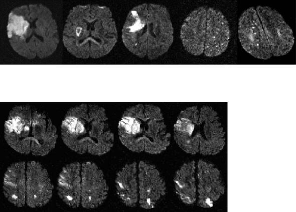
Fig 15.1. Diffusion-weighted imaging demonstrates different patterns of acute stroke in occlusive internal carotid artery disease: 1, territorial stroke; 2, subcortical stroke; 3, territorial stroke with fragmentation; 4, disseminated small lesions; 5, borderzone infarction
Fig. 15.2. Diffusion-weighted imaging in a 54-year-old patient with acute onset of severe left-sided hemiplegia shows a territorial infarction in the right middle cerebral artery territory, as well as additional bilateral hemodynamic lesions. Ultrasound examination in this patient showed high-grade internal carotid artery stenosis on both sides

228 |
|
|
|
K. Szabo and M. G. Hennerici |
Table 15.1. Diffusion-weighted imaging in symptomatic occlusive carotid disease |
|
|||
|
|
|
|
|
Study |
Number of |
Stenosis |
Lesions identified |
Conclusions |
|
patients |
grade |
|
|
Kastrup |
107 |
> 70% or |
• Single lesion (n=18) |
et al. (2002) |
|
occlusion |
• Multiple lesions (n=37) |
|
|
|
• No lesion (retinal or hemispheric |
TIA, n=52)
The majority of patients with symptomatic high-grade stenosis had multiple, frequently very small DWI lesions
Kang |
35 |
> 70% or |
• Territorial lesion (n=21) |
et al. (2002) |
|
occlusion |
• Borderzone lesion with or without |
|
|
|
a territorial lesion (n=10) |
|
|
|
• Bilateral hemispheric lesions (n=4) |
Szabo |
102 |
> 50 or |
• Territorial stroke (n=30) |
et al. (2001) |
|
occlusion |
• Subcortical stroke (n=13) |
|
|
|
• Territorial infarction with frag- |
mentation (n=11)
• Disseminated small lesions (n=15)
• Borderzone lesions (n=33)
Acute ischemic lesion in ICA occlusive disease is mainly multiple; borderzone infarction was mostly associated with territorial infarction
The degree of ICA stenosis may favor certain stroke patterns. In patients with high-grade stenosis the highest frequency of lesions occurs in the hemodynamic risk zones
phenomenon also confirmed by early experimental studies with primates (Brierley and Excell 1966). Borderzone infarcts were identified in brains of patients who died shortly after cardiac surgery and ascribed to episodes of abrupt arterial hypotension (Adams et al. 1966). In patients with occlusive ICA disease alike stroke lesions have been described and are believed to be caused by a similar, yet gradual compromise of cerebral perfusion, especially under the conditions of limited collateral circulation (Bogousslavsky 1986b). These so-called borderzones are divided into the superficial or cortical borderzones wedged between the territory of ACA and the MCA, or between the MCA and posterior cerebral artery (PCA) territory, and the deep or subcortical borderzone located in the vascular territory between deep and superficial arterial systems (Waterston et al. 1990; Bogousslavsky 1986a; Torvik 1984).
More recently, not only hemodynamic compromise, but also embolism was found to play an important role in the pathogenesis of borderzone infarcts. Caplan and Hennerici (1998) postulated the coexistence of hypoperfusion and intra-arterial embolism in borderzone infarcts and suggested that emboli lodged in hypoperfused regions may contribute to local ischemia and infarction. A similar theory was expressed by Pollanen and Deck (1989) who proposed that small thromboemboli may not be randomly distributed in the cerebral artery supply, but may be preferentially distributed to the small arterial branches of the borderzone due to anatomical features of vessel branches. Supporting the theory of microembolism, using the Doppler ultrasound phenomenon of “high intensity transient signals” (HITS) for detecting microemboli in the middle cerebral artery in patients with occlusive carotid disease, HITS were significantly more often
associated with severe (> 70%) (23.5%) than with moderate (50%–70%) internal carotid artery stenosis (3.4%) (Ries et al. 1996).
Yet, since the territories of the major cerebral arteries have been shown to possibly vary considerably, lesion localization alone may not be enough to identify borderzone lesions. Van der Zwaan and Hillen (1991) described different variations of the cortical distributions in all 25 human brains obtained at autopsy through injecting different colored substances into the six major arteries of the human brain. He also noted an inter-individual difference with remarkable asymmetrical territorial distribution in both hemispheres. With the clinical use of novel MRI techniques, however, it is possible to identify acute ischemic lesions in individual hemodynamic risk zones.
15.3.2
Diffusion-weighted and Perfusion Imaging Correlates of Hemodynamic Stroke
DWI is sensitive to changes of water molecule mobility and detects reduced proton mobility due to cytotoxic cell swelling, an early event in the cascade of ischemic tissue change, before T2-weighted MRI shows abnormality (LeBihan 1991; Sakuma et al. 1991; Tanner 1983). The regional reduction of diffusion is visible as hyperintensity on DWI images and as hypointensity on quantitative maps of the apparent diffusion coefficient (ADC) (see Chap. 7). DWI images provide a higher lesion-to-background contrast than conventional MRI facilitating lesion detection in early stages of stroke and allowing differentiation of acute and chronic tissue change. Current
Hemodynamic Infarcts and Occlusive Carotid Disease |
229 |
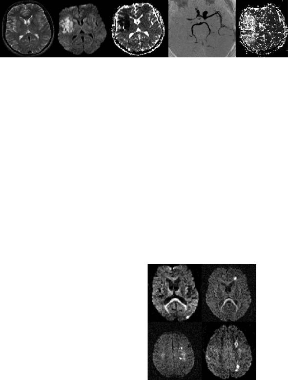
Fig. 15.3. Acute stroke MRI in a 34-year-old patient with acute-onset (1.5 h) right side hemiparesis. From left to right: the T2weighted image shows no definite parenchymal abnormality, only subtle signs such as lack of flow void in the MCA branches of the temporal region; the DWI image demonstrates the acute ischemic lesion involving the insular region of the right MCA territory with corresponding reduction of the ADC; the MIP-TOF MRA depicts signal loss in the occluded proximal right MCA and distal ICA, while the perfusion map (time-to-peak) demonstrates different grades of hypoperfused tissue in the complete MCA territory
evidence suggests that reduced diffusion is caused by cell swelling associated with cytotoxic edema formation, caused by failure of energy dependent pumps and influx of sodium and calcium, possibly accentuated by metabolic stress due to periinfarct depolarization. It is likely that both the narrowing of extracellular space and intracellular changes contribute to reduced diffusion. At present single shot echo planar (EP) MRI techniques are most widely used for DWI (Edelmann et al. 1994; Gass et al. 1999). Only a single nuclear spin excitation is required and a whole head (e.g., 24 slices, slice thickness of 5 mm) DWI study can typically be generated within 4–5 s. EP images are prone to specific artifacts (susceptibility artifact, N/2 or Nyquist ghost artifact, chemical shift artifact, eddy current artifact) that also need to be considered when interpreting DWI. Figure 15.3 shows an example of acute stroke MRI and the benefit of the different MRI techniques.
Figure 15.4 shows typical examples of hemodynamic acute stroke patterns in four different patients with carotid disease as identified by DWI. Lesions can be either located in the anterior or posterior superficial hemodynamic risk zone or in both, they can be located in the subcortical borderzones or, as in Fig. 15.5, they can affect all areas considered to be regions vulnerable to hemodynamic compromise. Commonly, these lesions are small and dot-like or confluent distributed in a chain-like fashion and can be either single or multiple. It is important to acknowledge that some of these lesions would probably be missed using conventional imaging techniques. Even though these stroke patterns bear very characteristic features and may allow an experienced clinician to suspect hemodynamic pathogenesis, with lesions located in the paraventricular region it is sometimes difficult to distinguish between superficial perforating artery and internal borderzone lesions. In
an interesting study Lee et al. (2003) compared MRI characteristics of these topographically welldefined types of deep cerebral infarcts and found that borderzone infarcts were larger, appeared in a chain-like distribution and had a higher degree of stenosis or occlusion of the ICA or the MCA, while superficial perforating artery strokes were more frequently accompanied by cortical spotty lesions and more frequent potential cardio-embolic sources.
For the exact description of hemodynamic stroke and identification of individual hemodynamic risk zones, as mentioned earlier, the assessment of the hemodynamic situation is essential. PI offers a means to obtain semiquantitative hemodynamic information in cerebral ischemia with relatively high resolution and short acquisition times covering all cerebral vascular territories. In acute cerebral ischemia dynamic susceptibility contrast
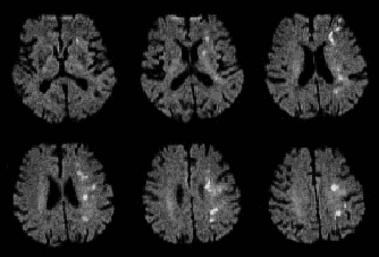
Fig. 15.4. DWI shows examples of acute borderzone lesions, namely in the posterior (upper row left) or anterior (upper row right) hemodynamic risk zones and in the subcortical or deep borderzone (bottom row)
230 |
K. Szabo and M. G. Hennerici |
Fig. 15.5. As in this 68-year-old man with a high-grade stenosis of the left internal carotid artery presenting with a fluctuating mild left-hemispheric syndrome, acute ischemic lesions can affect all areas considered to be hemodynamic risk zones
(DSC) MRI is the most widely evaluated technique (see Chap. 6). It uses information from transient local changes of magnetic field homogeneity (susceptibility effects) induced by the bolus passage of a paramagnetic contrast agent in dynamic T2*- weighted MRI acquisitions (Rosen et al. 1990). The extent of hemodynamically compromised tissue is readily provided by the temporal characteristics of the contrast bolus arrival in ‘time to peak’ or ‘mean transit time’ maps (Ostergaard et al. 1996; Röther et al. 1996). In the first few hours after stroke onset, PI abnormalities are often larger than the DWI lesions. This pattern (PI > DWI) is frequently associated with lesion growth into the PI/ DWI mismatch region and is therefore suggested to show “tissue at-risk” and is often found in critical carotid artery stenosis or occlusion (NeumannHaefelin et al. 2000). It is well established that impaired hemodynamics can be demonstrated with PI in patients with ICA disease. Compromise of autoregulation in patients with high-grade carotid stenosis has been demonstrated (Guckel et al. 1996; Maeda et al. 1999). As a compensatory mechanism of high-grade carotid stenosis, cerebral blood volume (CBV) maps may show a local increase of CBV. PI has been a reliable method for assessing perfusion changes in patients with unilateral carotid stenosis and patients with good collateral supply have been distinguished from those with poor collateral supply (Reith et al. 1997). Recently, large territorial perfusion deficits have been found with a predisposition to borderzone infarction in high-grade ICA stenosis (Chaves et al. 2000). The authors examined the MRI data of 17 patients with borderzone infarcts and identified
three patterns of perfusion abnormalities associated with the diffusion lesions. They found that the perfusion abnormality varied according to the mechanism of the borderzone infarction: (1) Transient perfusion deficits occurring with hypotension in the absence of significant large artery disease may not be revealed by PI; (2) embolism may cause some cases of small borderzone perfusion deficits; (3) critical large artery disease may cause large territorial perfusion deficits and predispose to borderzone infarction.
Frequently, visual analysis of DWI images allows the experienced reader to identify hemodynamic stroke patterns. However, this is not always reliable due to the fact that lesions are sometimes punctuated and small and it can be difficult to detect helpful anatomical landmarks on DWI. Sometimes only with help from PI is it possible to recognize lesions in the hemodynamic risk zones. Figure 15.6 gives an example of computerized superimposing of DWI and PI, to determine where the diffusion lesion is located with respect to the perfusion deficit. After aligning the time-to-peak and DWI images, it becomes clear that the lesions are indeed located in the borderzone area where the extent of the perfusion deficit is the greatest. Further studies to characterize these “dot-like” lesions with regard to the hypoperfused region are in the process and should shed new light on the role of microembolism in hemodynamically compromised regions. Figure 15.7 demonstrates two theoretical concepts of stroke in hemodynamic risk zones – microembolic lesions in the most distal arterial branches and the infarction of the compromised borderzone territory.