

Книги по МРТ КТ на английском языке / Magnetic Resonance Imaging in Ischemic Stroke - K Sartor R 252 diger von Kummer Tobias Back
.pdf210 |
J. M. Ferro |
does not include imaging information. The correspondence between the clinical classification and the imaging findings is not perfect, the positive predictive value ranging from 71 to 83 (Mead et al. 2000). Nevertheless the OCSP is very useful clinically because it is easy to communicate and predicts etiology (Mead et al. 1998), case fatality, functional recovery and risk of recurrence of stroke patients. TACIs have a highest mortality while LACIs have the lowest. The risk of recurrence is higher for PACI and POCI. The OSCP classification was validated in other populations (Wardlaw et al. 1996) and has a good inter-observer concordance (Lindley et al. 1993). The OSCP can also predict medical and neurological complications, therapeutic interventions and average cost of hospitalization (Pinto et al. 1998).
Impaired consciousness at onset or at presentation is the most powerful predictor of early death. Other predictors of death and disability include increasing age, male gender, previous dependency, dysphagia, pupil abnormality, gaze paresis, extensor plantars, dense hemiplegia, urine incontinence, breathing abnormalities, hypotension, hypertension, hypoglycemia, hyperglycemia (> 7 mmol/L), high hematocrit (> 50%), congestive heart failure, atrial fibrillation, medical complications, poststroke seizures, early deterioration, stroke severity as measured by a stroke scale (NIHSS 4–7 < 8–14 < 15–22), TACI subtype, extensive [> 1/3 middle cerebral artery (MCA) territory] early infarct size and mass effect on admission CT (Ebrahim and Harwood 1999a,b; Moulin et al. 2000; Warlow et al. 2001; Wolf 2002).
The TOAST classification is commonly used to classify ischemic stroke subtypes. However, the accuracy of the early clinical diagnosis of stroke subtypes is below 2/3 and is not significantly improved by CT findings which are often unrevealing. Early DWI has a higher detection rate of ischemic lesions than other imaging modalities. Ischemic lesion patterns on DWI can indicate a specific etiology such as carotid occlusion or cardioembolism. There is also an overall significant relationship between DWI lesion patterns and TOAST stroke subtypes (Kang et al. 2003): cor- tico-subcortical single lesions, multiple lesions in anterior and posterior circulation, multiple lesions in multiple vascular territories are associated with cardioembolism; unilateral multiple lesions in the anterior circulation and small scattered lesions in one vascular territory are related to large artery atherosclerosis. A total of 50% of subcortical
lesions being 15 mm or larger are cryptogenic or have a classical lacunar syndrome without cortical hypoperfusion.
In the first part of this chapter we will review the clinical-imaging patterns of acute territorial infarcts and their clinical relevance.
In the second part we will describe the clinical MR features of embolic stroke, with emphasis on cardioembolic stroke.
14.2
Clinical MR Patterns of Acute Territorial Infarcts
14.2.1
Middle Cerebral Artery (MCA) Infarcts
Middle cerebral artery (MCA) territorial infarcts are the most common type of ischemic stroke. They can be divided in superficial (involving the cortex and the underlying white matter), deep (involving the basal ganglia, the internal capsule and the deep white matter) and combined (Bogousslavsky and
Caplan 2001; Gorelick 1996).
MCA infarcts are mainly caused by cardioembolism, internal carotid artery (ICA) thrombosis, dissection or embolism and rarely (in Caucasians) by intrinsic MCA disease. MCA atherothrombotic territory infarctions related to intrinsic MCA disease often cause concomitant small cortical (territorial or borderzone) and subcortical infarcts (Min et al. 2000).
14.2.1.1
Superficial Middle Cerebral Artery (MCA) Infarcts
The most common stroke patterns of superficial (pial) MCA infarcts are those due to complete pial infarction and infarcts in the distribution of the anterior division and posterior division of the MCA. Clinical presentation differs depending on whether the left (dominant for language) or the right (nondominant) hemisphere is involved. In left hemispheric stroke the clinical picture is dominated by oral and written language disturbances, while after right MCA hemispheric strokes neglect is almost always present. The MCA has in general 12 pial branches and the syndromes corresponding to the infarcts in the distribution of such branches are also identifiable clinically.
Territorial and Embolic Infarcts
14.2.1.2
Large Middle Cerebral Artery (MCA) Infarcts
Large supratentorial infarcts covering at least two subterritories of the MCA (deep, superficial, anterior or superior, and posterior or inferior) carry an unfavorable vital and functional prognosis and produce a severe neurological deficit (gaze deviation, hemiplegia, global aphasia or neglect with anosognosia, hemianopia) including reduced consciousness from onset. They are caused by cardioembolism, ICA occlusion or ICA dissection (Heinsius et al. 1998). Cardioembolic stroke tends to involve the three subterritories simultaneously. Poor collateral blood flow is an additional predictor of lethal outcome (Schwarz et al. 2001).
14.2.1.3
MCA Anterior or Superior Division Infarcts
They produce a contralateral hemiparesis with predominant faciobrachial deficit, hemisensory loss, gaze deviation towards the lesion or decreased visual exploration toward the opposite side. Left-sided infarcts also produce a non-fluent aphasia, ranging from mutism to typical Broca’s aphasia and to articulatory, syntactical and naming difficulties. Buccofacial apraxia is very common. Neglect with anosognosia is an inconstant finding in right hemispheric strokes.
14.2.1.4
MCA Posterior or Inferior Division Infarcts
Motor deficits are absent, transient or mild. Hemisensory loss is more common and visual field defects (homonymous hemianopia or upper quadrantanopia) are usually present. In left-sided strokes, fluent aphasia of variable severity dominates the clinical picture. In the more severe form it is a Wernicke’s aphasia with anosognosia and behavioral disturbances including persecutory delusions. In right hemispheric stroke, neglect, anosognosia and an agitated confusional state usually occur. Constructional apraxia can be demonstrated.
14.2.1.5
Cortical Branch Syndromes
These infarcts can occur isolated or in combination. Infarcts in the distribution of the orbitofrontal and prefrontal arteries can produce a frontal syn-
211
drome. Left prefrontal infarcts can result in transcortical aphasia, while right prefrontal infarcts can produce motor neglect.
Precentral artery territory infarcts result in a hemiparesis with predominant proximal arm involvement. In left sided strokes, non-fluent aphasia, usually transcortical motor aphasia, apraxia and agraphia are present, while in right sided strokes, neglect and motor impersistence can be found.
Central sulcus or rolandic artery territory infarcts produce contralateral motor and sensory defects with face and arm predominance when the occlusion is proximal. If the occlusion is distal, motor deficit may be restricted to the arm or distal upper extremity. Cheiro-oral sensory loss may result from predominant involvement of the post central gyrus. Mild Broca’s aphasia (left sided strokes) combined with dysarthria and transient tongue, palatal and pharyngeal as well as faciobrachial weakness are a typical pattern of a central sulcus artery territory infarct. Bilateral central sulcus infarcts cause the cortical form of the anterior opercular syndrome (Foix-Chavany-Marie), with bilateral voluntary paresis of the facial, masticatory, lingual and pharyngeal muscles, featuring bilateral lower facial weakness, anarthria or severe dysarthria and dysphagia.
There are three parietal MCA branches: anterior, angular and posterior. Anterior parietal or postcentral sulcus artery infarct causes a contralateral sensory loss, with upper limb predominance (pseudothalamic syndrome) with involvement of the touch, pain, temperature and vibration senses. Pain and hyperpathia and parietal ataxia can also be present. Conduction aphasia, which is a fluent form of aphasia with disproportionate impairment of repetition, anomia, agraphia and apraxia are present in left hemispheric infarcts while neglect follows in right hemispheric ones.
Posterior parietal artery territory infarcts are rare and usually occur in combination with angular gyrus territory artery infarcts. These infarcts cause lower quadrantanopia, cortical sensation dysfunction with astereognosia and impairment of position sense, two-point discrimination and graphesthesia, but with sparing of light touch, vibration, pain and temperature senses. On the left side, fluent aphasia with alexia, Gerstmann’s syndrome (agraphia, acalculia, digitoagnosia and left–right disorientation) and ideomotor apraxia occur, while on the right, neglect and constructional apraxia result. If the upper parietal lobe is involved the patient shows elements of Balint’s syndrome, including disturbance in manual reaching (optic or visuo-motor ataxia), visual attention and
212 |
J. M. Ferro |
“psychic paralysis of gaze”, as they have difficulty in directing their gaze towards targets of interest and lose fixation easily.
There are usually five temporal branches of the MCA. Temporal MCA infarcts are often combined with lower posterior parietal infarcts. They cause upper quadrantanopia. Vertigo has occasionally been reported in association with superior temporal or insular infarcts. On the left side, the clinical picture is dominated by Wernicke’s aphasia. On the right, limited infarcts can be pauci-symptomatic. Extended infarcts cause amusia, neglect, acute agitation and confusion and sometimes delusions and manic symptoms.
14.2.2
Subcortical Hemispheric Infarcts
These include infarcts in the territory of: (1) the deep perforators of the MCA, anterior cerebral artery (ACA) and posterior cerebral artery (PCA), posterior communicating artery (PcomA), the lenticulostriate arteries and the anterior choroidal artery; (2) the superficial perforators (white matter medullary branches) of the superficial pial arteries; (3) borderzone or junctional infarcts between 1 and 2; (4) combined infarcts. Small (< 1.5 mm infarcts – lacunes) are usually caused by single perforator disease while larger infarcts have a more diverse pathophysiology including embolism and MCA stenosis (Bang et al. 2002).
MCA stenosis causes subcortical stroke either by occlusion of a single penetrating artery to produce a small lacunar infarct or by artery to artery embolism with impaired clearance of emboli that produces multiple small cerebral infarcts, mainly in borderzone regions (Wong et al. 2002).
Large striato-capsular infarcts produce a clinical picture similar to that of pial infarcts overlying their territory, including non-fluent aphasia and neglect. These cortical signs may be due to cortical diaschisis, cortical hypoperfusion or scattered cortical lesions not apparent on CT. These additional lesions may be evident on MR, in particular in DWI (Singer et al. 1998). In the MCA territory, the most common combination is a deep small striato-capsular infarct and a distal small cortical infarct. In large subcortical infarcts, cortical perfusion defects are apparent on MR perfusion imaging (PI) (Gerraty et al. 2002), while they are not detected in single perforator infarcts (lacunes). However, patients with lacunes can have subsidiary cortical lesions in DWI.
Subcortical white matter infarcts may mimic a superficial MCA infarct causing a partial anterior circulation syndrome or present as a lacunar syndrome (pure motor, ataxic hemiparesis or sensori motor stroke). Superficial perforating artery infarcts (medullary branches) are often accompanied by cortical spotty lesions. Borderzone and white matter medullary branches infarctions are usually caused by hypoperfusion due lo large vessel occlusion or stenosis (Bogousslavsky 1993; Donnan and Yasaka
1998), but white matter medullary branches infarction can also be caused by cardioembolism (Lee et al. 2003).
14.2.2.1
Anterior Choroidal Artery Infarcts
Aphasia and neglect can be found following respectively dominant and non-dominant anterior choroidal artery infarcts. Anterior choroidal artery infarcts usually cause the classical 3H syndrome: hemiparesis, hemihypesthesia, hemianopia. Pure motor hemiparesis and isolated hemianopia can also occur (Han et al. 2000). Anterior choroidal artery territory infarcts are rarely caused by small vessel occlusion. In general they are caused by cardioembolism or large artery disease with occlusion or artery-to-artery embolism (Leys et al. 1994).
Recently, using color-coded diffusion tensor imaging five different patterns of corticospinal tract stroke were identified that fall into two clinical subgroups with either little recovery or good recovery. Patients with poor motor recovery had lesions centered in the pyramidal tract (anterior choroidal artery). Patients with good recovery had either very small lesions or lesions located anteriorly or medially (Lie et al. 2004).
14.2.2.2 Thalamic Infarcts
There are four main types of large thalamic infarcts:
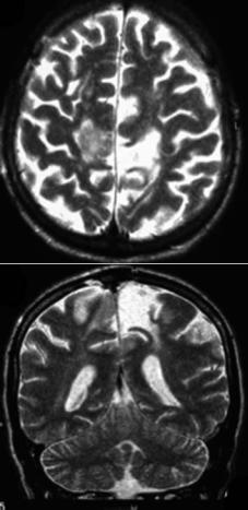
(1) Tuberothalamic (anterior or polar), in the territory of the tuberothalamic artery that arises from the posterior communicating artery; (2) Paramedian (thalamoperforate or posterior thalamic-sub- thalamic), in the territory of the paramedian artery that originates from the P1 segment of the PCA (Fig. 14.1); (3) Inferolateral (thalamogeniculate), in the territory of the inferolateral artery that originates from the P2 segment of the PCA; (4) Posterior

Territorial and Embolic Infarcts |
213 |
Fig. 14.1. Left paramedian thalamic infarct
(posterior choroidal artery) which arises from the P2 segment of PCA (Bogousslavsky et al. 1988; Schmahmann 2003). Sometimes two territories are involved simultaneously. Bilateral infarcts, in particular tuberothalamic or paramedian, are not infrequent. They may be due to simultaneous emboli to a common unilateral origin of the arterial branches supplying the anterior and the paramedian thalamus. The four major topographical patterns of bilateral thalamic infarcts are: bilateral paramedian, bilateral inferolateral, combined unilateral paramedian and inferolateral and combined unilateral tuberothalamic and inferolateral. Panthalamic infarcts, involving the four arterial territories of the thalamus are exceptional. Occlusion of the PCA in the absence of an ipsilateral posterior communicating artery may explain this unusual type of arterial infarct (Studer et al. 2003).
Inferolateral or thalamogeniculate infarcts usually present as a hemisensory loss with pain or dysesthesia, plus hemiataxia (Melo et al. 1992) [hemi- ataxia-hypesthesia (Melo and Bogousslavsky 1992), hypesthetic ataxic hemiparesis and ataxic hemiparesis] and involuntary movements (chorea or dystonia). Tuberothalamic infarcts usually cause drowsiness, apathy, executive deficits, personality changes, amnesia and aphasia or neglect (depending on the involved side). In paramedian infarcts drowsiness is prominent. Impaired memory and learning, altered social skills and personality are also apparent. Oculomotor troubles may be present (Bogousslavsky and Caplan 1993;
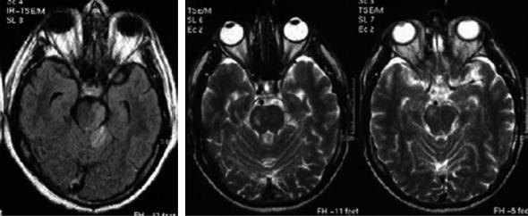
Fig. 14.2. Complete right posterior cerebral artery (PCA) territorial infarct. Notice the anterolateral mesencephalic and the inferolateral thalamic infarcts. Old left striatocapsular infarct
Bogousslavsky et al. 1988). Posterior choroidal artery infarcts result in visual field defects, variable sensory loss, hemiparesis, dystonia, hand tremor and occasionally amnesia and aphasia. Visual field defects, commonly quadrantanopia or hemianopia, are found if the lateral posterior choroidal arteries branches are involved in isolation. Unusual field defects such as homonymous horizontal sectoranopia or wedge shaped homonymous hemianopia, may also be found and may be explained by the dual blood supply to the lateral geniculate body by
214 |
J. M. Ferro |
the lateral posterior choroidal arteries and the anterior choroidal artery (Saeki et al. 1999). Bilateral thalamic infarcts cause a typical and devastating clinical picture of drowsiness, amnesia, abulia and vertical eye movement disorders. The neuropsychological and psychic changes tend to be permanent, causing a strategic infarct vascular dementia syndrome (Kumral et al. 2001).
14.2.3
Posterior Cerebral Artery (PCA) Infarcts
The predominant clinical findings of PCA infarcts are hemianopia or other visual field defects. Headache is rather frequent, in general unilateral and may be severe. PCA infarcts can be superficial (cortical) (Cals et al. 2002) or combined with lateral thalamic or rostral mesencephalic infarcts (Fig. 14.2). The thalamic infarct produces a sensory defect in variable combination with ataxia or involuntary movements. The midbrain lesion is usually limited and responsible for a transient motor deficit. More extensive midbrain lesions will produce vigilance and oculomotor troubles. Sensory abnormalities in PCA stroke are associated with ventrolateral thalamic infarcts in the thalamogeniculate or lateral posterior choroidal arteries or less frequently to ischemia to the white matter tracts to the somatosensory motor cortex (Georgiadis et al. 1999). The presence of hemiparesis in PCA infarcts can make the clinical differential diagnosis with MCA infarcts difficult. Hemiparesis can be due, as mentioned above, to the infarction of the cerebral peduncle and less frequently to infarction of the anterior segment of the posterior limb of the internal capsule, due to impaired perfusion in the distribution of the inferolateral thalamic branches of the PCA, which can include that part of the internal capsule (Montavont et al. 2003). The inferior striate cortex is more susceptible to ischemia due to poor collateral circulation. Therefore superior quadrantanopia is more common than inferior one. Neuropsychological manifestations are common and, when the dominant (usually left) hemisphere is damaged, include: (1) transcortical sensory aphasia or anomic aphasia, (2) alexia with or without agraphia; the lesion producing alexia without agraphia combines damage to the left calcarine cortex and to splenium of the corpus callosum or to the outflow of the callosum originating from the right visual cortex, (3) visual or color agnosia; infarcts causing persistent associative visual agnosia are usually large and
involve the parahippocampal and fusiform gyrus. On the non-dominant side neglect, prosopagnosia (often transient) and topographical amnesia can be found. On either side visual perseverations, visual illusions or hallucinations and agitation can occur. Declarative memory defects, concerning verbal (left sided infarcts), visual (right sided infarcts) material or both are usually present. Such infarcts usually involve the hippocampus. Bilateral occipitotemporal infarcts can cause cortical blindness without or with anosognosia (Anton’s syndrome), associative or apperceptive visual agnosia and severe memory defect (Brandt et al. 2000; Cals et al. 2002). PCA infarcts are due to cardiac embolism in about 1/3 of patients. Significant vertebrobasilar atheroma with occlusion or artery-to-artery embolism accounts for additional 25%. Local PCA stenosis or occlusions are much less frequent. In many PCA infarcts, embolism is suspected but cannot be confirmed and the cause remains undetermined. Complete infarction of the posterior branches of the PCA and hemorrhagic transformation are more frequent in cardioembolic strokes (Steinke et al. 1997). Migraine account for a few cases (migrainous infarcts). MELAS stroke-like events are predominantly located in the occipital lobes, but do not respect the territorial borders of PCA distribution. Anatomical variations may predispose to PCA infarct. Exclusive supply of the PCA territory via the carotid system; a patent posterior communicating artery with anteroposterior flow are less common in patients with PCA infarcts than in controls (Jongen et al. 2004).
14.2.4
Anterior Cerebral Artery (ACA) Infarcts
Strokes in the ACA territory are uncommon (< 2% in stroke registries). Left sided infarcts cause mutism, transcortical motor aphasia, hemiparesis and occasionally left arm apraxia and other callosal disconnection syndromes. Right-sided infarcts cause acute confusional state, hemiparesis and motor neglect (Kumral et al. 2002d). Hemiparesis predominates in the lower limb when the precentral gyrus is involved. If the infarction extends more posteriorly sensory loss in the leg can be found. In some cases the paresis may be proportional and the ACA stroke pattern will be indistinguishable from an MCA pattern. Proportional hemiparesis occurs when there is occlusion of the recurrent artery of Heubner that supplies the internal capsule. Abulia can be present with unilateral or bilateral infarcts that involve the
Territorial and Embolic Infarcts |
215 |
cingulum and the supplementary motor area. Bilateral infarcts may produce akinetic mutism (Wolff et al. 2002), gait apraxia (Della Sala et al. 2002), paraparesis and sphincter dysfunction and carry a bad prognosis for functional recovery (Fig. 14.3). Grasp reflex, utilization behavior (Boccardi et al. 2002) and executive deficits may be more or less prominent. Hand grasping correlates with orbitofrontal lesions. An expanding ACA infarct was recently reported (Ay et al. 2002).
14.2.5
Combined Hemispheric Infarcts
Combined ACA-MCA infarcts can result from embolic occlusion of the distal ICA (carotid T). Combined MCA-PCA infarcts may be due to simultaneous embolism to the two territories, to a fetal (internal carotid) origin of the PCA or to compression of the PCA by a herniating hemisphere at the edge of the tentorial foramen.
These combined infarcts, as well as massive MCA infarcts, are often complicated within 24 to 96 h from onset by edema which will produce mass effect. In young and middle-aged patients, this may cause clinical deterioration and death due to brain stem compression (malignant MCA infarct). Predictors of fatal brain edema include > 50% MCA hypodensity, involvement of additional vascular territories (ACA; PCA; anterior choroidal artery), hypertension and heart failure and increased white blood cell count (Kasner et al. 2001). Such patients are candidates to aggressive live saving treatments such as decompressive hemicraniectomy and hypothermia.
14.2.6
Brain Stem Infarcts
14.2.6.1 Mesencephalic Infarcts
The mesencephalon has four arterial territories: anteromedial (paramedian branches of the basilar artery; anterolateral (branches from the P2 segment of the PCA); lateral (branches from P2 segment of PCA and from posterior choroidal arteries) and dorsal (branches from P1 segment of PCA and superior cerebellar artery). Isolated mesencephalic infarcts are rare because the arteries supplying blood to the mesencephalon (basilar artery, posterior cerebral artery and superior cerebellar artery)
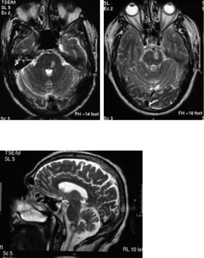
Fig. 14.3. Bilateral (acute and non-recent) anterior cerebral artery infarcts causing paraparesis
also serve other infratentorial and supratentorial anatomical sites (Fig. 14.4). Midbrain infarcts are more likely to be accompanied by infarcts in other structures than to occur alone (Martin et al. 1998; Kumral et al. 2002b). Patients with mesencephalic infarcts can be classified into four groups:
(1) those with isolated mesencephalic infarcts, and those whose mesencephalic infarct is accompanied by (2) proximal infarcts involving the medulla and the posterior inferior cerebellar artery (PICA) territory (Kumral et al. 2002a), by (3) “middle” infarcts including the pons and the anterior inferior cerebellar artery (AICA), and by (4) distal lesions comprising the thalamus and the PCA territory (Martin et al. 1998). “Middle” infarcts are the most common. Patients with isolated mesencephalic infarcts have a combination of unsteadiness/dizziness, diplopia due to ipsilesional 3rd cranial nerve palsy, vertical gaze palsy, contralateral or four-limb ataxia, hemiparesis
216 |
J. M. Ferro |
Fig. 14.4. Mesencephalic infarcts: anterolateral (right); extensive combined anteromedial and anterolateral involving also the vermis (left)
or hypesthetic ataxic hemiparesis or a combination of both and involuntary movements (tremor and chorea-like). Patients with isolated upper or lower midbrain infarcts have no localizing clinical findings, but patients with middle midbrain infarcts had a localizing clinical picture mainly with III nerve palsies that often occur in isolation (Bogousslavsky et al. 1994). The palsy of the 3rd cranial nerve can be nuclear or fascicular. A unilateral 3rd cranial nerve nuclear lesion causes bilateral ptosis and superior rectus weakness, divergent strabismus and mydriasis. In fascicular lesions there is unilateral findings with divergent strabismus, unilateral ptosis and mydriasis. The classic clinical picture of Claude’s syndrome (ipsilesional 3rd cranial nerve palsy and contralateral ataxia) or Benedikt’s syndrome (3rd cranial nerve palsy plus contralateral involuntary movements) are very rare. “Distal” mesencephalic infarcts are associated with disturbances of consciousness, gait ataxia, oculomotor disturbances and visual field defects. “Middle” strokes have disturbances of consciousness, dysarthria, horizontal oculomotor disorders and paresis, while “proximal” infarcts produce acute unsteadiness, vertigo, dysphagia, dysphonia, limb ataxia and paresis. Patients with mesencephalic infarcts can present involuntary repetitive stereotyped movements that may be confused with seizures (Saposnik and Caplan 2001; Lee et al. 2002). About 25% of midbrain strokes are bilateral (Kumral et al. 2002b). Such bilateral infarcts have a poorer outcome than unilateral infarcts. Large artery disease, producing artery-to- artery embolism or in situ thrombosis are the most
common mesencephalic infarct mechanisms. Cardioembolism and small vessel disease account for 25% of the cases each.
14.2.6.2 Pontine Infarcts
Pontine infarcts (Kumral et al. 2002c) can have five main clinical patterns: anteromedial, anterolateral, tegmental, unilateral multiple and bilateral. (Fig. 14.5). The anteromedial and anterolateral territories are supplied from the basilar artery, while the lateral territory also receives blood from the superior and anterior inferior cerebellar arteries. The small posterior territory is supplied by the superior cerebellar artery. The most frequent syndrome is the anteromedial pontine syndrome presenting with motor deficit with dysarthria, ataxia plus mild tegmental symptoms in 1/3 of the patients. The second most common is the anterolateral pontine syndrome, featuring motor-sensory deficits associated with tegmental signs in more than half of the patients. Tegmental pontine syndromes (mild motor and sensory deficits together with eye movement and vestibular symptoms/signs) are less common. Unilateral multiple pontine infarcts are always associated with severe sensorimotor deficits and tegmental signs. Bilateral infarcts cause an ominous clinical pattern of transient loss of consciousness, tetraparesis, pseudobulbar palsy and, in some cases, locked-in syndrome. The main stroke mechanisms of pontine infarcts are basilar artery branch and
Territorial and Embolic Infarcts |
217 |
Fig. 14.5. Pontine infarcts: anteromedial (right) and tegmental (left)
Fig. 14.6. Lateral medullary infarct causing a Wallenberg’s syndrome
small artery disease occlusions (Kumral et al. 2002a).
14.2.6.3 Medullary Infarcts
Medullary infarcts can be medial, lateral or combined (Fig. 14.6). The medial territory is supplied by penetrating vessels from the anterior spinal artery and the distal vertebral artery. The lateral territory main arterial supply comes from penetrating arteries from the distal vertebral artery and the posterior inferior cerebellar artery. The small posterior territory is supplied by the posterior spinal artery and the posterior inferior cerebellar artery. Medial
medullary infarcts have four major clinical patterns:
(1) Dejerine’s syndrome (contralateral hemiparesis and pain/thermal sensory loss plus ipsilateral lingual palsy, (2) sensorimotor stroke without lingual palsy, (3) hemiparesis, often combined with nystagmus, (4) bilateral syndromes. Lateral infarcts produce a more or less complete Wallenberg’s syndrome: ipsilateral paralysis of the 9th and 10th cranial nerves, loss of pain and temperature sense on the face, ataxia, vestibular signs (nystagmus, ipsilesional lateropulsion) and Horner’s syndrome, and contralateral dissociated hemianesthesia. Nausea and vomiting, hiccups and headache are frequent. Diplopia and oscillopsia are common complaints. Medial medullary infarcts are often accompanied by a cerebellar infarct. The most common mechanisms are vertebral artery thrombosis or dissection, and embolism. Combined (hemi-medullary) infarcts cause a Babinski-Nageotte syndrome (all the components of the Wallenberg syndrome plus ipsilateral tongue palsy and contralateral hemiplegia). They are usually secondary to occlusion of the vertebral artery.
14.2.6.4
Stroke Patterns in Basilar Artery Occlusion
The most feared vertebrobasilar stroke is occlusion of the basilar artery. Patients with lesions in the basilar artery are five times more likely to have a poor outcome independent of other factors (Glass et al. 2002). The importance of its early recognition is the possibility of performing intra-arterial throm-
218 |
J. M. Ferro |
bolysis. Basilar artery thrombosis may present as a locked-in syndrome, as a midbrain syndrome, a top- of-the-basilar syndrome or as partial syndromes. Basilar artery occlusion can have a sudden onset without warning signs, causing within minutes a dramatic and disabling neurological picture. More often it has a subacute onset with heralding TIAs or a progressive course with worsening or additive neurological deficits. The final neurological picture is reached within 6 h in about 1/3 of the patients, in 6–24 h in another 1/3 and up to 72 h in the remaining. The most common initial symptoms are motor weakness, dysarthria, vertigo, nausea/vomiting, and headache. The most common initial signs are motor deficits, facial palsies, eye movement abnormalities, lower cranial nerve deficits, altered level of consciousness and bilateral extensor plantar responses (von Campe et al. 2003). The prognosis of basilar artery occlusion is diverse. The outcome is invariably poor in patients with disorders of consciousness or a combination of dysarthria, pupillary disorders and involvement of the lower cranial nerves, while if these factors are absent only 11% have a poor outcome (Devuyst et al. 2002). The typical syndrome of basilar artery embolism (Schwarz et al. 1997) is an acute loss of consciousness followed by multiple brainstem symptoms and infarcts at several levels (thalamus, PCA, midbrain, pons). Usually, clinical symptoms improve quickly and even completely. Outcome correlates with the intensity of consciousness disorder and the number of infarcts.
14.2.7
Cerebellar Infarcts
Cerebellar infarcts can be grouped in territorial (superior cerebellar artery, anterior inferior cerebellar artery, posterior inferior cerebellar artery and combined), borderzone and lacunar. They are often combined with brain stem infarcts and with superficial posterior cerebral artery or thalamic infarcts. The most common isolated cerebellar infarcts are located in the superior cerebellar artery and posterior inferior cerebellar artery territories (Amarenco 1993; Amarenco et al. 1993, 1994).
14.2.7.1
Superior Cerebellar Artery (SCA)
by PCA or brainstem infarcts. They can be edematous and have a malignant course. Partial infarcts are often pure cerebellar infarcts and have a benign course. Both are usually cardioembolic. Their clinical presentation ranges from severe, dramatic patterns such as coma with tetraplegia and oculomotor disturbances due to simultaneous brain stem infarct, or top-of-the-basilar syndrome with concomitant involvement of the mesencephalon and PCA territories, to more limited and benign presentations. The classical Mills-Guillain syndrome (ipsilateral cerebellar ataxia and Horner’s syndrome and contralateral pain/temperature sensory loss and 4th cranial nerve palsy) is rare and in fact due to concomitant involvement of the pontine territory of the SCA. Tremor and other involuntary movements can be present and are due to ischemia of the superior cerebellar peduncle. Other patients have only a cerebello-vestibular syndrome. Dysarthria is almost always present. Lateral SCA infarcts involve the anterior rostral cerebellum and cause ipsilateral limb ataxia and lateropulsion and dysarthria. Medial SCA infarcts are less well described and cause ataxia and dysarthria.
14.2.7.2
Anterior Inferior Cerebellar Artery (AICA)
Symptoms of AICA territory infarct are related to damage to the inferolateral pons, the middle cerebellar peduncle and the flocculus. AICA infarcts are small and less frequent than the other cerebellar territorial infarcts. They almost always have a concomitant pontine infarct and their main cause is basilar artery occlusion, hence the importance of their recognition. Their clinical presentation has four major patterns: (1) coma with tetraplegia due to pontine infarct, (2) the classic AICA syndrome, which is the most frequent presenting pattern featuring ipsilateral involvement of the 5th, 7th and 8th cranial nerves with vertigo, vomiting, tinnitus, facial palsy and facial sensory loss, Horner’s syndrome and appendicular ataxia and contralateral temperature/pain sensory loss, (3) pure vestibular syndrome, including exceedingly rare cases of isolated vertigo, and (4) isolated cerebellar signs.
14.2.7.3
Posterior Inferior Cerebellar Artery (PICA)
The SCA territory has two divisions: lateral and medial. Full SCA infarcts are usually accompanied
The classic clinical pattern of PICA territorial infarct is the Wallenberg’s syndrome. However, symptoms
Territorial and Embolic Infarcts |
219 |
and signs in Wallenberg’s syndrome are due to the lateral medullary infarct, which can also be due to occlusion of branches arising directly from the vertebral artery. The clinical pattern of PICA infarcts is therefore dependent on whether or not the medulla is also involved. Infarcts restricted to the cerebellum are in general small and have a benign course. They can involve the medial and less often the lateral PICA subterritories. PICA infarcts combined with AICA or SCA can assume a pseudotumoral form or present as coma with tetraplegia. If the medulla is involved, a more or less complete dorsal lateral medullary syndrome results: ipsilesional vestibular (vertigo, vomiting, nystagmus, lateropulsion), 5th, 9th, 10th cranial nerve palsies, Horner’s syndrome and appendicular ataxia with contralesional loss of pain and temperature sensation. Hiccups are frequent and prolonged. If the medulla is spared, patients present with headache, vertigo, nystagmus, ipsilateral axial lateropulsion, gait and appendicular ataxia. There are a few reported cases of infarcts involving the nodulus and causing an isolated acute vertigo mimicking a vestibular disease. Medial PICA infarcts have a triangular shape on the dorsomedial rostral cerebellum with a ventral top pointing towards the 4th ventricle. They can produce: isolate vertigo; vertigo, axial lateropulsion and dysmetria; Wallenberg’s syndrome, if the medulla is also involved. Lateral PICA infarcts are very rare and present with vertigo and ipsilateral dysmetria.
14.2.7.4
Pseudotumoral Cerebellar Infarcts
Some large cerebellar infarcts can produce mass effect and lead to clinical deterioration and eventual death. These patients usually have combined territorial cerebellar infarcts, a full territorial PICA or SCA infarct, or infarcts confined to the medial vermian branches of the PICA or SCA. Isolated lateral hemispheric branches of the PICA or SCA and infarcts confined to the AICA territory do not produce clinically relevant mass effect. Pseudotumoral cerebellar infarcts are usually large (> 1/3 of the cerebellar hemisphere). The most frequent mechanisms of infarction are large vessel occlusion due to large artery disease or cardioembolism with reperfusion. Such patients progress from a cerebellar syndrome to a stage where brain stem signs develop and the level of consciousness fluctuates. If untreated, the patient will progress to coma. These infarcts with mass effect cause increased intracra-
nial pressure in the posterior fossa with compression of the basal cisterns and brain stem, hydrocephalus and/or downward or more rarely upward herniation. Clinical deterioration may start hours or days after stroke onset. Decompressive surgery is life-saving (Amarenco 1991; Hornig et al. 1994;
Rieke et al. 1993; van Horn and Hawes 1982; Koh et al. 2000).
14.2.8
Acute Multiple Brain Infarcts (AMBI)
DWI revealed that acute multiple infarcts were much more frequent than expected clinically or on CT (Altieri et al. 1999). In the vertebro-basilar circulation the commonest form of acute multiple infarcts is the top-of-the basilar syndrome due to embolism of the junction of the basilar and the PCA producing occipital, thalamic and midbrain (or midbrainsuperior cerebellar) lesions. Multi-level vertebrobasilar strokes can be related to cardioembolism or artery-to-artery embolism. Multiple, bilateral infarcts strongly suggest cardioembolism and are occasionally the herald of infective endocarditis (Timsit et al. 1992). Unilateral combined MCA-PCA or MCA-ACA infarcts are more often due to internal carotid artery (ICA) occlusion or embolism. Certain clinical syndromes are suggestive of simultaneous double ipsilateral infarction secondary to ICA occlusion: ipsilateral ocular ischemia (retinal or anterior) plus ipsilateral hemispheric infarct (optocerebral syndrome); hemiplegia-hemianopia syndrome and conduction aphasia with hemiparesis, due to combined anterior or deep MCA plus posterior MCA affection; mixed transcortical aphasia (isolation of the speech areas) results from the combination of anterior and posterior borderzone infarcts. Acute global aphasia without hemiparesis results from embolism to the anterior and posterior speech areas. Acute anarthria and pseudobulbar syndrome can be secondary to bi-opercular infarcts related to bilateral ICA occlusion (Kumral 2001).
Different topographical patterns of AMBI are associated with different vascular pathologies. Hemorheologic abnormalities or vascular anatomic variations may be contributing factors of AMBI in both hemispheres or in both the anterior and the posterior circulation (Roh et al. 2000). A scattered lesion pattern on DWI in patients with an initial negative CT is indicative of an arterial or embolic source and associated with favorable clinical outcome (Koennecke et al. 2001). Occlusion of the